

STIM1 gates TRPC channels, but not Orai1, by electrostatic interaction
- PMID: 18995841
- PMCID: PMC2586614
- DOI: 10.1016/j.molcel.2008.09.020
STIM1 gates TRPC channels, but not Orai1, by electrostatic interaction
Abstract
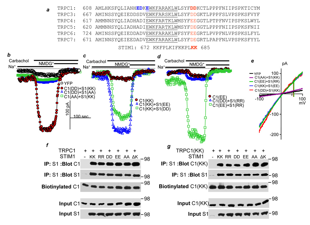
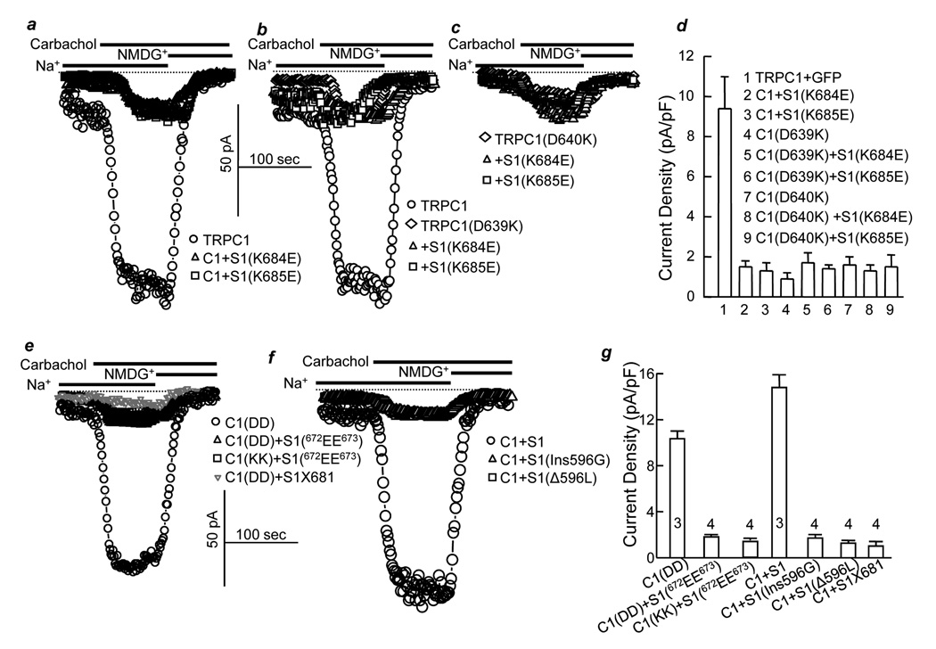
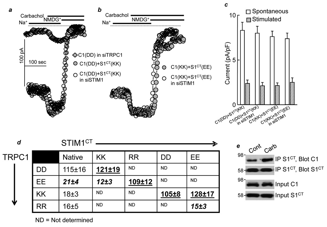
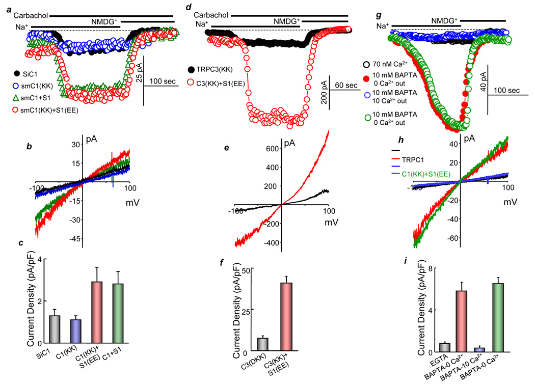
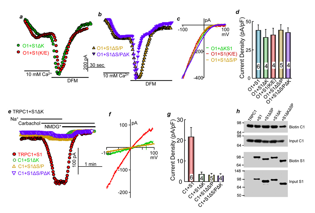
The receptor-evoked Ca(2+) signal includes activation of the store-operated channels (SOCs) TRPCs and the Orais. Although both are gated by STIM1, it is not known how STIM1 gates the channels and whether STIM1 gates the TRPCs and Orais by the same mechanism. Here, we report the molecular mechanism by which STIM1 gates TRPC1, which involves interaction between two conserved, negatively charged aspartates in TRPC1((639)DD(640)) with the positively charged STIM1((684)KK(685)) in STIM1 polybasic domain. Charge swapping and functional analysis revealed that exact orientation of the charges on TRPC1 and STIM1 are required, but all positive-negative charge combinations on TRPC1 and STIM1, except STIM1((684)EE(685))+TRPC1((639)RR(640)), are functional as long as they are reciprocal, indicating that STIM1 gates TRPC1 by intermolecular electrostatic interaction. Similar gating was observed with TRPC3((697)DD(698)). STIM1 gates Orai1 by a different mechanism since the polybasic and S/P domains of STIM1 are not required for activation of Orai1 by STIM1.
Figures
Similar articles
-
Molecular determinants mediating gating of Transient Receptor Potential Canonical (TRPC) channels by stromal interaction molecule 1 (STIM1).J Biol Chem. 2014 Mar 7;289(10):6372-6382. doi: 10.1074/jbc.M113.546556. Epub 2014 Jan 24. J Biol Chem. 2014. PMID: 24464579 Free PMC article.
-
STIM1 carboxyl-terminus activates native SOC, I(crac) and TRPC1 channels.Nat Cell Biol. 2006 Sep;8(9):1003-10. doi: 10.1038/ncb1454. Epub 2006 Aug 13. Nat Cell Biol. 2006. PMID: 16906149
-
TRPC channels as STIM1-regulated SOCs.Channels (Austin). 2009 Jul-Aug;3(4):221-5. doi: 10.4161/chan.3.4.9198. Epub 2009 Jul 15. Channels (Austin). 2009. PMID: 19574740 Review.
-
Local Ca²+ entry via Orai1 regulates plasma membrane recruitment of TRPC1 and controls cytosolic Ca²+ signals required for specific cell functions.PLoS Biol. 2011 Mar;9(3):e1001025. doi: 10.1371/journal.pbio.1001025. Epub 2011 Mar 8. PLoS Biol. 2011. PMID: 21408196 Free PMC article.
-
The TRPCs-STIM1-Orai interaction.Handb Exp Pharmacol. 2014;223:1035-54. doi: 10.1007/978-3-319-05161-1_13. Handb Exp Pharmacol. 2014. PMID: 24961979 Review.
Cited by
-
TRPC4 as a coincident detector of Gi/o and Gq/11 signaling: mechanisms and pathophysiological implications.Curr Opin Physiol. 2020 Oct;17:34-41. doi: 10.1016/j.cophys.2020.06.008. Epub 2020 Jul 2. Curr Opin Physiol. 2020. PMID: 32851198 Free PMC article.
-
P2Y receptor subtypes evoke different Ca2+ signals in cultured aortic smooth muscle cells.Purinergic Signal. 2012 Dec;8(4):763-77. doi: 10.1007/s11302-012-9323-6. Epub 2012 Jul 6. Purinergic Signal. 2012. PMID: 22767215 Free PMC article.
-
Orai1 calcium channels in the vasculature.Pflugers Arch. 2012 Apr;463(5):635-47. doi: 10.1007/s00424-012-1090-2. Epub 2012 Mar 9. Pflugers Arch. 2012. PMID: 22402985 Free PMC article. Review.
-
B-lymphocyte calcium influx.Immunol Rev. 2009 Sep;231(1):265-77. doi: 10.1111/j.1600-065X.2009.00822.x. Immunol Rev. 2009. PMID: 19754903 Free PMC article. Review.
-
Stromal Interaction Molecule 1 (STIM1) Regulates ATP-sensitive Potassium (KATP) and Store-operated Ca2+ Channels in MIN6 β-Cells.J Biol Chem. 2017 Feb 10;292(6):2266-2277. doi: 10.1074/jbc.M116.767681. Epub 2016 Dec 21. J Biol Chem. 2017. PMID: 28003364 Free PMC article.
References
-
- Ambudkar IS, Ong HL, Liu X, Bandyopadhyay B, Cheng KT. TRPC1: the link between functionally distinct store-operated calcium channels. Cell Calcium. 2007;42:213–223. - PubMed
-
- Berridge MJ, Bootman MD, Roderick HL. Calcium signalling: dynamics, homeostasis and remodelling. Nat Rev Mol Cell Biol. 2003;4:517–529. - PubMed
-
- Chang WC, Di Capite J, Singaravelu K, Nelson C, Halse V, Parekh AB. Local Ca2+ influx through Ca2+ release-activated Ca2+ (CRAC) channels stimulates production of an intracellular messenger and an intercellular pro-inflammatory signal. The Journal of biological chemistry. 2008;283:4622–4631. - PubMed
-
- Chang WC, Nelson C, Parekh AB. Ca2+ influx through CRAC channels activates cytosolic phospholipase A2, leukotriene C4 secretion, and expression of c-fos through ERK-dependent and -independent pathways in mast cells. Faseb J. 2006;20:2381–2383. - PubMed
-
- Chang WC, Parekh AB. Close functional coupling between Ca2+ release-activated Ca2+ channels, arachidonic acid release, and leukotriene C4 secretion. The Journal of biological chemistry. 2004;279:29994–29999. - PubMed
Publication types
MeSH terms
Substances
Grants and funding
- DA00266/DA/NIDA NIH HHS/United States
- R37 DA010309/DA/NIDA NIH HHS/United States
- R01 DE013902-07/DE/NIDCR NIH HHS/United States
- R01 DA010309/DA/NIDA NIH HHS/United States
- DE12309/DE/NIDCR NIH HHS/United States
- R01 DK038938-23/DK/NIDDK NIH HHS/United States
- MH068830/MH/NIMH NIH HHS/United States
- DA10309/DA/NIDA NIH HHS/United States
- P50 DA000266/DA/NIDA NIH HHS/United States
- R01 DE013902/DE/NIDCR NIH HHS/United States
- P50 MH068830/MH/NIMH NIH HHS/United States
- R01 DE012309/DE/NIDCR NIH HHS/United States
- R01 DK038938/DK/NIDDK NIH HHS/United States
- DK38938/DK/NIDDK NIH HHS/United States
LinkOut - more resources
Full Text Sources
Molecular Biology Databases
Miscellaneous
