. 2009 Feb 15;69(3):249-62.
doi: 10.1002/pros.20877.

Activation of beta-Catenin in mouse prostate causes HGPIN and continuous prostate growth after castration

Affiliations

Activation of beta-Catenin in mouse prostate causes HGPIN and continuous prostate growth after castration

Xiuping Yu et al. Prostate. .

Abstract

Background: The role of Wnt/beta-Catenin signaling in embryogenesis and carcinogenesis has been extensively studied in organs such as colon, lung and pancreas, but little is known about Wnt/beta-Catenin signaling in the prostate. Although stabilizing mutations in APC and beta-Catenin are rare in primary prostate tumors, recent studies suggest that cytoplasmic/nuclear beta-Catenin is associated with advanced, metastatic, hormone-refractory prostate carcinoma.

Methods: To better understand the role of beta-Catenin in prostatic development and carcinogenesis, we studied Wnt expression during prostate development and activated Wnt/beta-Catenin signaling in the developing and adult prostate.

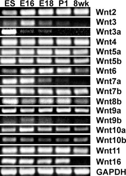
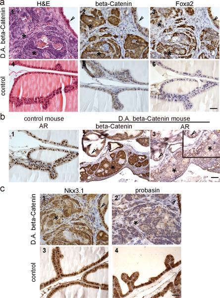
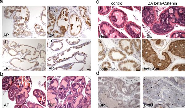
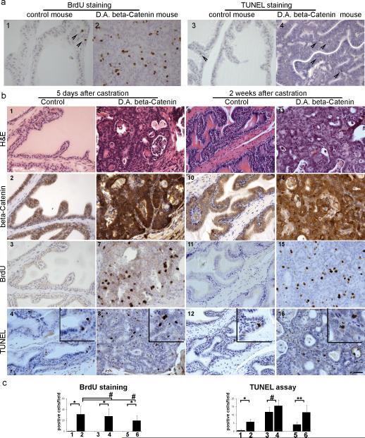
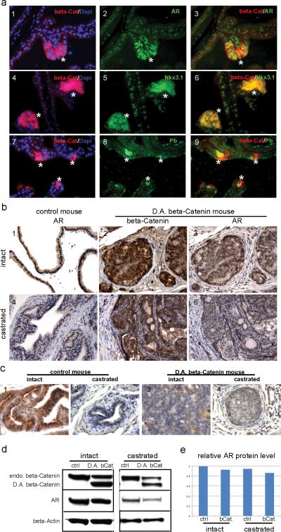
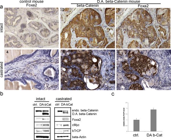
Results: Our results demonstrated that during prostate development Wnt ligands display a dynamic expression pattern. Activation of beta-Catenin during prostate development caused epithelial hyperplasia followed by prostatic intraepithelial neoplasia (PIN) in prostate. In the adult prostate, activation of beta-Catenin resulted in high grade PIN (HGPIN) and continuous prostatic growth after castration. As a result of activation of beta-Catenin, AR was first up-regulated with the emergence of epithelial hyperplasia, but was later down-regulated when HGPIN developed. Furthermore, activation of beta-Catenin induced Foxa2 re-expression in adult prostate which normally is only expressed in the embryonic budding stage during prostate development.

Conclusions: The results from this study strongly suggest that Wnt/beta-Catenin signaling is involved in the regulation of prostate development and confirm that constitutive activation of this pathway enables the mouse prostate to grow after castration.


Figures

Fig. 1
Fig. 1
Expression of Wnts in prostate. Expression of Wnts during prostate development was analyzed by RT-PCR using total RNA extracted from embryo day 16 (E16) and day 18 (E18) UGS, postnatal day 1 (P1) and 8 week dorsolateral prostate (8 wk). RNA extracted from embryo stem cells (ES) served as positive control. 16 out of 19 Wnts were expressed in prostate.
Fig. 2
Fig. 2
Constitutive activation of β-Catenin during prostate development caused prostatic intraepithelial neoplasia, induced Foxa2 re-expression, and reduced AR and AR signaling pathway. E16 to E18 UGS from Nkx3.1Cre/ CatnbΔ(ex3) and litter mate control male embryos were rescued and grafted under renal capsules of adult male nude mice. a, activation of β-Catenin caused PIN and Foxa2 re-expression. 1-3: serial sections of rescued prostate with Nkx3.1-Cre/ CatnbΔ(ex3) genotype; 4-6: serial sections of rescued prostate with Nkx3.1-Cre genotype. Asterisks in panel 1-3 indicate cytoplasmic/nuclear β-Catenin staining areas, and arrowheads indicate normal areas that show only membrane β-Catenin staining. At the areas where β-Catenin was activated (2, asterisks), PIN lesions developed (1, asterisks). While Foxa2 was not expressed in rescued control prostate (panel 6), Foxa2 expression was detected in β-Catenin accumulating prostate epithelia (panel 2-3). b, activation of β-Catenin reduced AR level. 1, AR staining performed on section from rescued control prostate. 2 and 3, β-Catenin or AR staining performed on serial sections from a rescued Nkx3.1Cre/ CatnbΔ(ex3) mouse prostate. At the area where β-Catenin was activated (2 and 3, asterisks), AR level was reduced compared with adjacent normal histology area (2 and 3, arrowheads) or with control prostate (1). inset: higher magnification. c, AR target genes (Nkx3.1 and probasin) were down-regulated in active β-Catenin mouse prostates. 1 and 2: immunostaining of Nkx3.1 or probasin performed on serial sections (same as those used in Fig2a, panel 1-3) from Nkx3.1-Cre/ CatnbΔ(ex3) prostate. 3 and 4: immunostaining performed on sections from rescued control prostate. Nkx3.1 antibody nicely stained the epithelial cell nuclei from control prostate (panel 3), or from the normal histology region of Nkx3.1-Cre/ CatnbΔ(ex3) prostate (panel 1, arrowhead), but at the areas that show activated β-Catenin (serial section from panel 2 in Fig.2a, asterisk), the Nkx3.1 staining was diffused (panel 1, asterisk) and the level was much lower when compared with adjacent normal histology area (panel 1, arrowhead) or with control prostate. Another AR target gene-probasin was lost at the area where β-Catenin was activated (panel 2, asterisk), while control prostate showed strong probasin staining. Scale bars represent 50 μm.
Fig. 3
Fig. 3
Activation of β-Catenin in adult prostate resulted in epithelial hyperplasia followed by progression to HGPIN. a, immunostaining for β-Catenin performed on a 12 week old PBCre4/ CatnbΔ(ex3) mouse prostate. With the deletion of exon 3, cytoplasmic/ nuclear β-Catenin accumulated focally in prostate epithelial cells at 12 weeks. AP, anterior prostate; DP, dorsal prostate; LP, lateral prostate; VP, ventral prostate. b, H&E staining performed on a 12 week old PBCre4/ CatnbΔ(ex3) mouse prostate. The accumulation of β-Catenin in cytoplasm/nucleus resulted in prostatic hyperplasia at 12 weeks. c, histology of PBCre4/ CatnbΔ(ex3) mouse prostate at 9 months. 1-2: H&E staining of a 9 month old control or PBCre4/ CatnbΔ(ex3) mouse prostate. 3-4: β-Catenin staining performed on serial sections from the above mouse prostates. At this age, prostatic epithelial cells displayed extensive cytoplasmic and nuclear β-Catenin (panel 4), which resulted in HGPIN to develop. Highly proliferating epithelial cells expanded and filled prostatic acini but were still restricted within stroma, and prostate basement membranes were still intact. d, BrdU staining. BrdU staining was performed on a 23 week old PBCre4/ CatnbΔ(ex3) (panel 2) or an age matched control mouse prostate (panel 1). BrdU incorporation in active β-Catenin prostate was significantly increased compared with control prostate (p<0.01). Scale bars represent 50 μm.
Fig. 4
Fig. 4
Activation of β-Catenin caused continuous prostate growth after castration. PBCre4/ CatnbΔ(ex3) and litter mate control mice were castrated at 22-23 weeks and sacrificed 5 days or 2 weeks later. a, histology of control and PBCre4/ CatnbΔ(ex3) mouse prostates without castration. 1 and 2: BrdU staining; 3 and 4: TUNEL staining. Arrow heads indicate positive staining. b, histology of control and PBCre4/ CatnbΔ(ex3) mouse prostates 5 days or 2 weeks post castration. 1-4, serial sections from a control mouse prostate 5 days after castration; 5-8, serial sections from a PBCre4/ CatnbΔ(ex3) mouse prostate 5 days after castration; 9-12, serial sections from a control mouse prostate 2 weeks after castration; 13-16, serial sections from a PBCre4/ CatnbΔ(ex3) mouse prostate 2 weeks after castration. Panel 1, 5, 9, and 13: H&E staining. Activation of β-Catenin resulted in HGPIN in PBCre4/ CatnbΔ(ex3) mouse prostates. Panel 2, 6, 10, and 14: β-Catenin staining. While β-Catenin can only be detected on cell membranes of control prostates (panel 2 and 10), β-Catenin accumulated in cytoplasm/nucleus of PBCre4/ CatnbΔ(ex3) mouse prostates (panel 6 and 14). Panel 3, 7, 11, and 15: BrdU staining. While control prostate stopped grow after 5 days and 2 weeks castration, activation of β-Catenin enabled PBCre4/ CatnbΔ(ex3) mouse prostate to continuously proliferate after castration as marked by the extensive BrdU incorporation in panel 7 and 15. Panel 4, 8, 12, and 16: TUNEL assay by ApopTag staining (inset for higher magnification). Both control and PBCre4/ CatnbΔ(ex3) mouse prostates showed apoptosis after castration. c, quantification of BrdU and TUNEL staining. column 1: prostate from control mouse without castration; 2: prostate from PBCre4/ CatnbΔ(ex3) mouse without castration; 3: prostate from control mouse 5 days post castration; 4, prostate from PBCre4/ CatnbΔ(ex3) mouse 5 days post castration; 5, prostate from control mouse 2 weeks post castration; 6, prostate from PBCre4/ CatnbΔ(ex3) mouse 2 weeks post castration. * p< 0.01; ** p< 0.05, # p> 0.05. Scale bars represent 50 μm.
Fig. 5
Fig. 5
AR signaling in PBCre4/ CatnbΔ(ex3) mouse prostate. a, immunofluorescence staining performed on prostate sections from a 12 week old PBCre4/ CatnbΔ(ex3) mouse prostate. 1-3: dual staining of β-Catenin and AR. 4-6: dual staining of β-Catenin and Nkx3.1. 7-9: dual staining of β-Catenin and probasin (Pb). β-Catenin was in red; AR, Nkx3.1 and probasin were in green. At the areas where β-Catenin accumulated in cytoplasm/nucleus (indicated by asterisks), AR was up-regulated compared with other areas that showed only membrane β-Catenin staining. Concurrently, AR target genes-Nkx3.1 and probasin were up-regulated with the activation of β-Catenin. b, immunostaining performed on prostate sections from 22 week old intact (panel 1-3) or castrated (panel 4-6) control and PBCre4/ CatnbΔ(ex3) mice. At this age, AR level was slightly decreased in PBCre4/ CatnbΔ(ex3) mouse prostates compared with control under both intact and castration condition. c, probasin staining. d, western blot to analyze the AR level in intact or castrated control and PBCre4/ CatnbΔ(ex3) mouse prostates. e, quantification of western blot results. Scale bars represent 50 μm.
Fig. 6
Fig. 6
genes altered in PBCre4/ CatnbΔ(ex3) mouse prostates. a, immunostaining performed on sections from intact (1-3) or castrated (4-6) mouse prostates. 1 and 4: Foxa2 staining performed on control prostates. Foxa2 was not detected in these prostates under either intact or castration condition. 2 and 3: serial sections from intact PBCre4/ CatnbΔ(ex3) mouse prostates. 5 and 6: serial section from castrated PBCre4/ CatnbΔ(ex3) mouse prostates. With the activation of β-Catenin, Foxa2 was induced under both intact and castration condition. b, western blot. Protein lysis was prepared from intact or castrated control and PBCre4/ CatnbΔ(ex3) mouse prostates. The deletion of exon 3 of β-Catenin resulted in truncated β-Catenin protein that was labeled as D.A. beta-Catenin. The endogenous β-Catenin band was labeled as endo. beta-Catenin. With the accumulation of D.A. β-Catenin, endogenous β-Catenin level was decreased. The induction of Foxa2 in PBCre4/ CatnbΔ(ex3) mouse prostates was confirmed in western blot. Protein levels of cMyc and βTrCP increased with the activation of β-Catenin. c, real-time RT-PCR to analyze βTrCP level.

Similar articles

Cited by

References

    1. Miller JR. The Wnts. Genome Biol. 2002;3 REVIEWS3001. - PMC - PubMed
    1. Mulholland DJ, Dedhar S, Coetzee GA, Nelson CC. Interaction of nuclear receptors with the Wnt/beta-catenin/Tcf signaling axis: Wnt you like to know? Endocr Rev. 2005;26:898–915. - PubMed
    1. Mulholland DJ, Cheng H, Reid K, Rennie PS, Nelson CC. The androgen receptor can promote Beta -catenin nuclear translocation independently of adenomatous polyposis coli. J Biol Chem. 2002;277:17933–43. - PubMed
    1. Edlund S, Lee SY, Grimsby S, Zhang S, Aspenström P, Heldin CH, Landström M. Interaction between Smad7 and beta-catenin: importance for transforming growth factor beta-induced apoptosis. Mol Cell Biol. 2005;25:1475–88. - PMC - PubMed
    1. He TC, Sparks AB, Rago C, Hermeking H, Zawel L, da Costa LT, Morin PJ, Vogelstein B, Kinzler KW. Identification of c-MYC as a target of the APC pathway. Science. 1998;281:1509–12. - PubMed

Publication types

MeSH terms