Review
. 2008 Oct:1142:266-309.
doi: 10.1196/annals.1444.017.

Cerebral white matter: neuroanatomy, clinical neurology, and neurobehavioral correlates

Affiliations
Review

Cerebral white matter: neuroanatomy, clinical neurology, and neurobehavioral correlates

Jeremy D Schmahmann et al. Ann N Y Acad Sci. 2008 Oct.

Abstract

Lesions of the cerebral white matter (WM) result in focal neurobehavioral syndromes, neuropsychiatric phenomena, and dementia. The cerebral WM contains fiber pathways that convey axons linking cerebral cortical areas with each other and with subcortical structures, facilitating the distributed neural circuits that subserve sensorimotor function, intellect, and emotion. Recent neuroanatomical investigations reveal that these neural circuits are topographically linked by five groupings of fiber tracts emanating from every neocortical area: (1) cortico-cortical association fibers; (2) corticostriatal fibers; (3) commissural fibers; and cortico-subcortical pathways to (4) thalamus and (5) pontocerebellar system, brain stem, and/or spinal cord. Lesions of association fibers prevent communication between cortical areas engaged in different domains of behavior. Lesions of subcortical structures or projection/striatal fibers disrupt the contribution of subcortical nodes to behavior. Disconnection syndromes thus result from lesions of the cerebral cortex, subcortical structures, and WM tracts that link the nodes that make up the distributed circuits. The nature and the severity of the clinical manifestations of WM lesions are determined, in large part, by the location of the pathology: discrete neurological and neuropsychiatric symptoms result from focal WM lesions, whereas cognitive impairment across multiple domains--WM dementia--occurs in the setting of diffuse WM disease. We present a detailed review of the conditions affecting WM that produce these neurobehavioral syndromes, and consider the pathophysiology, clinical effects, and broad significance of the effects of aging and vascular compromise on cerebral WM, in an attempt to help further the understanding, diagnosis, and treatment of these disorders.


Conflict of interest statement

Conflicts of Interest

The authors declare no conflicts of interest.

Figures
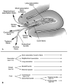
Figure 1
Figure 1
Diagram (A) and schema (B) of the principles of organization of white matter fiber pathways emanating from the cerebral cortex. Long association fibers are seen end-on as the stippled area within the white matter of the gyrus. In their course, these fibers either remain confined to the white matter of the gyrus or travel deeper in the white matter of the hemisphere. Short association fibers, or U-fibers, link adjacent gyri. Neighborhood association fibers link nearby regions, usually within the same lobe. Striatal fibers intermingle with the association fibers early in their course, before coursing in the subcallosal fascicle of Muratoff or in the external capsule. Cord fibers segregate into commissural fibers that arise in cortical layers II and III, and the subcortical bundle, which further divides into fibers destined for thalamus arising from cortical layer VI, and those to brain stem and spinal cord in the pontine bundle arising from cortical layer V.
Figure 2
Figure 2
Course of the cingulum bundle (CB). (A) Surface views of the ventral (top), medial (middle), and lateral (lower) convexities of the cerebral hemisphere of a rhesus monkey show the trajectory of the CB reflected onto the cortical surface, and the cortical areas that it links, as determined by autoradiographic tract tracing. (B) CB fibers in the monkey are shown in this sagittal dimension by using diffusion spectrum magnetic resonance imaging (DSI). CB fibers that intersect a disc (shown by the arrow) course between rostral and caudal cingulate regions and link the cingulate gyrus with the prefrontal and parietal areas. Fibers in the ventral limb of the CB course to the parahippocampal region. (C) The course of the CB in human brain is demonstrated using diffusion tensor imaging (DTI), remarkably similar to the findings in monkey.
Figure 3
Figure 3
Location of long association fiber pathways in the monkey. The coronal sections in (A) and (B) are taken at the corresponding levels shown on the figure of the lateral hemisphere (top). The fiber bundles are colored for ease of identification. Fiber pathways: AF, arcuate fasciculus; CBd, cingulum bundle dorsal component; CBv, cingulum bundle ventral component; EmC, extreme capsule; FOF, fronto-occipital fascicle; ILF, inferior longitudinal fascicle; MdLF, middle longitudinal fascicle; SLF (I, II, III), superior longitudinal fascicle, subcomponents I, II, and III; UF, uncinate fasciculus. Cerebral sulci: AS, arcuate sulcus; CS, central sulcus; Cing S, cingulate sulcus; IPS, intraparietal sulcus; LF, lateral fissure; PS, principal sulcus; OTS, occipitotemporal sulcus; STS, superior temporal sulcus.
Figure 4
Figure 4
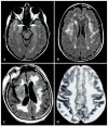
MRI appearance of (A) X-linked adrenoleukodystrophy (X-ALD), T1-weighted image post-gadolinium; (B) metachromatic leukodystrophy (MLD), FLAIR image; (C) globoid cell leukodystrophy (GLD), T2-weighted image; and (D) vanishing white matter disease (VWMD), T1-weighted image.
Figure 5
Figure 5
Imaging and pathology in a patient with adult-onset leukodystrophy with neuroaxonal spheroids. (A) FLAIR MRI in the axial plane showing confluent high signal in the periventricular, deep, and subcortical white matter of the frontal and parietal lobes extending through the splenium of the corpus callosum. (B) Gross pathology of a coronal section of the cerebral hemisphere, showing gliosis in the centrum semiovale (arrow) and internal capsule (arrowhead). (C) Several neuroaxonal spheroids on microscopic analysis of frontal white matter (original magnification, ×20; Luxol fast blue hematoxylin and eosin stain). (D) Neurofilament immunostain of white matter reveals mild loss of axons and an axonal spheroid (original magnification, ×20).
Figure 6
Figure 6
FLAIR MRI in a patient with mitochondrial encephalopathy with lactic acidosis and stroke-like episodes (MELAS).
Figure 7
Figure 7
MRI features of fragile X–associated tremor ataxia syndrome (FXTAS). White matter pallor is seen in the cerebellar parenchyma (A), as well as in the middle cerebellar peduncles (B).
Figure 8
Figure 8
FLAIR MRI in multiple sclerosis. (A) White matter hyperintensity perpendicular to the lateral ventricle (Dawson’s finger), shown by the arrow. (B) In a second case, the focal area of hyperintensity (arrow) corresponded to the initial clinical presentation.
Figure 9
Figure 9
MRI features of acute disseminated encephalomyelitis (ADEM). (A) Coronal T1-weighted postgadolinium image showing enhancing lesions in the right more than left hemispheres. (B) Axial zero-B MRI demonstration of the multiple lesions. (C) FLAIR MRI 6 months after marked clinical recovery shows much improved areas of hyperintensity.
Figure 10
Figure 10
FLAIR MRI showing hyperintensities in prefrontal white matter in a patient with HIV and cognitive impairment.
Figure 11
Figure 11
MRI features of progressive multifocal leukoencephalopathy (PML). (A) T2-weighted image shows involvement of white matter of the right occipital region (arrow), accounting for the hemianopsia in this HIV-positive patient. (B) FLAIR MRI in a patient with systemic lymphoma and PML, demonstrating confluent prefrontal white matter lesion spreading across the genu of the corpus callosum (arrow), and additional lesions affecting local association fibers of the right prefrontal and parieto-occipital cortices (arrowheads). (C, D) Axial FLAIR images in an HIV-positive patient showing confluent subcortical and deep white matter involvement by PML. (Panels A and B are from reference .)
Figure 12
Figure 12
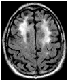
FLAIR MRI in the axial plane of a patient with cognitive decline after receiving methotrexate.
Figure 13
Figure 13
T2-weighted MRI appearance in the axial plane of toluene encephalopathy in two patients (A, B).
Figure 14
Figure 14
MRI scans after heroin inhalation, known colloquially as “chasing the dragon.” FLAIR images in the axial plane (AD). Corresponding 1H MRS imaging spectra in two of the images show characteristic lactate peak and decreased NAA.
Figure 15
Figure 15
Axial MRI in delayed leukoencephalopathy after hypoxic–ischemic insult. (A) FLAIR image shows extensive, symmetric white matter hyperintensities with relative sparing of subcortical white matter. (B) Diffusion-weighted imaging shows restricted diffusion of the white matter abnormalities, confirmed on (C), apparent diffusion coefficient mapping.
Figure 16
Figure 16
Coronal T1-weighted image in a patient with gliomatosis cerebri. Note the spread of tumor along white matter planes.
Figure 17
Figure 17
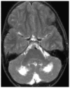
T2-weighted axial MRI in a patient with Langerhans cell histiocytosis, showing hyperintense signal abnormality in the white matter of the cerebellum.
Figure 18
Figure 18
Leukoaraiosis is visible as (A) white matter hypodensity on CT and (B) white matter hyperintensity on FLAIR MRI in the same patient.
Figure 19
Figure 19
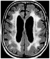
FLAIR MRI of a patient with Binswanger’s encephalopathy. Hyperintense signal abnormality is seen at periventricular zones, white matter immediately beneath cortex, splenium of the CC, and internal and external/extreme capsule regions. Multiple hypodensities consistent with lacunar infarcts are also seen in the basal ganglia and thalamus.
Figure 20
Figure 20
MRI appearance of white matter changes in axial sections of patients with CADASIL. (A, B) FLAIR MRI in an asymptomatic 39-year-old, notch 3 gene positive with family history of early stroke, whose imaging findings were incidentally noted. Characteristic temporal lobe white matter involvement is highlighted (arrows). (C) FLAIR MRI in a patient with clinically established CADASIL. (D) T2-weighted MRI in a patient with notch 3 gene and pathologically proven disease.
Figure 21
Figure 21
MRI in the axial plane in cerebral amyloid angiopathy. (A) Gradient echo MRI demonstrating multiple punctuate areas of hemorrhage (microbleeds, arrow) at the cortico–subcortical junctions. (B) MRI FLAIR sequence in a patient with lobar intraparenchymal hemorrhage in the left occipital lobe (double arrows), as well as periventricular WMH (single arrow) and subcortical WMH (arrowheads).
Figure 22
Figure 22
Focal WM lesions with neurobehavioral manifestations. (A) Lacune in the genu of the right internal capsule (arrow) on CT presenting with hemineglect., (B) Diagram of the WM lesion responsible for parietal pseudothalamic pain syndrome, thought to disrupt the second somatosensory cortex from thalamus. (C) FLAIR MRI of posterior reversible encephalopathy syndrome producing visual loss. (D, E) Focal WM lesion consisting of metastatic melanoma with surrounding edema, producing alexia without agraphia.
Figure 23
Figure 23
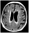
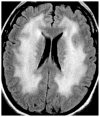
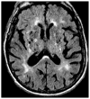
FLAIR MRI in the axial plane of an 80-year-old man with slowly evolving WM dementia. No single cause has been identified for the cognitive decline or WM hyperintensities.

Similar articles

Cited by

References

    1. Neuburger M. Die historische Entwicklung der experimentellen Gehirn- und Rückenmarksphysiologie vor Flourens. Ferdinand Enke Verlag, Stuttgart. Translated and edited, with additional material, by Edwin Clarke (1981) The Historical Development of Experimental Brain and Spinal Cord Physiology before Flourens. Johns Hopkins University Press; Baltimore and London: 1897.
    1. Schmahmann JD, Pandya DN. Fiber Pathways of the Brain. Oxford University Press; New York: 2006.
    1. Schmahmann JD, Pandya DN. Cerebral white matter–historical evolution of facts and notions concerning the organization of the fiber pathways of the brain. J Hist Neurosci. 2007;16:237–267. - PubMed
    1. Geschwind N. Disconnexion syndromes in animals and man. I. Brain. 1965;88:237–294. - PubMed
    1. Geschwind N. Disconnexion syndromes in animals and man. II. Brain. 1965;88:585–644. - PubMed

Publication types