. 2009 Jun;40(6):643-53.
doi: 10.1165/rcmb.2008-0217OC. Epub 2008 Nov 6.

Hepatocyte growth factor inhibits epithelial to myofibroblast transition in lung cells via Smad7

Affiliations

Hepatocyte growth factor inhibits epithelial to myofibroblast transition in lung cells via Smad7

Manasi N Shukla et al. Am J Respir Cell Mol Biol. 2009 Jun.

Abstract

Idiopathic pulmonary fibrosis is a lethal parenchymal lung disease characterized by denudation of the lung epithelium, fibroblast proliferation, and collagen deposition. Cellular changes underlying disease progression involve injury to alveolar epithelial cells, epithelial to mesenchymal transition, proliferation of alpha-smooth muscle actin (alpha-SMA)-expressing myofibroblasts and of fibroblasts resulting in enhanced deposition of extracellular matrix proteins. Hepatocyte growth factor (HGF) inhibits progression of bleomycin-induced pulmonary fibrosis in mice. The mechanism underlying the inhibitory effect of HGF was investigated in an in vitro model. We show that HGF markedly antagonizes basal and transforming growth factor (TGF)-beta-induced expression of myofibroblast markers such as alpha-SMA, collagen type 1, and fibronectin in rat alveolar epithelial cells. HGF also inhibited TGF-beta-induced alpha-SMA expression in primary murine alveolar epithelial cells. Since TGF-beta is known to regulate alpha-SMA expression, the effect of HGF on components of TGF-beta signaling was investigated. HGF induced expression of Smad7, an inhibitor of TGF-beta signaling, in a mitogen-activated protein kinase-dependent manner. HGF also induced the nuclear export of Smad7 and Smad ubiquitin regulatory factor 1 (Smurf1) to the cytoplasm. HGF-dependent decrease in alpha-SMA was abolished with specific siRNAs targeted to Smad7. Thus, induction of Smad7 by HGF serves to limit acquisition of the myofibroblast phenotype in alveolar epithelial cells.


Figures

<b>Figure 1.</b>
Figure 1.
The alveolar epithelial cells rat lung epithelial (RLE)-6TN express basal myofibroblast marker α-smooth muscle actin (α-SMA) and epithelial cell markers E-cadherin, aquaporin-5, and ZO-1. RLE cells were plated on glass slides for 24 hours, fixed in 2% paraformaldehyde, and immunostained for α-SMA (green) and ZO-1 (red). Nuclei were stained with DRAQ5 (blue). A–C are representative images captured at a ×40 magnification. Note the fibrous strands present across these cells. A number of α-SMA and ZO-1 double-positive cells are evident along with cells expressing ZO-1 alone. (D) RLE cells were analyzed by Western blotting methods for epithelial cell markers E-cadherin and aquaporin-5, and the myofibroblast marker α-SMA. β-actin was assessed for equal protein loading. Cells were plated at a low density for samples in lane 1 and high density for samples in lane 2 harvested after 24 hours in culture. Results shown are representative of two independent experiments.
<b>Figure 2.</b>
Figure 2.
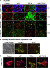
Treatment of primary pneumocytes and RLE cells with hepatocyte growth factor (HGF) results in inhibition of transforming growth factor (TGF)-β–mediated EMT. (A) RLE cells treated with or without HGF ± TGF-β were fixed and immunofluorescently stained for (i) α-SMA (green) and ZO-1 (blue); (ii) collagen type I (green); (iii) fibronectin (green); or (iv) E-cadherin (green). Nuclei were stained with DRAQ5 (red). (B) Primary murine alveolar epithelial cells in culture were observed by phase contrast (left panel); or immunofluorescence microscopy for pro–surfactant protein C (green, middle panel) or CC-10 (green, right panel) expression. Nuclei were stained with DRAQ5 (red). (C) Primary murine alveolar epithelial cells were cultured as shown and were fixed and stained for ZO1 (red), α-SMA (green), and nuclei (blue). Results shown represent at least two independent experiments. All images were digitally captured at ×40 magnification.
<b>Figure 3.</b>
Figure 3.
The TGF-β type I receptor kinase inhibitor, SB-431542, significantly reduces α-SMA protein expression in RLE-6TN cells. Western blot analysis of RLE cells incubated without or with 0.5, 1, or 5 μM inhibitor for 24 hours. (A) Blots were probed for α-SMA and β-actin protein expression. The blots were analyzed by densitometry and α-SMA expression was normalized to β-actin. (B) Blots were re-probed with pSmad2 and total Smad2. Blots were analyzed by densitometry and levels of pSmad2 were normalized to total Smad2. The Western blots are representative of at least three independent experiments.
<b>Figure 4.</b>
Figure 4.
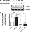
HGF decreases basal and TGF-β–induced α-SMA protein in RLE cells. (A) RLE cells were incubated without or with 100 ng/ml HGF alone or in combination with 2.5 ng/ml TGF-β. After 24 hours, cells were lysed and samples containing equal amounts of total protein were resolved by SDS-PAGE. Expression of α-SMA and β-actin proteins was assessed by immunoblotting using specific monoclonal antibodies. (B) Relative expression of α-SMA was evaluated by densitometric analysis by calculating α-SMA to β-actin ratios for each sample. Statistical significance was established using Mann-Whitney t test. Results shown are representative of at least three independent experiments. *P < 0.05; **P < 0.01.
<b>Figure 5.</b>
Figure 5.
HGF-mediated reduction of α-SMA proceeds through the MEK-ERK1/2 signaling pathway. (A) Stimulation of RLE cells with 100 ng/ml HGF for 5 and 25 minutes results in activation of Akt, ERK1/2, JNK, and p38 as shown by western blotting with phospho-specific antibodies. Total Akt, ERK1/2, JNK, and p38 protein levels were also assessed as controls for protein loading. (B) RLE cells were pre-treated for 30 minutes without or with vehicle (DMSO) or specific pharmacologic inhibitors for the kinases AKT (AKT inhibitor, 10 μM), p38 (SB203580, 20 μM),or MEK (PD98059, 50 μM). Cells were then treated with HGF (100 ng/ml) for 24 hours. Analysis of α-SMA expression was performed using immunoblotting and densitometric analysis of α-SMA/β-actin ratios. Results shown are representative of at least three independent experiments. *P < 0.05 compared with control (CTL) using Mann Whitney t test.
<b>Figure 6.</b>
Figure 6.
HGF treatment increases Smad7 protein in RLE cells. (A) RLE cells were treated with HGF for 0 to 24 hours. Western blotting demonstrates Smad7 protein increases in a time-dependent manner after HGF treatment with protein levels peaking at 3 hours and remaining elevated through 24 hours. The blot was analyzed by densitometry and data are shown as percentage of control levels (0 h) as a ratio to β-actin. Standard error was calculated from the average of three independent experiments. (B) Thirty minutes of pretreatment with 50 μM PD98059 antagonized the HGF-dependent increase in Smad7 protein. Each blot was analyzed by densitometry and data are shown as percentage of control levels (0 h time-point) as a ratio to β-actin. Standard error was calculated from the average of three independent experiments. pSmad2 and pSmad3 levels were determined in cells treated with 50 μM PD98059 for 30 minutes and HGF for 1 or 3 hours. Nuclear (N) and cytoplasmic (C) extracts were collected and Western blots generated. pSmad2 levels were increased in response to HGF treatment with the MEK inhibitor antagonizing the activation. The MEK inhibitor had no effect on pSmad3 levels.
<b>Figure 7.</b>
Figure 7.
HGF induces export of Smad7 from the nucleus to the cytoplasm. RLE cells were plated on glass slides. After 24 hours, cells were treated with 100 ng/ml HGF for 3 hours, fixed with 2% paraformaldehyde for 10 minutes at room temperature, and immunostained for Smad7 (green) and E-cadherin (red). Nuclei (blue) were stained with DRAQ5. Representative images were captured using ×40 magnification. HGF-treated cells exhibit predominantly cytoplasmic Smad7 staining. Average intensity of the nuclear region was determined by using the nuclear stain to define the areas of interest and then performing intensity analysis with MetaMorph software. Intensity was calculated on a per-cell basis, averaged, and presented for control and HGF-treated cells. Five fields containing a total of 100 cells were analyzed from images captured using a ×40 objective.
<b>Figure 8.</b>
Figure 8.
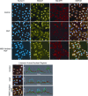
HGF-dependent translocation of Smad7/Smurf1. RLE cells were plated on glass microscope slides and treated with 50 μM PD98059 (MEK inhibitor), 100 ng/ml HGF, or the combination for 3 hours. Cells were fixed in 2% paraformaldehyde and immunostained with antibodies against Smad7 (green) and Smurf1 (red). Nuclei (blue) were stained with DRAQ5. Nuclear intensities of Smad7 and Smurf1 proteins were determined as described in the legend to Figure 6. Linescan profiles of representative cells in MetaMorph correspond to results of image analysis and show a dramatic reduction in the nuclear presence of both Smad7 and Smurf1 in response to HGF which was antagonized by the MEK inhibitor.
<b>Figure 9.</b>
Figure 9.
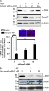
Smad7 siRNA antagonizes the HGF-dependent decrease in α-SMA protein in RLE cells. (A) RLE cells were untransfected or transfected with 10 or 50 nM Smad7-specific siRNA or (B) 50 nM nonspecific siRNA followed by treatment with 100 ng/ml HGF for 24 hours. Cell extracts were collected and Western blotting was performed for detection of α-SMA, Smad7, and β-actin protein. Each blot was analyzed by densitometry, and averages and standard errors were determined from three independent experiments. The level of α-SMA protein was normalized to β-actin and the untreated control and presented as a percentage of the untreated control. **P < 0.05; ***P < 0.01. Inset: RLE cells transfected with fluorescently-tagged siRNA (siGLO) or mock transfected with delivery reagent only (Control) exhibited approximately 100% transfection efficiency.

Similar articles

Cited by

References

    1. Selman M. Idiopathic pulmonary fibrosis challenges for the future. Chest 2001;120:8–10. - PubMed
    1. Selman M, Pardo A. Role of epithelial cells in idiopathic pulmonary fibrosis: from innocent targets to serial killers. Proc Am Thorac Soc 2006;3:364–372. - PubMed
    1. Thannickal VJ, Toews GB, White ES, Lynch JP III, Martinez FJ. Mechanisms of pulmonary fibrosis. Annu Rev Med 2004;55:395–417. - PubMed
    1. Gharaee-Kermani M, Gyetko MR, Hu B, Phan SH. New insights into the pathogenesis and treatment of idiopathic pulmonary fibrosis: a potential role for stem cells in the lung parenchyma and implications for therapy. Pharm Res 2007;24:819–841. - PubMed
    1. Khalil N, Bereznay O, Sporn M, Greenberg AH. Macrophage production of transforming growth factor beta and fibroblast collagen synthesis in chronic pulmonary inflammation. J Exp Med 1989;170:727–737. - PMC - PubMed

Publication types