

Hypoxia-inducible factor 1 alpha expression increases during colorectal carcinogenesis and tumor progression
- PMID: 18983642
- PMCID: PMC2584660
- DOI: 10.1186/1471-2407-8-320
Hypoxia-inducible factor 1 alpha expression increases during colorectal carcinogenesis and tumor progression
Abstract
Background: Hypoxia-inducible factor 1 alpha (HIF-1alpha) is involved in processes promoting carcinogenesis of many tumors. However, its role in the development of colorectal cancer is unknown. To investigate the significance of HIF-1alpha during colorectal carcinogenesis and progression we examined its expression in precursor lesions constituting the conventional and serrated pathways, as well as in non-metastatic and metastatic adenocarcinomas.
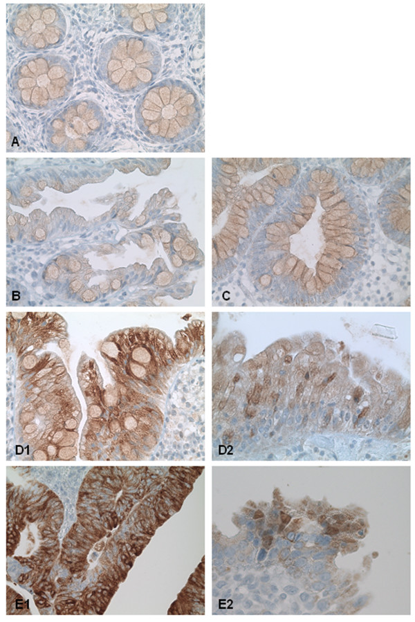
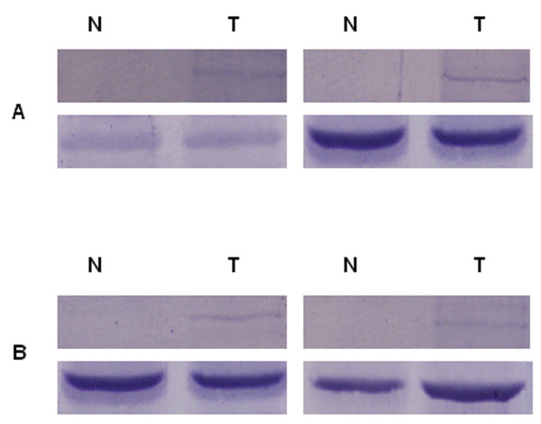
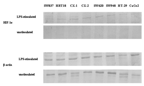
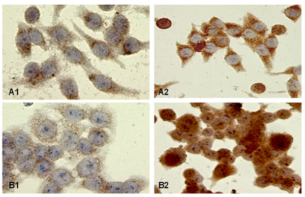
Methods: Immunohistochemistry and Western blot is used to analyse HIF-1alpha expression in normal colonic mucosa, hyperplastic polyps (HPP), sessile serrated adenomas (SSA), low-grade (TA-LGD) and high-grade (TA-HGD) traditional adenomas as well as in non-metastatic and metastatic colorectal adenocarcinomas. Eight colorectal carcinoma cell lines are tested for their HIF-1alpha inducibility after lipopolysaccharide (LPS) stimulation using western blot and immunocytochemistry.
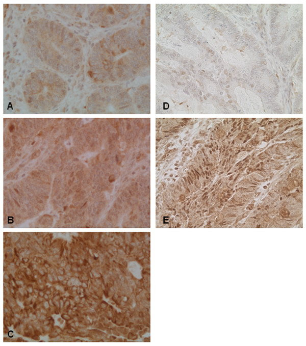
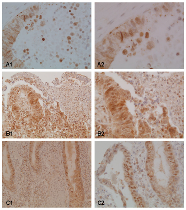
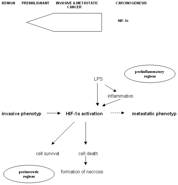
Results: In normal mucosa, HPP and TA-LGD HIF-1alpha was not expressed. In contast, perinuclear protein accumulation and nuclear expression of HIF-1alpha were shown in half of the examined SSA and TA-HGD. In all investigated colorectal carcinomas a significant nuclear HIF-1alpha overexpression compared to the premalignant lesions was observed but a significant correlation with the metastatic status was not found. Nuclear HIF-1alpha expression was strongly accumulated in perinecrotic regions. In these cases HIF-1alpha activation was seen in viable cohesive tumor epithelia surrounding necrosis and in dissociated tumor cells, which subsequently die. Enhanced distribution of HIF-1alpha was also seen in periinflammatory regions. In additional in vitro studies, treatment of diverse colorectal carcinoma cell lines with the potent pro-inflammatory factor lipopolysaccharide (LPS) led to HIF-1alpha expression and nuclear translocation.
Conclusion: We conclude that HIF-1alpha expression occurs in early stages of colorectal carcinogenesis and achieves a maximum in the invasive stage independent of the metastatic status. Perinecrotic activation of HIF-1alpha in invasive tumors underlines a dual role of HIF-1alpha by regulating both pro-survival and pro-death processes. HIF-1alpha up-regulation in response to LPS-mediated stimulation and periinflammatory expression in invasive carcinomas suggest its involvement in inflammatory events. These patterns of HIF-1alpha inducibility could contribute indirectly to the acquisition of a metastatic phenotype.
Figures
Similar articles
-
Expression levels and significance of hypoxia inducible factor-1 alpha and vascular endothelial growth factor in human colorectal adenocarcinoma.Chin Med J (Engl). 2004 Oct;117(10):1541-6. Chin Med J (Engl). 2004. PMID: 15498380
-
Up-regulation and stabilization of HIF-1alpha in colorectal carcinomas.Surg Oncol. 2007 Dec;16 Suppl 1:S25-7. doi: 10.1016/j.suronc.2007.10.014. Epub 2007 Nov 26. Surg Oncol. 2007. PMID: 18023174 Review.
-
Coordinate up-regulation of hypoxia inducible factor (HIF)-1alpha and HIF-1 target genes during multi-stage epidermal carcinogenesis and wound healing.Cancer Res. 2000 Nov 1;60(21):6189-95. Cancer Res. 2000. PMID: 11085544
-
Cyclooxygenase-2 and Hypoxia-Inducible Factor-1alpha protein expression is related to inflammation, and up-regulated since the early steps of colorectal carcinogenesis.Cancer Lett. 2009 Jul 8;279(2):221-9. doi: 10.1016/j.canlet.2009.02.001. Epub 2009 Mar 5. Cancer Lett. 2009. PMID: 19268443
-
Serrated polyps and colorectal cancer: new pathway to malignancy.Annu Rev Pathol. 2009;4:343-64. doi: 10.1146/annurev.pathol.4.110807.092317. Annu Rev Pathol. 2009. PMID: 19400693 Review.
Cited by
-
Impairment of vascularization of the surface covering epithelium induces ischemia and promotes malignization: a new hypothesis of a possible mechanism of cancer pathogenesis.Clin Transl Oncol. 2015 Jun;17(6):446-53. doi: 10.1007/s12094-014-1255-x. Epub 2014 Nov 19. Clin Transl Oncol. 2015. PMID: 25408194 Free PMC article.
-
Elevated d-2-hydroxyglutarate during colitis drives progression to colorectal cancer.Proc Natl Acad Sci U S A. 2018 Jan 30;115(5):1057-1062. doi: 10.1073/pnas.1712625115. Epub 2018 Jan 16. Proc Natl Acad Sci U S A. 2018. PMID: 29339485 Free PMC article.
-
The recent progress of the mechanism and regulation of tumor necrosis in colorectal cancer.J Cancer Res Clin Oncol. 2016 Feb;142(2):453-63. doi: 10.1007/s00432-015-1997-z. Epub 2015 Jun 21. J Cancer Res Clin Oncol. 2016. PMID: 26094047 Review.
-
Role of hypoxia in cancer therapy by regulating the tumor microenvironment.Mol Cancer. 2019 Nov 11;18(1):157. doi: 10.1186/s12943-019-1089-9. Mol Cancer. 2019. PMID: 31711497 Free PMC article. Review.
-
Cytokine-induced killer cells mediated pathways in the treatment of colorectal cancer.Cell Commun Signal. 2022 Mar 28;20(1):41. doi: 10.1186/s12964-022-00836-0. Cell Commun Signal. 2022. PMID: 35346234 Free PMC article. Review.
References
-
- Zhong H, De Marzo AM, Laughner E, Lim M, Hilton DA, Zagzag D, Buechler P, Isaacs WB, Semenza GL, Simons JW. Overexpression of hypoxia-inducible factor 1alpha in common human cancers and their metastases. Cancer Res. 1999;59:5830–5835. - PubMed
Publication types
MeSH terms
Substances
LinkOut - more resources
Full Text Sources
Medical
Research Materials
