

CLIP: crosslinking and immunoprecipitation of in vivo RNA targets of RNA-binding proteins
- PMID: 18982285
- PMCID: PMC4492529
- DOI: 10.1007/978-1-60327-475-3_6
CLIP: crosslinking and immunoprecipitation of in vivo RNA targets of RNA-binding proteins
Abstract
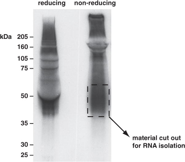
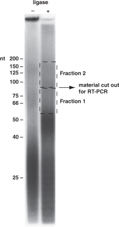
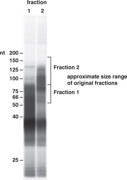
We present a newly developed method for fixing RNA-protein complexes in situ in living cells and the subsequent purification of the RNA targets. Using this approach, complex tissue such as mouse brain can be ultraviolet (UV) irradiated to covalently crosslink RNA-protein complexes. Once covalently bound, RNA-protein complexes can be purified under stringent conditions, allowing a highly specific purification scheme to be employed. After UV irradiation, the tissue is solubilized and the RNA partially digested, allowing a small fragment to remain attached to protein. RNA-protein complexes of interest are partially purified by immunoprecipitation and noncovalently associated RNA removed by sodium dodecyl sulfate polyacrylamide gel electrophoresis (SDS-PAGE). These purified RNA-protein complexes are isolated and treated with proteinase K, which removes protein but leaves intact RNA. This RNA is abundant enough, and competent for, RNA linker ligation, reverse transcriptase polymerase chain reaction (RT-PCR) amplification, and sequencing. Database matching of these short 70- to 100-nt RNA CLIP (crosslinking and immunoprecipitation of RNA-protein complexes) "tags," which mark the native binding sites of RNA binding proteins, potentially allows the entire target repertoire of an RNA binding protein to be determined.
Figures
Similar articles
-
Genome-Wide Profiling of RNA-Protein Interactions Using CLIP-Seq.Methods Mol Biol. 2016;1421:137-51. doi: 10.1007/978-1-4939-3591-8_12. Methods Mol Biol. 2016. PMID: 26965263 Free PMC article.
-
PAR-CliP--a method to identify transcriptome-wide the binding sites of RNA binding proteins.J Vis Exp. 2010 Jul 2;(41):2034. doi: 10.3791/2034. J Vis Exp. 2010. PMID: 20644507 Free PMC article.
-
3'-Linker Ligation and Size Selection by SDS-PAGE for Cross-Linking Immunoprecipitation (CLIP).Cold Spring Harb Protoc. 2018 Dec 3;2018(12). doi: 10.1101/pdb.prot097964. Cold Spring Harb Protoc. 2018. PMID: 30510127
-
[Advances in technologies for large-scale enrichment and identification of ribonucleic acid-protein complexes].Se Pu. 2021 Feb;39(2):105-111. doi: 10.3724/SP.J.1123.2020.07019. Se Pu. 2021. PMID: 34227341 Free PMC article. Review. Chinese.
-
Evaluation of Post-transcriptional Gene Regulation in Pancreatic Cancer Cells: Studying RNA Binding Proteins and Their mRNA Targets.Methods Mol Biol. 2019;1882:239-252. doi: 10.1007/978-1-4939-8879-2_22. Methods Mol Biol. 2019. PMID: 30378060 Review.
Cited by
-
Deep sequencing of cardiac microRNA-mRNA interactomes in clinical and experimental cardiomyopathy.Methods Mol Biol. 2015;1299:27-49. doi: 10.1007/978-1-4939-2572-8_3. Methods Mol Biol. 2015. PMID: 25836573 Free PMC article.
-
miRNA in pluripotent stem cells.Regen Med. 2010 Jul;5(4):545-55. doi: 10.2217/rme.10.34. Regen Med. 2010. PMID: 20632858 Free PMC article. Review.
-
PRC2 binds active promoters and contacts nascent RNAs in embryonic stem cells.Nat Struct Mol Biol. 2013 Nov;20(11):1258-64. doi: 10.1038/nsmb.2700. Epub 2013 Oct 20. Nat Struct Mol Biol. 2013. PMID: 24141703 Free PMC article.
-
Shaping the Innate Immune Response Through Post-Transcriptional Regulation of Gene Expression Mediated by RNA-Binding Proteins.Front Immunol. 2022 Jan 11;12:796012. doi: 10.3389/fimmu.2021.796012. eCollection 2021. Front Immunol. 2022. PMID: 35087521 Free PMC article. Review.
-
Protein-mRNA interactome capture: cartography of the mRNP landscape.F1000Res. 2016 Nov 3;5:2627. doi: 10.12688/f1000research.9404.1. eCollection 2016. F1000Res. 2016. PMID: 29098073 Free PMC article. Review.
References
-
- Ule J, Jensen KB, Ruggiu M, et al. CLIP identifies Nova-regulated RNA networks in the brain. Science. 2003;302:1212–1215. - PubMed
-
- Ule J, Stefani G, Mele A, et al. An RNA map predicting Nova-dependent splicing regulation. Nature. 2006;444:580–586. - PubMed
-
- Cameron V, Uhlenbeck OC. 3′-Phosphatase activity in T4 polynucleotide kinase. Biochemistry. 1977;16:5120–5126. - PubMed
MeSH terms
Substances
Grants and funding
LinkOut - more resources
Full Text Sources
Other Literature Sources
Research Materials
