. 2008 Nov 3;183(3):441-55.
doi: 10.1083/jcb.200807043.

P bodies promote stress granule assembly in Saccharomyces cerevisiae

Affiliations

P bodies promote stress granule assembly in Saccharomyces cerevisiae

J Ross Buchan et al. J Cell Biol. .

Abstract

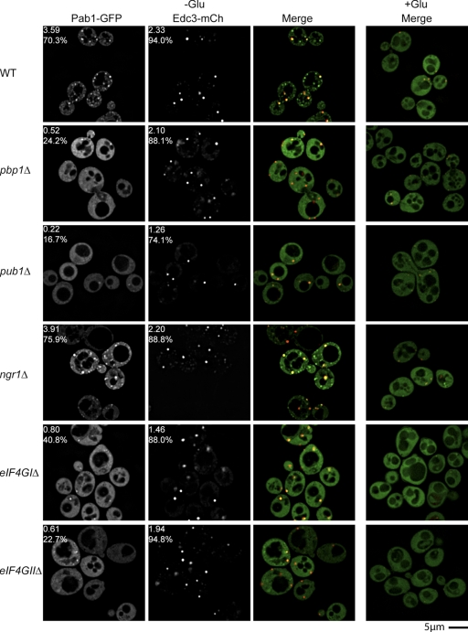
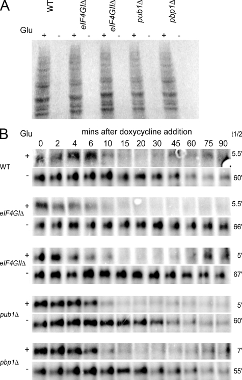
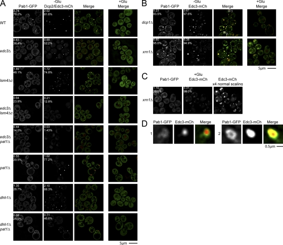
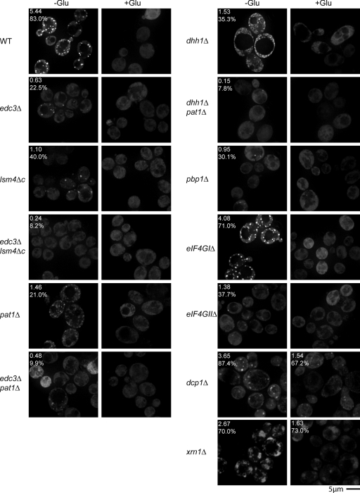
Recent results indicate that nontranslating mRNAs in eukaryotic cells exist in distinct biochemical states that accumulate in P bodies and stress granules, although the nature of interactions between these particles is unknown. We demonstrate in Saccharomyces cerevisiae that RNA granules with similar protein composition and assembly mechanisms as mammalian stress granules form during glucose deprivation. Stress granule assembly is dependent on P-body formation, whereas P-body assembly is independent of stress granule formation. This suggests that stress granules primarily form from mRNPs in preexisting P bodies, which is also supported by the kinetics of P-body and stress granule formation both in yeast and mammalian cells. These observations argue that P bodies are important sites for decisions of mRNA fate and that stress granules, at least in yeast, primarily represent pools of mRNAs stalled in the process of reentry into translation from P bodies.


Figures

Figure 1.
Figure 1.
Candidate yeast stress granule proteins form P body–distinct cytoplasmic foci during glucose deprivation. Log-phase wild-type cells expressing chromosomal GFP-tagged proteins and Edc3-mCh (pRP1574) were glucose deprived and examined. No GFP foci, and only faint P bodies were observed in +glucose control conditions, hence display of merge only.
Figure 2.
Figure 2.
Yeast stress granules are sensitive to cycloheximide, and stimulated by a constitutively active allele of the GCN2 kinase. (A) Log-phase yRP840 cells transformed with pRP1659 were glucose deprived and examined. Cycloheximide-treated cells (100 μg/ml) were preincubated with the drug for 1 min before harvest. (B) Cells identical to those in A were subject to the following stresses. Oxidative stress: shift to SC media containing 3 mM H2O2 or a mock treatment (H2O) for 15 min. Hyperosmotic stress: shift to SC media containing 1 M KCL for 15 min. Hypotonic stress: shift to H2O containing 2% dextrose for 15 min. SC media was used as a mock osmotic control. (C) Cells similar to those in A, but additionally transformed with either pRP1663 (Gcn2c) or pRS416 (empty vector control) were glucose deprived and examined for altered stress granule/P-body assembly.
Figure 3.
Figure 3.
Mutations that inhibit stress granule assembly do not significantly affect P-body assembly. Log-phase BY4741 wild-type and isogenic knockout strains were transformed with pRP1657, glucose deprived, and examined for altered stress granule/P-body assembly. Numbers indicate average foci number per cell, and percentage of cells with foci.
Figure 4.
Figure 4.
Stress granule assembly mutants are not deficient in their ability to both translationally repress and stabilize mRNA during stress. (A) Log-phase BY4741 wild-type and isogenic knockout strains were washed and incubated in media +/− glucose at 30°C for 10 min, followed by 35S-met/cys labeling for 5 min. Lysates were prepared and separated by SDS-PAGE for PhosphorImager analysis. (B) Log-phase BY4741 WT and isogenic knockout strains transformed with pRP1192 were resuspended in media +/− glucose, followed by doxycycline addition to transcriptionally repress the MFA2-pG mRNA reporter. Thus, only decay of existing MFA2-pG mRNA was examined. Time points were taken and analyzed via Northern blot. mRNA half-lives (right) are indicated.
Figure 5.
Figure 5.
Mutations that inhibit P-body formation also inhibit stress granule assembly. (A) Various yeast deletion strains and wild-type isogenic controls were transformed with pRP1657, pRP1658, or pRP1659, according to auxotrophies and genetic properties of the strains; bottom three panels feature Edc3-mCh as a P-body marker, others feature Dcp2-mCh. Glucose deprivation and quantitation as shown in Fig. 3. (B) Decapping (dcp1Δ) and 5′–3′decay (xrn1Δ) mutant strains were transformed with pRP1659. (C) Individual Pab1-GFP and Edc3-mCh images from +glucose xrn1Δ control cells, highlighting large and diffuse nature of constitutive Edc3-mCh foci (more visible at 4x scaling intensity) and occasional large and diffuse Pab1-GFP foci. (D) Zoom panels from dcp1Δ images in B (white boxes), showing examples of Pab1-GFP forming donut-like foci, with Edc3 foci overlapping the Pab1-GFP holes.
Figure 6.
Figure 6.
Formation of Pub1-mCh foci predominantly mirrors null strain sensitivity trends exhibited by Pab1-GFP. Various yeast deletion strains and wild-type isogenic controls were transformed with pRP1661 or pRP1662, according to strain auxotrophies. Log-phase strains were glucose deprived and examined for altered stress granule assembly. Images are collapsed Z-stacks.
Figure 7.
Figure 7.
Yeast temporal analysis reveals accumulation of Pab1 in P bodies before formation of P body–distinct stress granules. (A) Log-phase yRP840 strain, transformed with pRP1660, was glucose deprived for 5 min and followed over time. For technical reasons, identical cells at the 0-min time point are not shown; instead a typical image of this time point is shown. Orange arrowheads indicate initial accumulation of Pab1-GFP in preexisting P bodies. Purple arrowheads indicate appearance of P body–distinct stress granules. Images are typical of multiple independent experiments. Images were taken from Video 1 (available at http://www.jcb.org/cgi/content/full/jcb.200807043/DC1). (B) Zoom panels from the 19-min time point (white boxes). Arrowheads indicate common occurrence of faint P-body foci colocalizing with stress granules. Purple arrowhead indicates same foci in A, turquoise arrowheads indicate location of additional faint P bodies. Right: Dcp2-mCh image at 10x normal scaling intensity, indicating relative difference in Dcp2-mCh abundance between P bodies and stress granules. Asterisk indicates auto-fluorescent vacuolar signal.
Figure 8.
Figure 8.
Temporal analysis of arsenite-stressed HeLa cells reveals P bodies increase in size and number before formation of stress granules. (A) HeLa cells were fixed at several time points after arsenite stress, from 0 (no arsenite added) to 60 min. Endogenous Pabp (red) and Rck (green) were used as stress granule and P-body markers, respectively. DAPI staining (blue) reveals the nucleus. Images are typical of three independent experiments. (B) Representative images of HeLa cells after 45 and 60 min of arsenite stress, demonstrating a greater accumulation of Rck in stress granules at 60 min.
Figure 9.
Figure 9.
Model for predominant cytoplasmic flow of mRNAs through P-body and stress granule mRNP states. After exit from polysomes, mRNAs are bound by P-body components, forming a P-body mRNP state that could either target the mRNA for decay, or for a return to translation, initially via transition into a stress granule mRNP state. Factors affecting this decision process may include specific mRNA binding proteins (factor “X”) or the presence of a poly(A) tail. Transition into a stress granule aggregate is favored by initial accumulation and mRNP remodeling in a P-body aggregate, though direct mRNP remodeling not involving visible cytoplasmic aggregates may also occur (dashed arrow). Having achieved a stress granule mRNP state, mRNAs would acquire additional translational components (eIF2, eIF3, and 40S subunits) before reentering translation.

Similar articles

Cited by

References

    1. Abramoff, M.D., P.J. Magelhaes, and S.J. Ram. 2004. Image processing with ImageJ. Biophotonics Intl. 11:36–42.
    1. Anderson, P., and N. Kedersha. 2006. RNA granules. J. Cell Biol. 172:803–808. - PMC - PubMed
    1. Anderson, P., and N. Kedersha. 2008. Stress granules: the Tao of RNA triage. Trends Biochem. Sci. 33:141–150. - PubMed
    1. Beckham, C.J., H.R. Light, T.A. Nissan, P. Ahlquist, R. Parker, and A. Noueiry. 2007. Interactions between brome mosaic virus RNAs and cytoplasmic processing bodies. J. Virol. 81:9759–9768. - PMC - PubMed
    1. Beliakova-Bethell, N., C. Beckham, T.H. Jr. Giddings, M. Winey, R. Parker, and S. Sandmeyer. 2006. Virus-like particles of the Ty3 retrotransposon assemble in association with P-body components. RNA. 12:94–101. - PMC - PubMed

Publication types