. 2008 Nov 15;181(10):7367-79.
doi: 10.4049/jimmunol.181.10.7367.

Sunlight triggers cutaneous lupus through a CSF-1-dependent mechanism in MRL-Fas(lpr) mice

Affiliations

Sunlight triggers cutaneous lupus through a CSF-1-dependent mechanism in MRL-Fas(lpr) mice

Julia Menke et al. J Immunol. .

Abstract

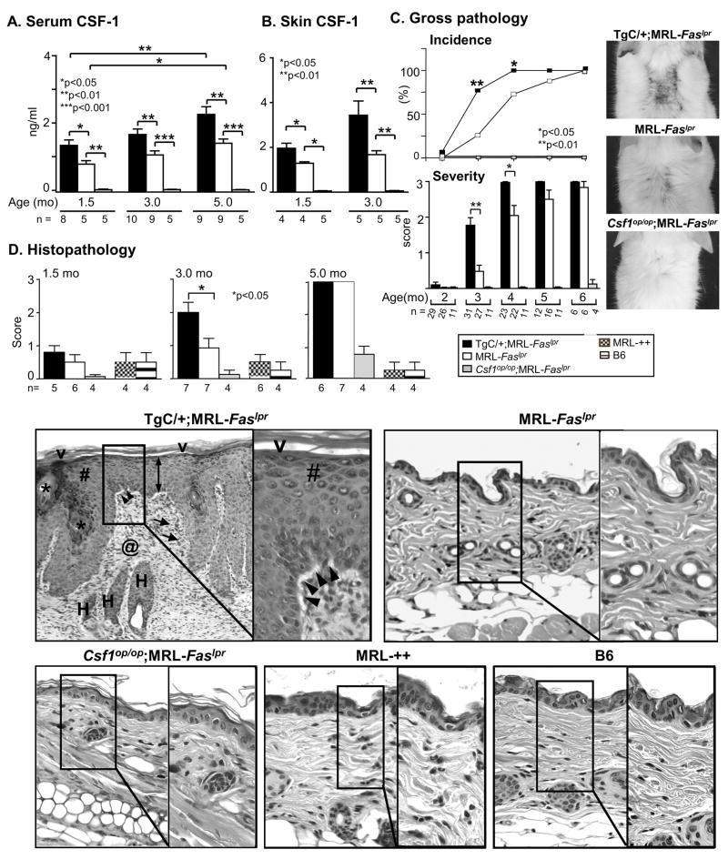
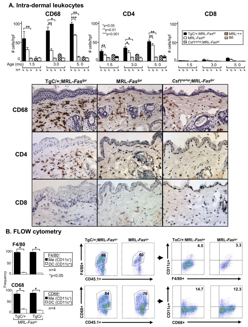
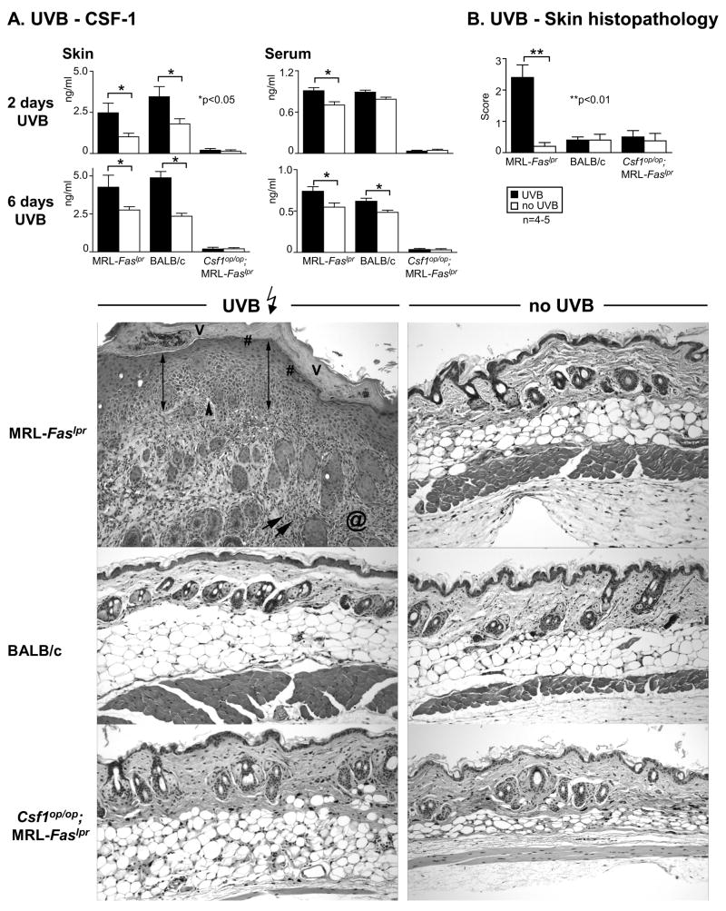
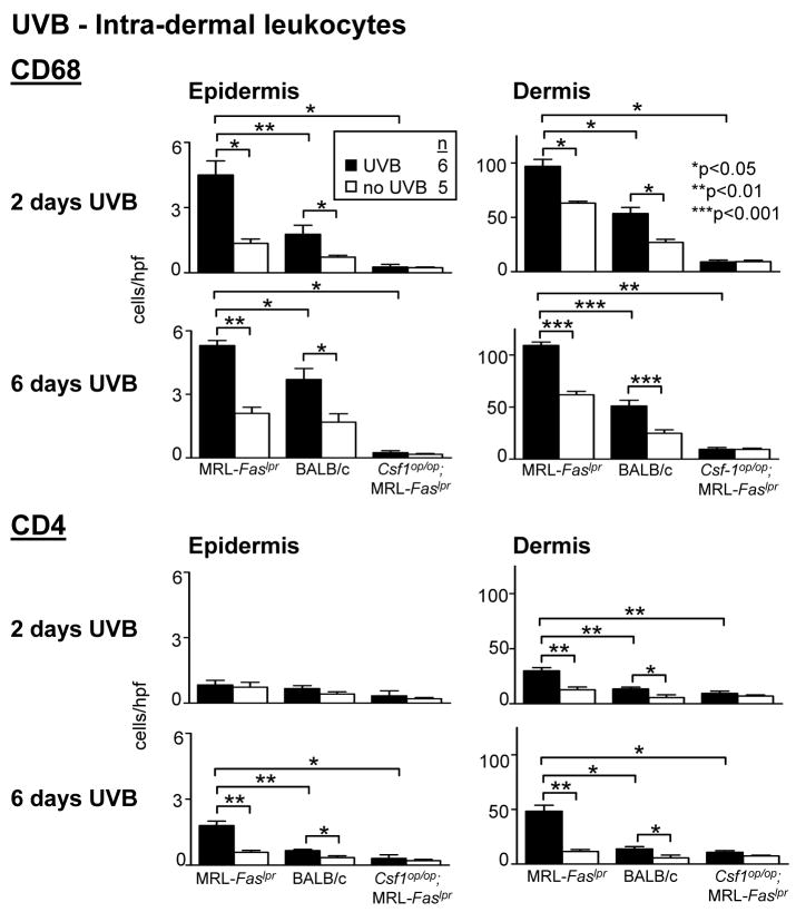
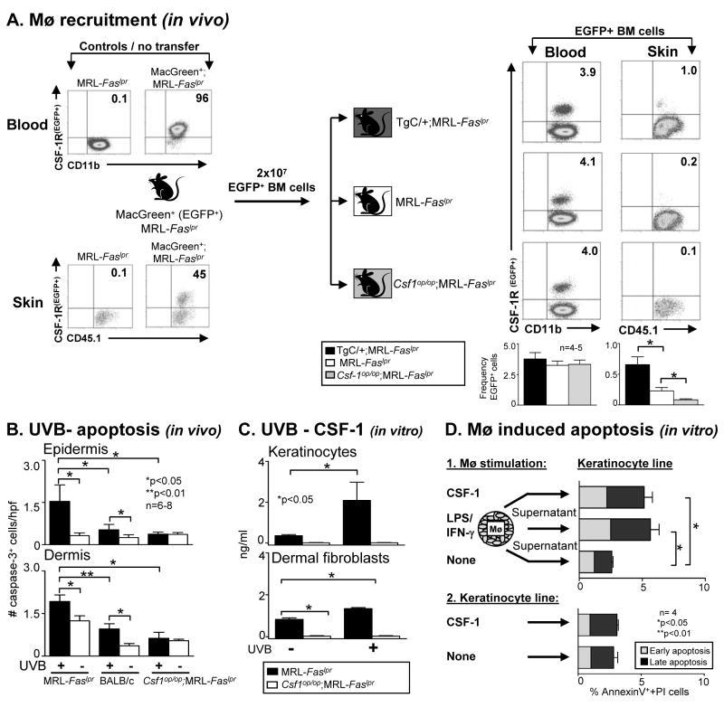
Sunlight (UVB) triggers cutaneous lupus erythematosus (CLE) and systemic lupus through an unknown mechanism. We tested the hypothesis that UVB triggers CLE through a CSF-1-dependent, macrophage (Mø)-mediated mechanism in MRL-Fas(lpr) mice. By constructing mutant MRL-Fas(lpr) strains expressing varying levels of CSF-1 (high, intermediate, none), and use of an ex vivo gene transfer to deliver CSF-1 intradermally, we determined that CSF-1 induces CLE in lupus-susceptible MRL-Fas(lpr) mice, but not in lupus-resistant BALB/c mice. UVB incites an increase in Møs, apoptosis in the skin, and CLE in MRL-Fas(lpr), but not in CSF-1-deficient MRL-Fas(lpr) mice. Furthermore, UVB did not induce CLE in BALB/c mice. Probing further, UVB stimulates CSF-1 expression by keratinocytes leading to recruitment and activation of Møs that, in turn, release mediators, which induce apoptosis in keratinocytes. Thus, sunlight triggers a CSF-1-dependent, Mø-mediated destructive inflammation in the skin leading to CLE in lupus-susceptible MRL-Fas(lpr) but not lupus-resistant BALB/c mice. Taken together, CSF-1 is envisioned as the match and lupus susceptibility as the tinder leading to CLE.


Figures

Figure 1
Figure 1
Cutaneous lesions in MRL-Faslpr mice share features with human discoid CLE. A. Gross cutaneous lesions in the intra-scapular area and ears in MRL-Faslpr mice. Histopathologically cutaneous lesions in MRL-Faslpr mice resemble the verrucoid variant of discoid lupus characterized by interface vacuolar changes (black arrowheads) with apoptotic keratinocytes (white open arrow), acanthotic epidermis (double headed arrow) with hyperkeratosis (V), hypergranulosis (#) and follicular plugging (*), superficial and deep, as well as perifollicular inflammatory infiltrates (arrows) and associated scarring alopecia with markedly reduced hair follicles (H) and dermal fibrosis (@). Representative photomicrographs from MRL-Faslpr mice (5 mo of age), H&E stained sections (n=18). Magnification: 20x, inset 40x. Inset in right panel: apoptotic keratinocytes, 60x. B. Positive lupus band (IgG and C3 deposits) at the dermal-epidermal junction in lesional and non-lesional skin of MRL-Faslpr mice (3 mo of age) determined by immunofluorescence staining. Representative of n=5–7. The pattern of IgM and IgG1 staining at the dermal-epidermal junction (data not shown) was more focal than IgG and C3. We did not detect a lupus band in BALB/c skin (data not shown). Stars indicate dermal-epidermal junction; dotted line the border of the epidermis to the environment. C. MRL-Faslpr skin is photosensitive. UVB ‚irradiation (6 consecutive days, 500J/m2) induces the lupus band (IgG, C3) as well as IgM and IgG1 (data not shown) at the dermal-epidermal junctions in MRL-Faslpr mice (2 mo of age), but not in BALB/c mice (data not shown). Representative of n = 4–6, experiment repeated twice.
Figure 2
Figure 2
Increasing CSF-1 expression in MRL-Faslpr mice accelerates the tempo of CLE. A. Serum CSF-1 levels in TgC/+;MRL-Faslpr, MRL-Faslpr and Csf1op/op;MRL-Faslpr mice at 1.5, 3.0, 5.0 mo of age and B. Skin CSF-1 levels in homogenates determined by ELISA. C. Time-related incidence and severity of skin lesions in TgC/+;MRL-Faslpr, MRL-Faslpr and Csf1op/op;MRL-Faslpr mice from 2–6 mo of age. Values are mean ± SEM. Representative photomicrographs from MRL-Faslpr at 3 mo of age. D. Skin (intrascapular) histopathology comparing TgC/+;MRL-Faslpr, MRL-Faslpr and Csf1op/op;MRL-Faslpr mice. MRL-++ and B6 mice with normal skin are controls. The cutaneous lesions in TgC/+;MRL-Faslpr mice include: interface vacuolar changes (black arrowheads), acanthotic epidermis (double arrow) with hyperkeratosis (V), hypergranulosis (#) and follicular plugging (*), superficial and deep, as well as perifollicular inflammatory infiltrates (arrows) and dermal scarring (@); Representative skin H&E stained sections (3 mo of age). Magnification: 20x, Inset 40x.
Figure 3
Figure 3
Intra-dermal leukocytes (CD68, CD4) increase in TgC/+;MRL-Faslpr mice compared with MRL-Faslpr mice. A. We evaluated the number of intra-dermal CD68+ cells (Mø, DC) and T cells (CD4+ and CD8+) from TgC/+;MRL-Faslpr, MRL-Faslpr, Csf1op/op;MRL-Faslpr mice, MRL-++ and B6 mice at 1.5, 3.0 and 5.0 mo of age. Representative photomicrographs are from mice at 3 mo of age. In addition, intra-epidermal leukocytes (CD68+ and CD4+) were increased (n= 4–7, data not shown). B. Intra-dermal CD68+ leukocytes in MRL-Faslpr mice are predominantly Mø. Flow cytometric analysis of intra-dermal Mø (F4/80+CD11c and CD68+CD11c) and DC (F4/80+CD11c+ and CD68+CD11c+) in TgC/+;MRL-Faslpr compared to MRL-Faslpr mice. Graphs depict the average frequency of CD11c and CD11c+ cells in the CD45.1+ F4/80+ and CD68+ populations. Values are mean ± SEM.
Figure 4
Figure 4
Increasing intra-dermal CSF-1 incites CLE in MRL-Faslpr mice. A.1) We verified that dermal fibroblasts from MRL-Faslpr and BALB/c mice genetically modified with a retroviral vector encoding CSF-1 constitutively express similar levels of CSF-1 (“CSF-1 carrier cells”),“empty vector carrier cells” serve as controls. A.2) CSF-1 was measured in the skin (area adjacent to infusion site) and serum of MRL-Faslpr and BALB/c mice following intra-dermal delivery of “carrier cells” at 3 days post-infusion. B. CLE is incited in the area of infusion and adjacent to infusion site 28 days post-infusion in MRL-Faslpr, but not BALB/c mice infused intra-dermally with “CSF-1 carrier cells” and characterized by interface vaculolar changes (black arrowheads) with hypergranulosis (#), and superficial, deep and peri-follicular inflammatory infiltrates (arrows) with scarring alopecia (@). Representative photomicrographs. Magnification: 20x, Inset 40x. C. Leukocyte-rich CLE incited by “CSF-1 carrier cells” in MRL-Faslpr, but not BALB/c mice. We evaluated intra-dermal Mø/DC (CD68+) and CD4+ and CD8+ T cells in the area adjacent to the “carrier cell” infusion. Data is representative of two separate experiments using dermal fibroblast and TEC “carrier cells” and an additional experiment infusing more (1.0 × 10⁷) “carrier cells”. Values are mean ± SEM.
Fig 5
Fig 5
UVB-irradiation increases CSF-1 expression and incites CLE in MRL-Faslp mice. A. We irradiated MRL-Faslpr, Csf1op/op;MRL-Faslpr and BALB/c mice daily (500J/m2 UVB) until we detected visible lesions (6 days). CSF-1 levels were measured (ELISA) in the skin and serum of MRL-Faslpr and BALB/c following 2 and 6 days of UVB-irradiation. UVB irradiated Csf1op/op;MRL-Faslpr mice served as negative controls. Values are mean ± SEM. B. Histopathologically, UVB incited CLE in MRL-Faslpr, but not BALB/c mice. The UVB incited features of CLE consisted of vacuolar interface changes (black arrowheads), apoptotic keratinocytes, acanthotic epidermis (double arrow) with hyperkeratosis (V), hypergranulosis (#), and superficial and deep, as well as perifollicular inflammatory infiltrates (arrows) with dermal fibrosis (@). Notably, UVB irradiated Csf1op/op;MRL-Faslpr mice did not develop CLE. Representative photomicrographs; Magnification: 20x. Data is representative of 2 separate experiments.
Figure 6
Figure 6
More CD68+ leukocytes are in the skin of MRL-Faslpr, Csf1op/op;MRL-Faslpr and BALB/c following UVB exposure. MRL-Faslpr, Csf1op/op;MRL-Faslpr and BALB/c mice (2 mo of age) exposed to UVB for 2 and 6 days. We analyzed the epidermis and dermis for CD68+ leukocytes and CD4+ T cells. Values are mean ± SEM.
Figure 7
Figure 7
CSF-1, generated by keratinocytes/dermal fibroblasts, recruits and activates Mø in the skin, which in turn, induce apoptosis in keratinocytes. A. CSF-1 recruits EGFP+ BM cells into the skin. We transferred BM cells from MacGreen (EGFP+);MRL-Faslpr mice into TgC/+;MRL-Faslpr, MRL-Faslpr and Csf1op/op;MRL-Faslpr mice and evaluated the recruitment of EGFP+ cells into the skin (flow cytometry). Data is representative of 3 separate experiments. B. UVB-induced apoptosis in epidermis and dermis is far more robust in MRL-Faslpr mice than in Csf1op/op;MRL-Faslpr and BALB/c mice. We evaluated the epidermis and dermis for apoptotic (cleaved-caspase-3+) cells following UVB-exposure (6 days). Sham-treated mice served as controls. Values are mean ± SEM. C. CSF-1 is up-regulated in mouse primary keratinocytes and dermal fibroblast (derived from MRL-Faslpr mice) irradiated with UVB one time with a dose of 500J/m2 in vitro. CSF-1 levels in the supernatant were analyzed 24 h after UVB-exposure (ELISA). Keratinocytes and dermal fibroblasts isolated from Csf1op/op;MRL-Faslpr mice served as negative controls. Data is representative of 2 separate experiments. D. Mø release mediators that induce apoptosis in keratinocytes. Supernatants from BMMø stimulated with CSF-1 (100μg/ml) released mediators that induced apoptosis in keratinocytes that was more robust than the apoptosis induced by supernatants from unstimulated BMMø (Annexin-V-FITC and PI using flow cytometry). Data is representative of 3 experiments. Values are mean ± SEM.
Figure 8
Figure 8
Scheme of UVB triggered CSF-1 and Mø mediated apoptosis during cutaneous lupus in MRL-Faslpr mice. 1) UVB induces CSF-1 expression in keratinocytes. 2) BMMø are recruited by CSF-1 to sites rich in CSF-1 in the skin (eg, keratinocytes). 3) CSF-1 activates Mø that release mediators and 4) induce apoptosis of adjacent cells (eg, keratinocytes).

Similar articles

Cited by

References

    1. Lin JH, Dutz JP, Sontheimer RD, Werth VP. Pathophysiology of cutaneous lupus erythematosus. Clin Rev Allergy Immunol. 2007;33:85–106. - PubMed
    1. Furukawa F, Yoshimasu T. Animal models of spontaneous and drug-induced cutaneous lupus erythematosus. Autoimmun Rev. 2005;4:345–350. - PubMed
    1. Werth VP. Cutaneous lupus: insights into pathogenesis and disease classification. Bull NYU Hosp Jt Dis. 2007;65:200–204. - PubMed
    1. Synkowski DR, Provost TT. Characterization of the inflammatory infiltrate in lupus erythematosus lesions using monoclonal antibodies. J Rheumatol. 1983;10:920–924. - PubMed
    1. Kanauchi H, Furukawa F, Imamura S. Characterization of cutaneous infiltrates in MRL/lpr mice monitored from onset to the full development of lupus erythematosus-like skin lesions. J Invest Dermatol. 1991;96:478–483. - PubMed

Publication types

MeSH terms

Substances