Comparative Study
. 2008 Oct 29;28(44):11174-85.
doi: 10.1523/JNEUROSCI.2093-08.2008.

Excitatory local connections of superficial neurons in rat auditory cortex

Affiliations
Comparative Study

Excitatory local connections of superficial neurons in rat auditory cortex

Dennis L Barbour et al. J Neurosci. .

Abstract

The mammalian cerebral cortex consists of multiple areas specialized for processing information for many different sensory modalities. Although the basic structure is similar for each cortical area, specialized neural connections likely mediate unique information processing requirements. Relative to primary visual (V1) and somatosensory (S1) cortices, little is known about the intrinsic connectivity of primary auditory cortex (A1). To better understand the flow of information from the thalamus to and through rat A1, we made use of a rapid, high-throughput screening method exploiting laser-induced uncaging of glutamate to construct excitatory input maps of individual neurons. We found that excitatory inputs to layer 2/3 pyramidal neurons were similar to those in V1 and S1; these cells received strong excitation primarily from layers 2-4. Both anatomical and physiological observations, however, indicate that inputs and outputs of layer 4 excitatory neurons in A1 contrast with those in V1 and S1. Layer 2/3 pyramids in A1 have substantial axonal arbors in layer 4, and photostimulation demonstrates that these pyramids can connect to layer 4 excitatory neurons. Furthermore, most or all of these layer 4 excitatory neurons project out of the local cortical circuit. Unlike S1 and V1, where feedback to layer 4 is mediated exclusively by indirect local circuits involving layer 2/3 projections to deep layers and deep feedback to layer 4, layer 4 of A1 integrates thalamic and strong layer 4 recurrent excitatory input with relatively direct feedback from layer 2/3 and provides direct cortical output.


Figures
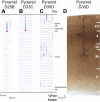
Figure 1.
Figure 1.
Spatial resolution of photostimulation technique. A, B, Voltage traces of layer 2/3 pyramidal neurons held in whole-cell current clamp indicate that action potentials in a neuron generally occur only when that neuron is photostimulated near its soma. Soma locations of patched cells are indicated by red triangles. Voltage traces (blue) are plotted at the corresponding site of laser stimulation. Laser duration of 10 ms is indicated by a horizontal line at each voltage plot. Scale bars correspond to either the voltage traces or distance within the slice. C, Neurons with extensive tufting in layer 1 also fire action potentials when stimulated near their tufts. A layer 5 pyramidal neuron recorded from its apical dendrite demonstrates not only stimulated action potentials near its soma but also near its extensive apical tuft in layer 1. For this reason, stimulation in layer 1 is not considered when evaluating input maps. D, Apical dendrite, nearly 1 mm long of layer 5 pyramidal neuron, is preserved within a single 80 μm section of the slice, indicating that intraslice projections are largely intact.
Figure 2.
Figure 2.
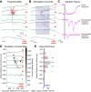
Photostimulation of a layer 2/3 pyramidal neuron. A, Drawing of pyramidal neuron dendrites (red) and axons (blue) as well as laminar boundaries (green). Inset shows current-clamp responses to depolarizing and hyperpolarizing currents and regular spiking behavior. B, Voltage-clamp current traces (blue) in response to the laser-scanning photostimulation grid reveal both large direct currents seen for stimulation near the soma that are disregarded and EPSCs seen for stimulation elsewhere in the slice. C, Expanded versions of sample traces (pink) reveal inward currents. These currents are brought about by (from top to bottom): direct stimulation near the soma, a strong monosynaptic connection and a weaker monosynaptic connection. D, EPSC areas in each trace of B are summed to create a charge transfer measurement for each stimulation site. Grayscale values in all current maps represent deciles from spontaneous activity to maximum stimulated response. E, Mean charge transfer for stimulation sites in each cortical layer reveal significant excitatory input from layers 2/3, 4, and 5a (***p < 0.001, ANOVA) and 5b (*p < 0.05).
Figure 3.
Figure 3.
Laminar input summaries for layer 2/3 pyramidal neurons. A, Mean laminar input Z scores computed for 35 layer 2/3 pyramidal neurons demonstrate significant excitatory input from layers 2–5a (Z > 1.96, p < 0.05). Additionally, the numbers of individual neurons receiving excitatory input from each layer (numbers associated with each horizontal bar) demonstrate significant input from layers 2–5a (***p < 0.001, binomial test). Input from layer 4 dominates in this neuronal population. B, Subdividing the layer 2/3 pyramidal neuron population by depth within layer 2/3 reveals that layer 2 pyramidal neurons receive substantially greater layer 4 input than layer 3 neurons (layer 2, black bars; layer 3, white bars). C, This trend can be seen more clearly by observing the excitatory input Z scores for each neuron as a function of depth within layer 2/3. Greater layer 4 input is apparent for many layer 2 neurons compared with layer 3. Division between layer 2 and 3 for the histograms of B is indicated by the horizontal line.
Figure 4.
Figure 4.
Laminar input correlations for layer 2/3 pyramidal neurons. A, Scatterplot showing the relationship between excitatory layer 4 and layer 2/3 input for layer 2/3 pyramidal neurons. A strong inverse trend is present in this population: more layer 4 input into a cell correlates with less layer 2/3 input. B, Relationship between excitatory layer 5 and layer 2/3 input. A weaker but similar inverse trend is present, such that more layer 5 input implies less layer 2/3 input. C, Relationship between excitatory layer 5 and layer 4 input. No significant trend is present, implying that the amount of layer 4 input poorly predicts the amount of layer 5 input.
Figure 5.
Figure 5.
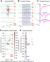
Photostimulation of a layer 4 pyramidal neuron. A, Drawing of pyramidal neuron dendrites (red) and axons (blue) as well as laminar boundaries (green). Inset shows regular spiking behavior. B, Voltage-clamp current traces (blue) in response to photostimulation. C, Expanded versions of sample traces (pink) reveal inward currents. D, EPSC areas in each trace of B are summed to create a charge transfer measurement for each stimulation site. Because of the large range of values for this neuron, many sites with low-amplitude stimulated events are mapped to the lowest decile, which yields a white pixel. E, Significant excitatory input derives from layers 4 and 5a (***p < 0.001) and layers 2/3 (**p < 0.01).
Figure 6.
Figure 6.
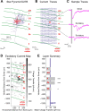
Photostimulation of a layer 4 star pyramid neuron. A, Drawing of star pyramid neuron dendrites (red) and axons (blue) as well as laminar boundaries (green). Inset shows regular spiking behavior. B, Voltage-clamp current traces (blue) in response to photostimulation. C, Expanded versions of sample traces (pink) reveal inward currents. D, EPSC areas in each trace of B are summed to create a charge transfer measurement for each stimulation site. E, Significant excitatory input derives from layers 4 and 5a (***p < 0.001), and layer 5b (*p < 0.05).
Figure 7.
Figure 7.
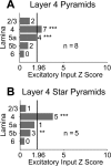
Laminar input summaries for layer 4 spiny neurons. A, B, Convention as in Figure 3A: Z > 1.96 corresponds to p < 0.05; ***p < 0.001; **p < 0.01. Input from layer 4 also dominates both of these neuronal populations.
Figure 8.
Figure 8.
Laminar input correlations for layer 4 spiny neurons. A–C, Scatterplots showing the pairwise relationships among excitatory layer 4, layer 2/3, and layer 5 input for layer 4 pyramidal neurons. The only trend present in this population is that increased layer 4 input implies decreased layer 5 input. D–F, Scatterplots showing the pairwise relationships among excitatory layer 4, layer 2/3, and layer 5 input for layer 4 star pyramid neurons. No correlation among input layers is evident in this population.
Figure 9.
Figure 9.
Photostimulation of layer 2/3 interneurons. A, Layer 2/3 fast-spiking interneuron charge transfer map. B, Layer 2/3 fast-spiking interneuron laminar charge transfer summary revealing significant excitatory input from layers 2/3 and 4 (***p < 0.001) and layer 5a (*p < 0.05). Inset shows fast-spiking behavior. C, Layer 2/3 adapting interneuron charge transfer map. D, Layer 2/3 adapting interneuron laminar charge transfer summary revealing significant excitatory input from layers 2/3 and 4 (***p < 0.001) and layer 5a (**p < 0.01).
Figure 10.
Figure 10.
Laminar input summaries for layer 2–4 aspiny neurons. A–D, Convention as in Figure 3A: Z > 1.96 corresponds to p < 0.05; ***p < 0.001; **p < 0.01; *p < 0.05.
Figure 11.
Figure 11.
Laminar input correlations for layer 2/3 aspiny neurons. A–C, Scatterplots showing the pairwise relationships among excitatory layer 4, layer 2/3, and layer 5 input for layer 2/3 fast spiking neurons. D–F, Scatterplots showing the pairwise relationships among excitatory layer 4, layer 2/3, and layer 5 input for layer 2/3 adapting neurons. Both of these neuronal populations appear to have a range of possible excitatory input patterns.
Figure 12.
Figure 12.
Laminar input proportions for all cell types. The mean fractional excitatory input from each of the laminar subdivisions demonstrates that layer 4 represents the single greatest laminar input into each neuronal type, and layer 4 neurons receive more deep input than do layer 2/3 neurons.

Similar articles

Cited by

References

    1. Agmon A, Connors BW. Correlation between intrinsic firing patterns and thalamocortical synaptic responses of neurons in mouse barrel cortex. J Neurosci. 1992;12:319–329. - PMC - PubMed
    1. Atzori M, Lei S, Evans DI, Kanold PO, Phillips-Tansey E, McIntyre O, McBain CJ. Differential synaptic processing separates stationary from transient inputs to the auditory cortex. Nat Neurosci. 2001;4:1230–1237. - PubMed
    1. Barbour DL, Callaway EM. Excitatory connections in superficial rat primary auditory cortex. Soc Neurosci Abs. 2004;30:987–12.
    1. Bender KJ, Rangel J, Feldman DE. Development of columnar topography in the excitatory layer 4 to layer 2/3 projection in rat barrel cortex. J Neurosci. 2003;23:8759–8770. - PMC - PubMed
    1. Binzegger T, Douglas RJ, Martin KA. A quantitative map of the circuit of cat primary visual cortex. J Neurosci. 2004;24:8441–8453. - PMC - PubMed

Publication types

LinkOut - more resources