. 2009 Jan;296(1):L57-70.
doi: 10.1152/ajplung.90411.2008. Epub 2008 Oct 24.

Lung alveolar integrity is compromised by telomere shortening in telomerase-null mice

Affiliations

Lung alveolar integrity is compromised by telomere shortening in telomerase-null mice

Jooeun Lee et al. Am J Physiol Lung Cell Mol Physiol. 2009 Jan.

Abstract

Shortened telomeres are a normal consequence of cell division. However, telomere shortening past a critical point results in cellular senescence and death. To determine the effect of telomere shortening on lung, four generations of B6.Cg-Terc(tm1Rdp) mice, null for the terc component of telomerase, the holoenzyme that maintains telomeres, were bred and analyzed. Generational inbreeding of terc-/- mice caused sequential shortening of telomeres. Lung histology from the generation with the shortest telomeres (terc-/- F4) showed alveolar wall thinning and increased alveolar size. Morphometric analysis confirmed a significant increase in mean linear intercept (MLI). terc-/- F4 lung showed normal elastin deposition but had significantly decreased collagen content. Both airway and alveolar epithelial type 1 cells (AEC1) appeared normal by immunohistochemistry, and the percentage of alveolar epithelial type 2 cells (AEC2) per total cell number was similar to wild type. However, because of a decrease in distal lung cellularity, the absolute number of AEC2 in terc-/- F4 lung was significantly reduced. In contrast to wild type, terc-/- F4 distal lung epithelium from normoxia-maintained mice exhibited DNA damage by terminal deoxynucleotidyltransferase (TdT)-mediated dUTP nick end labeling (TUNEL) and 8-oxoguanine immunohistochemistry. Western blotting of freshly isolated AEC2 lysates for stress signaling kinases confirmed that the stress-activated protein kinase (SAPK)/c-Jun NH(2)-terminal kinase (JNK) stress response pathway is stimulated in telomerase-null AEC2 even under normoxic conditions. Expression of downstream apoptotic/stress markers, including caspase-3, caspase-6, Bax, and HSP-25, was also observed in telomerase-null, but not wild-type, AEC2. TUNEL analysis of freshly isolated normoxic AEC2 showed that DNA strand breaks, essentially absent in wild-type cells, increased with each successive terc-/- generation and correlated strongly with telomere length (R(2) = 0.9631). Thus lung alveolar integrity, particularly in the distal epithelial compartment, depends on proper telomere maintenance.


Figures

Fig. 1.
Fig. 1.
Determination of telomere length on chromosomes isolated from wild-type (WT) F4 and terc−/− F2, F3, and F4 alveolar epithelial type 2 cells (AEC2). Mean terminal restriction size of telomeres on chromosomes from WT F4 (n = 7) and terc−/− F2 (n = 6), F3 (n = 4), and F4 (n = 5) AEC2 hybridized with a fluorescein-tagged PNA telomere probe is shown. Values are derived from histogram data plotted onto a standard curve generated by FITC-labeled Quantum MESF beads. By Spearman nonparametric rank correlation, **P = 0.001 for differences between WT and terc−/− samples, *P = 0.0142 for differences among terc−/− samples. Nonparametric rank testing was used to compare WT F4 to terc−/−samples: †P = 0.0455, WT F4 vs. terc−/−F2; #P = 0.014, WT F4 vs. terc−/−F3; §P = 0.007, WT F4 vs. terc−/− F4.
Fig. 2.
Fig. 2.
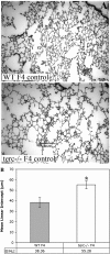
Morphometric analysis of distal lung tissue shows an increase in mean linear intercept (MLI) in terc−/− lung under normoxic conditions. A: lung tissue from terc−/− F4 and WT F4 mice maintained in normoxia. Lungs from terc−/− F4 and WT F4 mice were sectioned and stained with hematoxylin and eosin. Sections were prepared from lungs harvested from animals maintained in normoxia (control) and observed at ×20. Representative sections from the 8–12 observed for each sample are presented. B: MLI analysis of lung tissue samples from WT F4 and terc−/− F4 normoxia-maintained mice. Hematoxylin and eosin-stained lung sections from terc−/− F4 and WT F4 animals maintained in normoxia were analyzed for MLI. For each sample, n = 8–12. *P = 0.001.
Fig. 3.
Fig. 3.
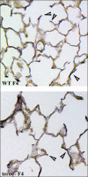
Elastin deposition in lung tissue from normoxia-maintained WT F4 and terc−/− F4 mice. Lung sections from WT F4 and terc−/− F4 lung were stained with Hart's resorcin-fuchsin, counterstained with tartrazine-acetic acid, mounted with Permount, and observed at ×20. Hart's staining produces dark brown elastin fibers. Elastin deposition was observed along alveolar walls and at the tips of alveolar septa (arrows).
Fig. 4.
Fig. 4.
Collagen content of lung tissue from normoxia-maintained WT F4 and terc−/− F4 mice. Whole lung from normoxia-maintained WT F4 and terc−/− F4 mice was analyzed for collagen content by acid hydrolysis. For each strain, n = 6. Mean insoluble collagen content in WT F4 lung was 207.32 (SE ±21.69) μg/g wet wt, while mean content of terc−/− F4 lung was 166.60 (SE ±28.43) μg/g wet wt. This difference was not significant. Mean soluble collagen content in WT F4 lung was 123.90 (SE ±6.34) μg/g wet wt, while mean content of terc−/−F4 lung was 67.53 (SE ±7.50) μg/g wet wt. By Student's t-test, this difference was highly significant (*P = 0.002).
Fig. 5.
Fig. 5.
Analysis of Clara cell secretory protein (CCSP)-positive airway cells, T1a-positive AEC1, and surfactant protein-C (SP-C)-positive AEC2 in normoxia-maintained WT F4 and terc−/− F4 samples in situ. A: lung tissue from WT F4 and terc−/− F4 mice maintained in normoxia was analyzed for CCSP and SP-C expression by immunohistochemistry. To control for nonspecific antibody staining, purified rabbit or hamster IgG was used at the same concentration as primary anti-CCSP, anti-T1a, and anti-SP-C antibodies (not shown). Sections were fixed and subjected to immunohistochemistry with the primary antibodies indicated and a Cy3-labeled anti-rabbit IgG secondary antibody. Top: CCSP-positive cells are shown lining the large airways in each sample. Middle: long, thin, T1a-positive AEC1 can be observed lining alveolar walls. Bottom: arrows indicate brightly staining SP-C-positive AEC2 scattered through lung parenchyma. In the WT F4 section, arrows point to a portion of all SP-C-positive cells, while in the terc−/− F4 section, arrows point to all SP-C-positive cells identified. All panels were observed at ×20. B: quantitation of SP-C positive AEC2 present in WT F4 and terc−/− F4 lung tissue. SP-C-labeled sections were observed microscopically, and the number of SP-C-positive cells was counted per microscopic field at ×20. For each sample, n = 8. The mean number of SP-C positive cells per field in normoxic WT lung was 42.6 (SE ±2.88), while in normoxic terc−/− F4 lung, the number was 19.2 (SE ±6.53). The 2-tailed P value for comparison of these populations was highly significant (*P = 0.0095) C: quantitation of SP-C-positive AEC2 present in WT F4 and terc−/−F4 lung tissue as % of total cell number. SP-C-labeled sections were observed microscopically, and the number of SP-C-positive cells was counted per total number of cells (identified by positive DAPI staining) per microscopic field at ×20. For each sample, n = 8. Mean % of SP-C positive cells per field in normoxic WT lung was 12.4% (SE ±0.89), while in normoxic terc−/−F4 lung, the mean was 10.6% (SE ±4.39). The 2-tailed P value for comparison of these populations was not significant (P = 0.6773).
Fig. 6.
Fig. 6.
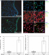
terc−/− F4 lung tissue exhibits markers for DNA damage under normoxic conditions. A: TUNEL in situ of normoxia-maintained WT F4 and terc−/− F4 samples. Lung tissue samples were subjected to TUNEL to detect cells carrying DNA strand breaks. FITC-labeled dUTP incorporated into damaged DNA appears green, while tissue cell nuclei were stained blue with DAPI. Sections were observed at ×20. B: quantitation of TUNEL in situ of normoxia-maintained WT F4 and terc−/− F4 samples. TUNEL-labeled sections were observed microscopically, and the number of TUNEL-positive cells was counted per total number of cells (by DAPI) in each microscopic field at ×20. Fields were counted from sections obtained the left lobe of each lung. For each sample, n = 3. *P = 0.0044 by Student's t-test. C: 8-oxoguanine (8-OHdG, 8-oxo-dG) immunohistochemical analysis of normoxia-maintained WT F4 and terc−/− F4 samples and colocalization with SP-C expression. Top: lung tissue from WT F4 and terc−/− F4 mice maintained in normoxia was analyzed for 8-OHdG expression by immunohistochemistry. Green arrows indicate brightly staining OHdG-positive cells. Middle: immunohistochemistry for 8-OHdG was combined with staining for T1a expression. T1a-positive and 8-OHdG double-positive cells are indicated by green arrows. Bottom: immunohistochemistry for 8-OHdG was combined with staining for SP-C expression. SP-C-positive cells and OHdG-positive cells are indicated by red and green arrows, respectively. Yellow arrows point to red, SP-C-positive cells with cytoplasmic expression (green) of 8-OHdG. Sections were observed at ×40. D: quantitation of 8-OHdG in situ of normoxia-maintained WT F4 and terc−/− F4 samples. 8-OHdG-labeled sections were observed microscopically, and the number of OHdG-positive cells was counted per whole left lobe section. Six sections from 2 individual animals were analyzed for each sample (n = 6). *P = 0.0042 by Student's t-test.
Fig. 7.
Fig. 7.
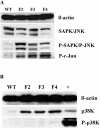
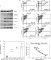
AEC2 isolated from normoxia-maintained terc−/− F2, F3, and F4 lung show that stress response signaling is exclusively activated in the stress-activated protein kinase (SAPK)/c-Jun NH2-terminal kinase (JNK) pathway. AEC2 freshly isolated from normoxia-maintained WT and terc−/− lungs were analyzed for protein expression by Western blotting. Data presented are representative of expression in AEC2 isolated from 3 separate animals from each generation (WT F4, terc−/− F2, F3, and F4). Equality of loading was determined by probing for β-actin expression. A: SAPK/JNK is phosphorylated and therefore activated to phosphorylate its downstream target, c-Jun, in normoxia-maintained terc−/− AEC2. The level of expression of stress signaling kinase proteins SAPK/JNK is similar in WT F4 AEC2 and all terc−/− samples. Phosphorylation, and therefore activation, of SAPK/JNK in normoxic AEC2 only occurs in terc−/− AEC2. No significant difference in the level activation was observed among terc−/− generations by densitometric scanning (not shown). Phosphorylation of the JNK downstream target, c-Jun, at Ser63 occurs only in terc−/− AEC2. Although the level of phosphorylation in terc−/− AEC2 trended higher with increasing generation and decreasing telomere length, the difference among terc−/− samples was not significant by densitometric analysis (not shown). B: the stress-activated protein kinase p38K is expressed but not activated in normoxia-maintained terc−/− AEC2. Blots for WT F4 and terc−/− AEC2 from 3 generations showed no expression of p38K in WT AEC2 but elevated expression in all normoxic terc−/− samples. Probing for p38K phosphorylated at Thr180/Tyr182 showed no activation of the kinase in either WT F4 or terc−/− samples. Specificity of the P-p38K antibody was confirmed by inclusion of a positive control sample (+).
Fig. 8.
Fig. 8.
AEC2 isolated from normoxia-maintained terc−/− F2, F3, and F4 lung exhibit altered expression of markers for DNA damage, oxidative stress, and apoptosis. A: expression of markers for apoptosis and stress in AEC2 isolated from normoxia-maintained terc−/− lung. AEC2 freshly isolated from normoxia-maintained WT and terc−/− lung were analyzed for protein expression by Western blotting. Data presented are representative of expression in AEC2 isolated from 3–5 separate animals from each generation (WT F4, terc−/− F2, F3, and F4). Equality of loading was determined by probing for β-actin expression. The levels of expression of apoptotic markers caspase-3 (uncleaved and cleaved), caspase-6, apoptotic agonist Bax, and stress marker heat shock protein (HSP)-25 were then analyzed. B: TUNEL FACS analysis of AEC2 isolated from normoxia-maintained WT and terc−/− lung. Fresh, uncultured AEC2 isolated from WT F4 and terc−/− F2, F3, and F4 lung were fixed and analyzed by FACS to ascertain level of TUNEL. A representative experiment from 3 repetitions is shown. C: quantitation of TUNEL-positive AEC2 isolated from normoxia-maintained WT and terc−/− lung. Data from 3 separate experiments were analyzed, and the mean numbers of TUNEL-positive AEC2 were compared. In WT F4 animals maintained in normoxia, mean % of TUNEL-positive cells was 2.82% (SE ±1.38). Under the same conditions, mean % of TUNEL-positive AEC2 in terc−/− F2 fresh isolates increased to 11.1% (SE ±2.15). In terc−/− F3 isolates, % was 16.91% (SE ±0.57), while % in terc−/− F4 AEC2 was 19.9% (SE ±4.04). By Spearman nonparametric rank correlation analysis of the difference between WT F4 and terc−/− samples, *P = 0.001. For differences among terc−/− samples as a group, **P = 0.011. D: correlation of telomere length to the level of TUNEL present in AEC2 isolated from normoxia-maintained WT and terc−/− lung. Mean % of AEC2 that were TUNEL positive from each generation were correlated with mean telomere length. Regression analysis gave an R2 value of 0.9631.
Fig. 9.
Fig. 9.
Schematic for possible mechanisms that underlie the differences observed in cellular integrity of WT AEC2 vs. terc−/− AEC2. In WT AEC2 either the normoxic environment does not induce stress signaling or minor stress events are modulated by both translocation of TERT to mitochondria and the stabilizing influence of normal-length telomeres. Both events feed back to inhibit the JNK signaling cascade, resulting in quiescence and maintenance of cellular integrity. In terc−/− AEC2 cells cannot respond to minor stress events by TERT translocation. Any resulting mitochondrial damage feeds back to upregulate stress signaling. These effects may be amplified in a dose-dependent manner by chronic instability induced by shortened telomeres, leading to loss of cellular integrity, the level of which correlates with telomere length.

Similar articles

Cited by

References

    1. Ahmed S, Passos JF, Birket MJ, Beckmann T, Brings S, Peters H, Birch-Machin MA, von Zglinicki T, Saretzki G. Telomerase does not counteract telomere shortening but protects mitochondrial function under oxidative stress. J Cell Sci 121: 1046–1053, 2008. - PubMed
    1. Albertine KH, Plopper CG. DNA oxidation or apoptosis: will the real culprit of DNA damage in hyperoxic lung injury please stand up? Am J Respir Cell Mol Biol 26: 381–383, 2002. - PubMed
    1. Alder JK, Chen JJ, Lancaster L, Danoff S, Su SC, Cogan JD, Vulto I, Xie M, Qi X, Tuder RM, Phillips JA 3rd, Lansdorp PM, Loyd JE, Armanios MY. Short telomeres are a risk factor for idiopathic pulmonary fibrosis. Proc Natl Acad Sci USA 105: 13051–13056, 2008. - PMC - PubMed
    1. Armanios MY, Chen JJ, Cogan JD, Alder JK, Ingersoll RG, Markin C, Lawson WE, Xie M, Vulto I, Phillips JA 3rd, Lansdorp PM, Greider CW, Loyd JE. Telomerase mutations in families with idiopathic pulmonary fibrosis. N Engl J Med 356: 1317–1326, 2007. - PubMed
    1. Atexier C POT of gold: modeling dyskeratosis congenita in the mouse. Genes Dev 22: 1731–1736, 2008. - PMC - PubMed

Publication types

MeSH terms

LinkOut - more resources