Review
. 2008 Oct;18(5):495-503.
doi: 10.1016/j.conb.2008.10.003. Epub 2008 Oct 27.

Axonal transport and the delivery of pre-synaptic components

Affiliations
Review

Axonal transport and the delivery of pre-synaptic components

Ann Y N Goldstein et al. Curr Opin Neurobiol. 2008 Oct.

Abstract

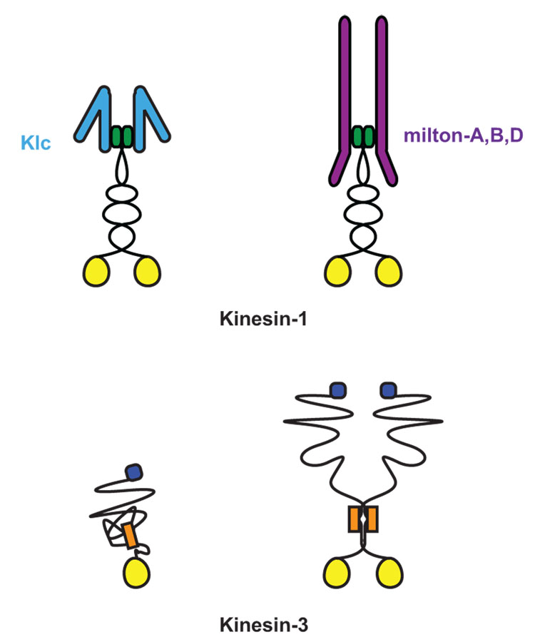
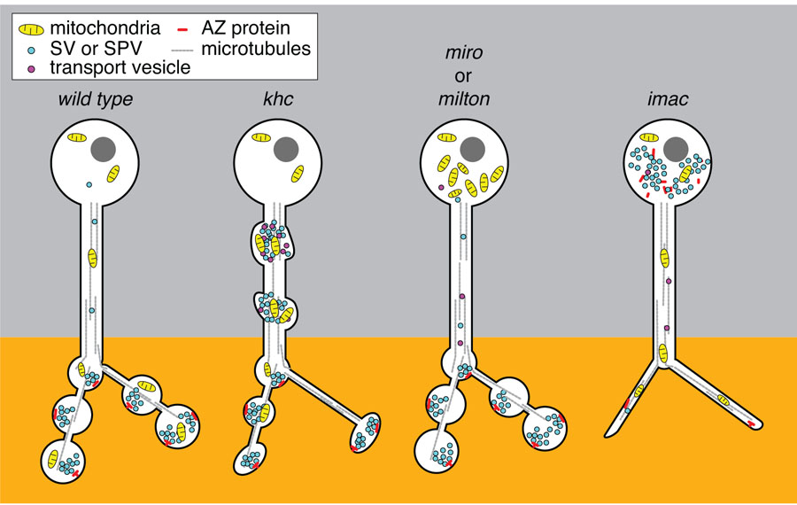
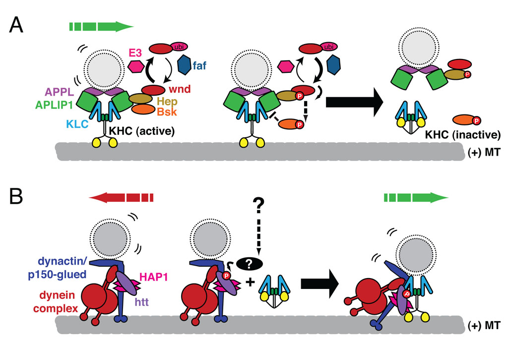
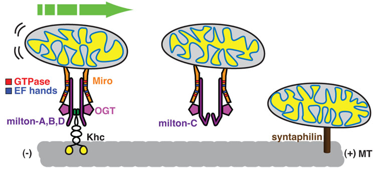
The mechanisms for delivering components to nerve terminals are diverse and highly regulated. The diversity of kinesin motors alone is insufficient to account for the specificity of delivery. Additional specificity and control are contributed by adaptor proteins and associated regulatory molecules. The interaction of cargos with these complexes can confer distinct behaviors on the transport of synaptic organelles. The rich regulatory mechanisms of transport that are only now emerging as the cargo-motor complexes are defined and subsequent local events that regulate their dynamic relationship are examined. Here we review recent studies of kinesin-related axonal transport of three crucial synaptic components, Piccolo-bassoon Transport Vesicles (PTVs), Synaptic Vesicle Precursors (SVPs), and mitochondria, and the mechanisms that modulate their transport.


Conflict of interest statement

Conflicts of interest

The authors have no conflicts of interest with the publication of this article.

Figures

Figure 1
Figure 1. Meet the family of Kinesin-1 and Kinesin-3
Kinesins are defined by their highly conserved ATP binding and microtubule binding motor domain (yellow circle) [8]. Both Kinesin-1 and Kinesin-3 family members have their motor domain at their N-termini and move toward the (+)-end microtubules. The Kinesin-1 family subfamily of KIF5, or kinesin heavy chain (KHC), is a homodimer that dimerizes via coiled-coil domains at its neck. KHC associates with two kinesin light chains (KLC) to link it to multiple cargo complexes [7]. Although initially believed to act as an obligate tetramer with KLC, KHC can also associate with cargos via a specialized adaptors independent of KLC, as is the case with the mitochondrial adaptor protein, Milton [•36]. In contrast, Kinesin-3 family members have been found as both monomers and dimers and are able to associate to vesicular cargo directly. Kinesin-3 family members share a conserved fork-head association domain (orange box) and multiple coiled-coil domains at the neck of the motor [7,8]. The defining motor of the Kinesin-3 family, Unc-104, has a pleckstrin homology domain (blue square) that is necessary for its association with synaptic vesicle precursors [24].
Figure 2
Figure 2. Distinct functions of Kinesin-1 and Kinesin-3 family motors for transport in the axon
The presynaptic compartment of the neuron is normally enriched with mitochondria and SVs in close apposition to a density of active zone proteins. Loss of the lone Kinesin-1 motor in the fly, KHC, leads to accumulations of mitochondria, SPV as well as post-golgi transport vesicles in the axon [7]. This is striking when compared to what occurs in the absence of either milton or Miro, the adaptor complex that associates KHC to mitochondria. Mitochondria are stranded in the cell body of both milton and miro mutants and do not generate traffic jams in the axonal processes or disrupt the distribution of SVs or active zone proteins [•36,38]. One interpretation of this discrepancy between the phenotype of Milton/Miro and KHC is that the traffic jams in the khc mutants are due to the mislocalization of additional cargos of KHC. For example, through binding of the JIP1 signaling complex associated with KLC and transport vesicles, decreased KHC dependent transport could compromise both the integrity of the cytoskeletal network as well as the signal for the proper sorting of cargos. In contrast, loss of the Kinesin-3 family motors Unc-104 or its fly homologue, Imac, leads to a severe reduction of synaptic vesicles (SVs) at terminals and an increase of SVs stranded in the cell body [7]. Mitochondrial distribution and axon guidance and growth remains normal in unc-104 and imac mutants demonstrating their specialized role for SVP transport. In addition, in the absence of Imac, morphologically mature synaptic endings do not form and active zone proteins are stranded in the cell body along with a notable reduction of active zones (AZ) at the synapse [••9]. Although loss of KHC does disrupt the normal trafficking of SVs and demonstrates a reduction of synapses, the severity of the imac phenotype implies that it is likely the primary motor to initiate the transport of SVs and as yet unidentified proteins critical for morphological synapse development.
Figure 3
Figure 3. Phosphorylation dependent regulation of transport
Two recent examples of how transport of vesicular cargos is modulated by adaptor complexes are illustrated above. (A) Recent work in the fly indicates that the interaction between the KHC/KLC kinesin complex and the JNK adaptor, JIP1 (APLIP1 in the fly) is modulated by the JNK signaling pathway [••14]. Synaptic material accumulates in axons when APLIP1 is overexpressed. These accumulations are alleviated by the overexpression of either the ubiquitin hydrolase, fat facets (faf) or the MAPKKK, wallenda (wnd). Transport defects are similarly apparent with the loss of wnd or the activity of its downstream signaling partners the MAPKK, hemipterous (Hep), or the JNK homologue, basket (Bsk), suggesting that JNK signaling is necessary for maintaining or initializing the transport of APLIP1 cargos, i.e. APPL- associated transport vesicles. The kinase activity of wnd promotes the activation of Hep, increased phospho-JNK, and decreased binding of APLIP1 with KLC. Thus the activity of the ubiquitin proteasome system may normally reduce wnd activity and thereby allow adaptor-cargo complexes to assemble. Increasing wnd activity, via modulation of the ubiquitination pathway, e.g via the E3 ubiquitin ligase Highwire, or direct activation of wnd would promote the dissociation of the motor and cargo and inactivation of the kinesin [64,65]. (B) A recent study helps to resolve how the Huntingtin protein (htt) may regulate the bidirectional movement of vesicles [18]. Htt associates with both the dynein/dynactin retrograde motor complex and kinesin-1, in part via huntingtin associated protein (HAP1) [•20,66]. Insulin growth factor, via the kinase Akt, phosphorylates htt at serine 421 and increased the association of KHC with vesicles, thereby promoting anterograde transport of BDNF-containing vesicles. In this model, phosphorylation of htt controls the balance between retrograde and anterograde forces on this cargo and others and places htt at the center of signaling pathways that may modulate transport normally and in disease states.
Figure 4
Figure 4. Mitochondria on the move
The KHC/milton/Miro complex is one of the best-defined motor/adaptor/cargo complexes. The association of milton splice isoforms that allow for binding to KHC (milton-A, B or D) links the motor with the mitochondrial membrane protein, Miro. In the presence of milton-C, miro and mitochondria do not associate with KHC, perhaps allowing for a competitive pathway to stop mitochondria when they reach a destination of high milton-C expression. Miro has GTPase domains as well as EF hands that may allow it to differentially bind to the Milton/KHC complex to link mitochondria to a microtubule transport pathway. Milton binds to and is glycosylated by O-GlcNAc transferase (OGT) binds. Glycosylation of milton has an unknown function but may serve as an additional signal links the metabolism of the cell with regulation of mitochondrial transport. Recent identification of a synaptic protein, synaptophilin, critical for the docking of mitochondria by linking it directly to microtubules adds a new layer of possibilities to how the motility of mitochondria are controlled in neurons.

Similar articles

Cited by

References

    1. De Vos KJ, Grierson AJ, Ackerley S, Miller CC. Role of Axonal Transport in Neurodegenerative Diseases. Annu Rev Neurosci. 2008;31:151–173. - PubMed
    1. Stokin GB, Goldstein LS. Axonal transport and Alzheimer's disease. Annu Rev Biochem. 2006;75:607–627. - PubMed
    1. Saxena S, Caroni P. Mechanisms of axon degeneration: from development to disease. Prog Neurobiol. 2007;83:174–191. - PubMed
    1. Schaefer AM, Sanes JR, Lichtman JW. A compensatory subpopulation of motor neurons in a mouse model of amyotrophic lateral sclerosis. J Comp Neurol. 2005;490:209–219. - PubMed
    1. Ibanez CF. Message in a bottle: long-range retrograde signaling in the nervous system. Trends Cell Biol. 2007;17:519–528. - PubMed

Publication types