. 2008 Nov 1;181(9):6427-34.
doi: 10.4049/jimmunol.181.9.6427.

Receptor-interacting protein homotypic interaction motif-dependent control of NF-kappa B activation via the DNA-dependent activator of IFN regulatory factors

Affiliations

Receptor-interacting protein homotypic interaction motif-dependent control of NF-kappa B activation via the DNA-dependent activator of IFN regulatory factors

William J Kaiser et al. J Immunol. .

Abstract

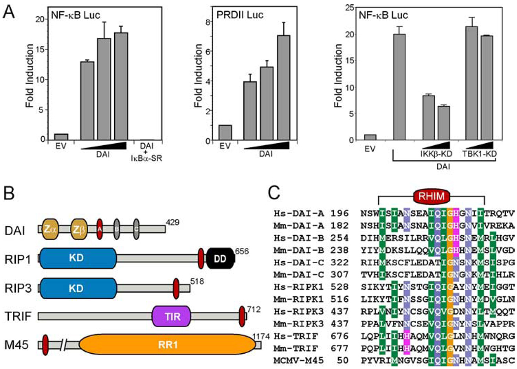
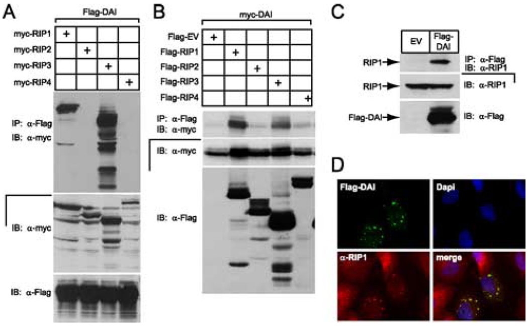
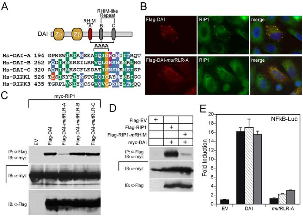
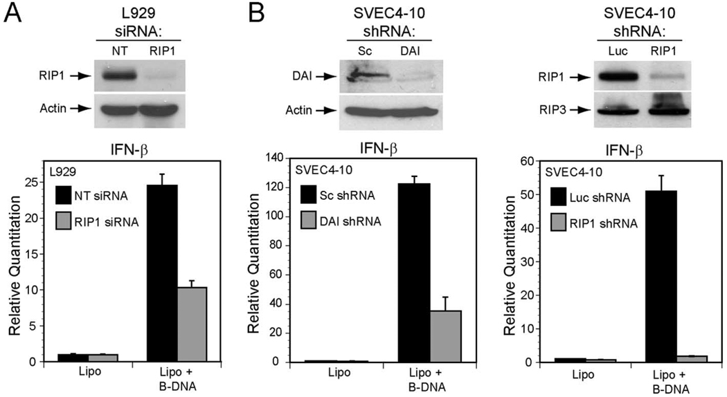
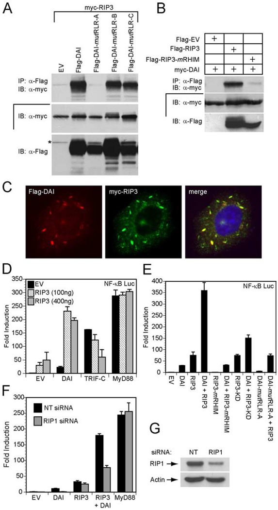
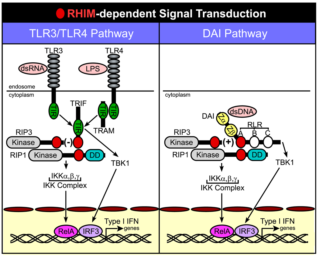
DNA-dependent activator of IFN regulatory factors (IRF; DAI, also known as ZBP1 or DLM-1) is a cytosolic DNA sensor that initiates IRF3 and NF-kappaB pathways leading to activation of type I IFNs (IFNalpha, IFNbeta) and other cytokines. In this study, induction of NF-kappaB is shown to depend on the adaptor receptor-interacting protein kinase (RIP)1, acting via a RIP homotypic interaction motif (RHIM)-dependent interaction with DAI. DAI binds to and colocalizes with endogenous RIP1 at characteristic cytoplasmic granules. Suppression of RIP1 expression by RNAi abrogates NF-kappaB activation as well as IFNbeta induction by immunostimulatory DNA. DAI also interacts with RIP3 and this interaction potentiates DAI-mediated activation of NF-kappaB, implicating RIP3 in regulating this RHIM-dependent pathway. The role of DAI in activation of NF-kappaB in response to immunostimulatory DNA appears to be analogous to sensing of dsRNA by TLR3 in that both pathways involve RHIM-dependent signaling that is mediated via RIP1, reinforcing a central role for this adaptor in innate sensing of intracellular microbes.


Conflict of interest statement

Disclosures

The authors have no financial conflict of interest.

Figures

FIGURE 1
FIGURE 1
DAI-mediated activation of NF-κB. A, 293T cells were transfected with 100 ng firefly luciferase reporter plasmid carrying five tandem NF-κB sites (left panel; NF-κB-Luc; Stratagene) or carrying two tandem NF-κB sites from the IFNβ promoter (middle panel; positive regulatory domain II-Luc) (28), together with 20, 100, or 500 ng (denoted by triangle) of Flag-DAI and 5 ng of a control noninducible Renilla luciferase expression vector, phRL-TK. IκBα-SR-mediated (400 ng) inhibition of DAI-dependent (100 ng) NF-κB-Luc activation is also shown in the left panel. 293T cells were transfected with 100 or 400 ng of plasmid expressing TBK1-KD or IKKβ-KD (28) together with 100 ng Flag-DAI (right panel). In all experiments, the total DNA transfected was held constant (605 ng) by adding empty vector (EV). Firefly and Renilla luciferase activities were assayed 18 h posttransfection. Firefly luciferase activity was divided by Renilla luciferase activity to normalize for transfection efficiency. Data shown are expressed as the mean relative stimulation in cells receiving DAI compared with the constitutive level present in cells transfected with EV ± SD (n = 3) from one representative experiment of a total of three performed. B, Schematic representation showing domains of RHIM-containing proteins. DAI contains an N-terminal Zα and Zβ domain and three RLR (labeled A, B, and C). The RLR-A motif in DAI is most similar to the RHIM (red oval). RIP1 and RIP3 each have an N-terminal kinase domain (KD), and RIP1 also contains a C-terminal DD. TRIF encodes a TIR domain, and M45 has a region of homology to a ribonucleotide reductase (RR1). C, The RHIM of human (Hs) and mouse (Mm) RIP1, RIP3, and TRIF and the MCMV protein M45 is aligned with each of the RLR motifs of DAI. The alignment was shaded with the BLOSUM62 matrix at a 60% identity threshold. The number adjacent to each sequence in the alignment indicates the starting amino acid within each full-length protein.
FIGURE 2
FIGURE 2
DAI interaction with the RHIM-containing RIP kinases, RIP1 and RIP3. A, Autoradiogram of IB and IP of 293T cell extracts following transfection of myc epitope-tagged RIP1, RIP2, RIP3, or RIP4 together with Flag epitope-tagged DAI vectors. B, Autoradiogram of IB and IP of 293T cell extracts following transfection of Flag epitope-tagged RIP1, RIP2, RIP3, and RIP4, or an EV together with myc epitope-tagged DAI vector. Cellular lysates were subjected to anti-Flag IP and analyzed by IB with anti-myc Ab. IB analysis of total cell lysate showed the relative expression of Flag and myc epitope-tagged proteins. C, Demonstration that Flag-DAI binds endogenous RIP1 via IB of 293T cells following transfection with Flag epitope-tagged DAI or an EV. IP was with anti-FLAG M2 beads and IB was with anti-RIP1 Ab. Total cell lysates were examined for the expression of RIP1 and Flag-DAI. D, Immunofluorescent localization of Flag-DAI with endogenous RIP1. HeLa cells were transfected with Flag-tagged DAI and then stained with rabbit anti-Flag Ab and with monoclonal anti-RIP1 Ab for endogenous RIP1. Flag-DAI was detected by an anti-rabbit Alexa-488-conjugated secondary Ab and RIP1 was detected by an anti-mouse Alexa-594-conjugated secondary Ab. Stained cells were examined by epifluorescent microscopy (×1000).
FIGURE 3
FIGURE 3
DAI-mediated, RIP1 RHIM-dependent activation of NF-κB. A, Diagram of RHIM mutations made by alanine substitution of the indicated aa (AAAA above the sequence alignment). B, Immunofluorescence analysis of Flag-DAI and Flag-DAI-mutRLR-A colocalization with endogenous RIP1. HeLa cells were transfected with Flag-tagged DAI or mutRLR-A and then stained with rabbit anti-Flag Ab and with monoclonal anti-RIP1 Ab for endogenous RIP1. Flag-DAI was revealed by an anti-rabbit Alexa-594-conjugated secondary Ab and RIP1 by an anti-mouse Alexa-488-conjugated secondary Ab. Stained cells were examined by microscopy (×1000). C, Autoradiogram following IP and IB to detect RHIM-dependent DAI interaction with RIP1. 293T cells were transfected with c-myc epitope-tagged RIP1 and Flag epitope-tagged DAI, DAI-mutRLR-A, DAI-mutRLR-B, DAI-mutRLR-C, or an EV. IP of cellular lysates was with anti-FLAG M2 beads and IB was with anti-myc Ab. IB analysis of total cell lysate determined the expression of myc-RIP1 and Flag-tagged proteins. D, Autoradiograph following IP and IB analysis to detect DAI-RIP1 interaction. 293T cells were transfected with myc epitope-tagged DAI and Flag epitope-tagged RIP1, RIP1-mRHIM, or an EV. IP of cellular lysates was followed by IB with anti-myc Ab. Anti-Flag Ab revealed the relative expression of epitope-tagged proteins in original lysates. E, Luciferase expression assay in 293T cells transfected with 100 ng of NF-κB-Luc together with 50 ng (black bar), 150 ng (hatched bar), or 450 ng (gray bar) of the indicated Flag-DAI expression vector containing either DAI or mutRLR-A DAI and 5 ng of the Renilla luciferase expression vector. In all samples, the amount of transfected DNA was kept constant with addition of EV plasmid DNA. Luciferase activity was evaluated as described in the legend to Fig. 1.
FIGURE 4
FIGURE 4
A, RIP1 knockdown attenuates B-DNA-mediated IFNβ gene induction in L929 and SVEC4-10 cells. Top, Autoradiogram of an IB for RIP1 protein in L929 cells 96 h following transfection with nontargeting (NT) siRNA or RIP1-specific siRNA. Actin expression control is shown below. Bottom, IFNβ gene induction in L929 cells assessed by quantitative RT-PCR 9 h after treatment with Lipofectamine 2000 (Lipo) alone or Lipo plus B-DNA (5 µg/ml). B, top left, Autoradiogram of an IB for DAI and actin control in SVEC4–10 cells stably expressing a scramble (Sc) shRNA or a shRNA to DAI. Top right, Autoradiogram of an IB for RIP1 and RIP3 protein in SVEC4–10 cells stably expressing a shRNA to luciferase (Luc) or to RIP1. Bottom, IFNβ gene induction in DAI shRNA (left) and RIP1 shRNA (right) SVEC4–10 cells was assessed by quantative RT-PCR following 5 h of Lipofectamine 2000 (Lipo) or Lipo plus B-DNA (5 µg/ml) stimulation.
FIGURE 5
FIGURE 5
DAI RHIM-dependent interaction with RIP3. A, Autoradiograph following IP and IB to detect mutant DAI interaction with RIP3. 293T cells were transfected with myc epitope-tagged RIP3 and Flag epitope-tagged DAI, DAI-mutRLR-A, DAI-mutRLR-B, DAI-mutRLR-C, or an EV. IP was with anti-Flag M2 beads and IB was with anti-myc Ab. IB analysis of total cell lysate confirmed the expression of myc-RIP3 and Flag-tagged proteins. The asterisk denotes residual signal of myc-RIP3. B, Autoradiograph following IP and IB to detect interaction of DAI and RIP3. 293T cells were transfected with myc epitope-tagged DAI and Flag epitope-tagged RIP3, RIP3-mRHIM, or an EV. IP used anti-Flag Ab and IB used anti-myc Ab. IB analysis of total cell lysate showed the relative expression of epitope-tagged proteins. C, Immunofluorescent localization of Flag-DAI with Myc-RIP3. Transfected HeLa cells were stained with rabbit anti-Flag Ab and with monoclonal 9E10 anti-myc Ab. Flag-DAI (red) was detected by an anti-rabbit Alexa-594-conjugated secondary Ab and Myc-RIP3 (green) was detected by an anti-mouse Alexa-488-conjugated secondary Ab. Stained cells were examined by epifluorescent microscopy (×1000). D, 293T cells were transfected with 100 ng of the pNF-κB luciferase reporter plasmid and 100 ng of Flag-DAI, Flag-TRIF, or Flag-MyD88 together with the indicated amount of Flag-RIP3 and 35 ng of the Renilla luciferase expression vectors. E, 293T cells were transfected with pNF-κB firefly luciferase reporter plasmid and 100 ng of the indicated Flag-DAI construct and/or Flag-RIP3 construct phRL-TK Renilla luciferase expression vector. F, RIP1 siRNA suppresses DAI-induced NF-κB activation. Nontargeting (NT) siRNA or RIP1 siRNA was transfected into 293T cells. At 72 h, cells were transfected with 100 ng of the pNF-κB luciferase reporter plasmid and 100 ng of Flag-DAI and/or Flag-RIP3, or Flag-MyD88 expression vector together with 5 ng of the Renilla luciferase expression vector. D–F, total DNA was held constant by adding EV. Luciferase activity assayed as described in the legend to Fig. 1. G, IB for RIP1 expression is shown using 293T cell extracts 72 h following transfection with the indicated siRNA.
FIGURE 6
FIGURE 6
Schematic diagram of RHIM-dependent signaling. Common steps in RHIM-dependent interactions are shown for TLR3/TLR4 signaling via TRIF (left), as summarized in (19), as well as for DAI (right), described in this report. When triggered, DAI and TRIF recruit RIP1 in a pathway that activates NF-κB by release of RelA from the IKK complex. TRIF and DAI also induce TBK1-mediated activation of IRF3. Together, RelA (NF-κB) and IRF3 promote Type I IFN gene expression. RIP3 regulates RIP1-dependent NF-κB activation in opposite ways, depressing RHIM-dependent TRIF-mediated events, but stimulating RHIM-dependent DAI-mediated events. Red circles indicate the RHIM of DAI, TRIF, RIP1, and RIP3. White circles on DAI indicate RLR-B and -C.

Similar articles

Cited by

References

    1. Takaoka A, Taniguchi T. Cytosolic DNA recognition for triggering innate immune responses. Adv. Drug Delivery Rev. 2007;60:847–857. - PubMed
    1. Ishii KJ, Akira S. Innate immune recognition of, and regulation by, DNA. Trends Immunol. 2006;27:525–532. - PubMed
    1. Kawai T, Akira S. Innate immune recognition of viral infection. Nat. Immunol. 2006;7:131–137. - PubMed
    1. Pichlmair A, Reis e Sousa C. Innate recognition of viruses. Immunity. 2007;27:370–383. - PubMed
    1. Kumagai Y, Takeuchi O, Akira S. TLR9 as a key receptor for the recognition of DNA. Adv. Drug Delivery Rev. 2008;60:795–804. - PubMed

Publication types

MeSH terms