. 2008 Oct 20;183(2):279-96.
doi: 10.1083/jcb.200806118.

Deconstructing Survivin: comprehensive genetic analysis of Survivin function by conditional knockout in a vertebrate cell line

Affiliations

Deconstructing Survivin: comprehensive genetic analysis of Survivin function by conditional knockout in a vertebrate cell line

Zuojun Yue et al. J Cell Biol. .

Abstract

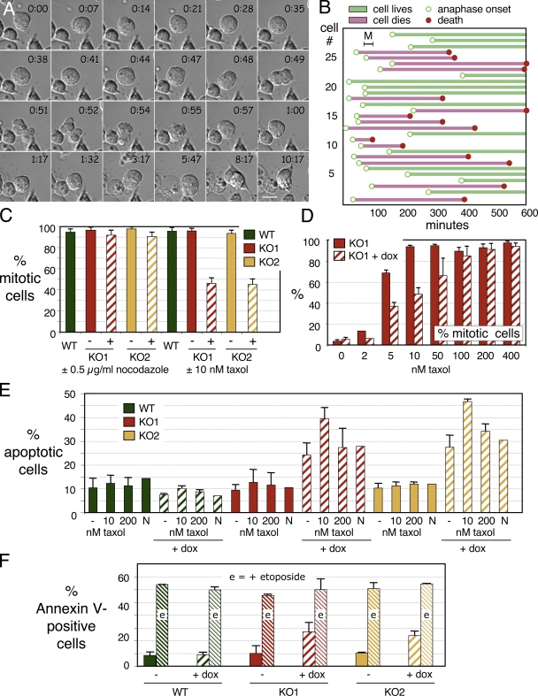
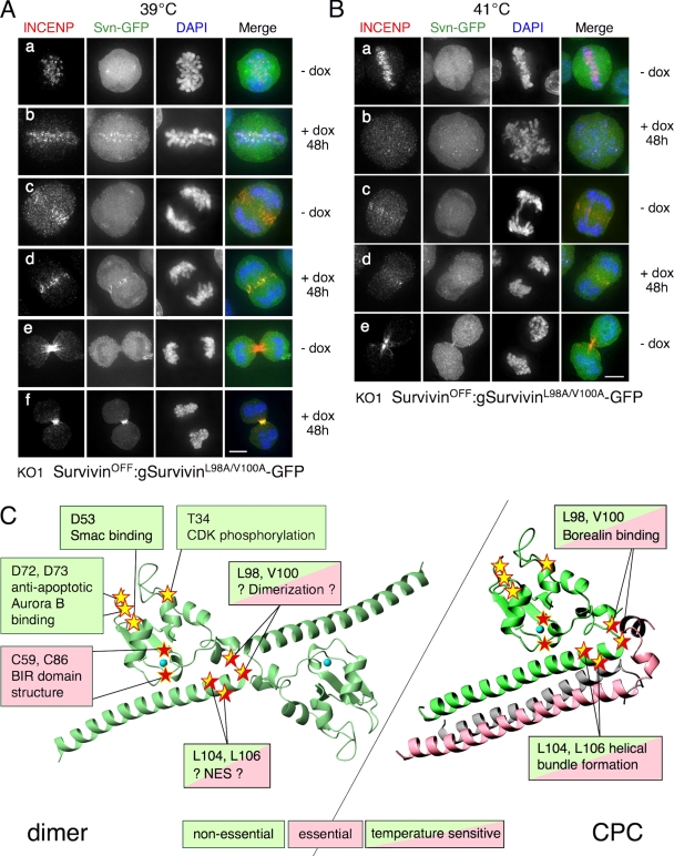
Survivin is a key cellular protein thought to function in apoptotic regulation, mitotic progression, or possibly both. In this study, we describe the isolation of two conditional knockouts of the survivin gene in chicken DT40 cells. DT40 cells lacking Survivin die in interphase after failing to complete cytokinesis. However, these cells show normal sensitivity to the chemotherapeutic agent etoposide. Expression of Survivin mutants against a null background to reassess the role of several key residues reveals that DT40 cells can grow normally if their sole Survivin is missing a widely studied cyclin-dependent kinase phosphorylation site or sites reportedly essential for binding to Smac or aurora B. Mutations in the nuclear export sequence or dimerization interface render cells temperature sensitive for growth. As an important caveat for other studies in which protein function is studied by transient transfection, three of the Survivin mutants fail to localize in the presence of the wild-type protein but do localize and indeed support life in its absence.


Figures

Figure 1.
Figure 1.
Generation of Survivin conditional knockout in DT40 cells. (A) Diagram of the survivin locus and targeting vectors used. Arrowheads, EcoRI cleavage sites. (B) Strategy for rescue and shutoff of Survivin expression. (C) Table showing constructs used for the two knockout cell lines. (D) Southern blot of wild-type, heterozygote, and Survivin-null clones. EcoRI-digested genomic DNA was hybridized with the 5′ external probe (red bar) shown in A. Superscripts 1 and 2 refer to heterozygotes from knockouts 1 and 2, respectively. (E) Repression of rescue Survivin mRNA confirmed by RT-PCR of total RNA from heterozygote and KO1 cells incubated with doxycycline. (F) Real-time PCR confirms survivin repression by doxycycline. Values were normalized relative to actin mRNA. Values for cells grown in doxycycline are shown as striped bars. Error bars indicate SD. (G) Immunoblotting analysis of Survivin repression for KO1. 20 μg of whole cell lysate from DT40 (wild type [WT]) and SurvivinOFF cells was subjected to 12.5% SDS-PAGE and probed with affinity-purified polyclonal anti-Survivin antibody. Loading control, anti–α-tubulin. (H) Immunoblotting analysis of Survivin repression for KO2 performed as for G.
Figure 2.
Figure 2.
Phenotype of cells after Survivin shutoff. (A) Distribution of Survivin (red), α-tubulin (green), and DNA (blue) in SurvivinON (A and A′) and SurvivinOFF (A″ and A‴) mitotic cells. Bar, 5 μm. (B) Phase-contrast images of SurvivinON and SurvivinOFF cultures (the latter after 60-h growth in doxycycline). Bar, 10 μm. (C) Cells from both survivin knockouts KO1 and KO2 die after exposure to doxycycline. (D) Annexin V–positive (apoptotic) cells appear 36 h after shutoff of survivin transcription in KO1. (E) INCENP (red) is diffuse, but kinetochores (CENP-H–GFP, green) appear normal in SurvivinOFF cells (bottom). Top, SurvivinON control. Bar, 5 μm. (F) INCENP and H3S10ph levels decrease shortly after Survivin levels drop after addition of doxycycline to KO1 cells. Loading control, anti–α-tubulin. WT, wild type. (G) Micrograph showing decreased H3S10ph staining (green) in SurvivinOFF cells (bottom). Top, SurvivinON control. Bar, 5 μm.
Figure 3.
Figure 3.
Survivin-depleted cells fail to complete cytokinesis. (A) The mitotic index of SurvivinOFF cells is increased for both KO1 and KO2 relative to wild-type (WT) DT40. (B) Shutoff of Survivin expression leads to an increase in multinucleated (primarily binucleated) cells. Values for cells grown in doxycycline are shown as striped bars. Error bars indicate SD. (C) Selected frames from videos of SurvivinON and SurvivinOFF cells. Merged images show differential interference contrast (red) and histone H2B-mRFP (green), with H2B-mRFP also shown in grayscale. Time is given in hours/minutes. A control SurvivinON cell (top) completes mitosis normally. The SurvivinOFF cell achieves a metaphase alignment (0:25) but exhibits lagging chromosomes in anaphase, and cytokinesis ultimately fails. Bar, 5 μm. (D) Synchronization of DT40 cells by centrifugal elutriation. (E) Cells harvested later in the cell cycle die before those harvested earlier in the cell cycle. (F) Cells harvested later in the cell cycle fail cytokinesis before those harvested earlier in the cell cycle.
Figure 4.
Figure 4.
Cells lacking Survivin die in interphase after failing to complete cytokinesis. (A) Selected frames from a video in which a SurvivinOFF cell fails to complete cytokinesis and dies by apoptosis in the subsequent interphase. Time is given in hours/minutes. Bar, 10 μm. (B) Analysis of cell death in SurvivinOFF cells from videos like those shown in A. Bars begin at anaphase onset (open circles) and terminate either at cell death (closed circles) or at the end of the video. Bar M shows the mean length of mitosis in DT40 (30 min). (C) SurvivinOFF cells exhibit a normal mitotic arrest in nocodazole but not in 10 nM taxol. (D) SurvivinOFF cells arrest in mitosis at taxol concentrations ≥100 nM. (E) The death of SurvivinOFF cells is exacerbated by 10 nM but not by 200 nM taxol. (F) SurvivinOFF and SurvivinON control cultures exhibit similar levels of apoptotic death after exposure to 10 μM etoposide. Values for cells grown in doxycycline are shown as striped bars. Error bars indicate SD. WT, wild type.
Figure 5.
Figure 5.
Functional analysis of Survivin protein in SurvivinON/OFF cells. (A) Alignment of human and chicken Survivin sequences showing conserved residues mutated in this study. (B and C) Chicken (g) and human (h) Survivin-GFP rescues the life of SurvivinOFF cells. Growth curves for cells expressing GFP fusion proteins are green. (D and E) Chicken and human Survivin-GFP colocalize normally with INCENP (red) in SurvivinOFF cells. (F) The location of residue T36 in the CPC and dimer structures of Survivin (Chantalat et al., 2000; Jeyaprakash et al., 2007). (G) Expression of SurvivinT36A/E-GFP and loss of wild-type (WT) Survivin expression from cultures grown in doxycycline. (H) Chicken SurvivinT36A-GFP rescues the life of SurvivinOFF cells. (I) gSurvivinT36A-GFP colocalizes normally with INCENP in SurvivinON/OFF cells. Bars, 5 μm.
Figure 6.
Figure 6.
The BIR domain is crucial for Survivin function. (A) Location of C86 in the CPC form of Survivin. (B) Expression of SurvivinC86A-GFP and loss of wild-type (WT) Survivin expression from cultures grown in doxycycline. (C) SurvivinC86A-GFP fails to rescue the life of SurvivinOFF cells; cells expressing this protein as their sole form of Survivin die with kinetics essentially identical to those of SurvivinOFF cells. (D) SurvivinC86A-GFP fails to localize in SurvivinON/OFF cells. INCENP localization is normal in SurvivinON cells, so SurvivinC86A-GFP fails to act as a dominant-negative mutant. Panel a shows the normal localization of Survivin-GFP in a control experiment. (E) Location of C59 in the Survivin dimer. (F) Expression of SurvivinC59A-GFP and loss of wild-type Survivin expression from cultures grown in doxycycline. (G) Survivin C59A-GFP fails to rescue the life of SurvivinOFF cells. (H) SurvivinC59A-GFP fails to localize in SurvivinON/OFF cells. INCENP localization is normal in SurvivinON cells, so SurvivinC59A-GFP also fails to act as a dominant-negative mutant. Bars, 5 μm.
Figure 7.
Figure 7.
Smac and aurora B binding are not essential for Survivin function. (A) Location of D55 in the Survivin dimer. (B) Expression of SurvivinD55A-GFP and loss of wild-type (WT) Survivin expression from cultures grown in doxycycline. (C) SurvivinD55A-GFP rescues the life of SurvivinOFF cells. (D) SurvivinD55A-GFP colocalizes normally with INCENP in SurvivinON/OFF cells. (E) Location of D72 and D73 in the Survivin dimer. (F) Expression of SurvivinD72A/D73A-GFP and loss of wild-type Survivin expression from cultures grown in doxycycline. (G) SurvivinD72A/D73A-GFP fails to localize in SurvivinON cells; INCENP localization is normal (a and b). SurvivinD72A/D73A-GFP fails to localize in SurvivinOFF cells at prometaphase and metaphase; INCENP localization at centromeres is also compromised (c). SurvivinD72A/D73A-GFP and INCENP localize normally in SurvivinOFF cells at anaphase/telophase (d). (H) SurvivinD72A/D73A-GFP rescues the life of SurvivinOFF cells. Bars, 5 μm.
Figure 8.
Figure 8.
Survivin linker mutants are temperature sensitive. (A) Location of L98, V100, L104, and L106 in the CPC. (B) Expression of SurvivinL98A/V100A-GFP and SurvivinL104A/L106A-GFP and loss of wild-type (WT) Survivin expression from cultures grown in doxycycline at 39 or 41°C. (C) SurvivinL98A/V100A-GFP rescues the life of SurvivinOFF cells at 39°C. (D) SurvivinL104A/L106A-GFP rescues the life of SurvivinOFF cells at 37°C. (E and F) Cells expressing solely SurvivinL98A/V100A-GFP and SurvivinL104A/L106A-GFP die at 41°C, albeit with slightly delayed kinetics.
Figure 9.
Figure 9.
A Survivin linker mutant localizes aberrantly. (A) SurvivinL98A/V100A-GFP fails to localize at 39°C in the presence of wild-type rescue Survivin (a, c, and e). In the absence of wild-type rescue Survivin, a subset of SurvivinL98A/V100A-GFP localizes normally (b, d, and f). INCENP localizes normally in all cells. (B) SurvivinL98A/V100A-GFP fails to localize at 41°C in the presence or absence of wild-type rescue Survivin. INCENP localizes normally only in the presence of wild-type rescue Survivin (a, c, and e). (C) Summary of the mutational analysis of Survivin. Bars, 5 μm.

Similar articles

Cited by

References

    1. Adams, R.R., S.P. Wheatley, A.M. Gouldsworthy, S.E. Kandels-Lewis, M. Carmena, C. Smythe, D.L. Gerloff, and W.C. Earnshaw. 2000. INCENP binds the Aurora-related kinase AIRK2 and is required to target it to chromosomes, the central spindle and cleavage furrow. Curr. Biol. 10:1075–1078. - PubMed
    1. Adams, R.R., H. Maiato, W.C. Earnshaw, and M. Carmena. 2001. Essential roles of Drosophila inner centromere protein (INCENP) and Aurora B in histone H3 phosphorylation, metaphase chromosome alignment, kinetochore disjunction, and chromosome segregation. J. Cell Biol. 153:865–880. - PMC - PubMed
    1. Altieri, D.C. 2006. The case for survivin as a regulator of microtubule dynamics and cell-death decisions. Curr. Opin. Cell Biol. 18:609–615. - PubMed
    1. Ambrosini, G., C. Adida, and D.C. Altieri. 1997. A novel anti-apoptosis gene, survivin, expressed in cancer and lymphoma. Nat. Med. 3:917–921. - PubMed
    1. Ambrosini, G., C. Adida, G. Sirugo, and D.C. Altieri. 1998. Induction of apoptosis and inhibition of cell proliferation by survivin gene targeting. J. Biol. Chem. 273:11177–11182. - PubMed

Publication types

MeSH terms