. 2008 Oct 20;183(2):223-39.
doi: 10.1083/jcb.200805092.

Identifying specific protein interaction partners using quantitative mass spectrometry and bead proteomes

Affiliations

Identifying specific protein interaction partners using quantitative mass spectrometry and bead proteomes

Laura Trinkle-Mulcahy et al. J Cell Biol. .

Abstract

The identification of interaction partners in protein complexes is a major goal in cell biology. Here we present a reliable affinity purification strategy to identify specific interactors that combines quantitative SILAC-based mass spectrometry with characterization of common contaminants binding to affinity matrices (bead proteomes). This strategy can be applied to affinity purification of either tagged fusion protein complexes or endogenous protein complexes, illustrated here using the well-characterized SMN complex as a model. GFP is used as the tag of choice because it shows minimal nonspecific binding to mammalian cell proteins, can be quantitatively depleted from cell extracts, and allows the integration of biochemical protein interaction data with in vivo measurements using fluorescence microscopy. Proteins binding nonspecifically to the most commonly used affinity matrices were determined using quantitative mass spectrometry, revealing important differences that affect experimental design. These data provide a specificity filter to distinguish specific protein binding partners in both quantitative and nonquantitative pull-down and immunoprecipitation experiments.


Figures

Figure 1.
Figure 1.
Protocols used for SILAC-based analysis of protein interaction partners in pull-down experiments. (A) HeLa cells expressing a GFP-tagged protein are metabolically labeled by culturing in “heavy” media containing 13C-isotopes of arginine and lysine, while the parental HeLa cells are grown in “light” media containing the 12C-isotopes of arginine and lysine. Whole cell extracts can be prepared or, as shown here, cells can be fractionated for preparation of separate cytoplasmic and nuclear extracts. In this case, extracts are pre-cleared on Sepharose beads and then mixed in equal amounts before affinity purification of the GFP-tagged protein using the GFP binder (1 h incubation). Proteins are eluted from the beads and separated by 1D SDS-PAGE for digestion and LC-MS/MS analysis. (B) For SILAC analysis of an endogenous protein, two populations of HeLa cells are grown in light and heavy media, respectively, before harvesting and preparation of cellular extracts. Equal total protein amounts of each extract are subjected to separate immunoaffinity experiments, either using an antibody to the protein of interest or a control antibody covalently bound to beads at an equivalent concentration. The separate immunoprecipitates are mixed carefully to minimize variability and the proteins eluted and analyzed as described above.
Figure 2.
Figure 2.
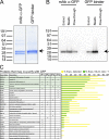
GFP as a tag in immunoaffinity experiments. Although a commercial monoclonal anti-GFP antibody is capable of isolating significant amounts of free GFP from a stable HeLa cell line, the GFP binder is more efficient, as demonstrated both by Coomassie staining of protein eluted from the affinity matrices (A) and Western blotting using anti-GFP antibodies (B). Whether the mAb or GFP binder is used to purify GFP, there are very few proteins that bind nonspecifically to this tag (C). Four independent experiments were performed to identify proteins that may copurify with GFP, as indicated by SILAC ratios greater than 1 (IP1: whole cell extract, GFP binder; IP2: whole cell extract, monoclonal anti-GFP antibody; IP3: cytoplasmic extract, monoclonal anti-GFP antibody; IP4: nuclear extract, monoclonal anti-GFP antibody). No one protein was identified in every experiment, and most of them (in bold) have been identified as binding nonspecifically to the Sepharose bead matrix. This list was then screened against a set of 18 independent GFP protein immunoaffinity experiments performed using the GFP binder for purification and parental cells as the negative control. Proteins were scored for the percentage of experiments in which they were detected (yellow), and for the percentage of experiments in which they were detected and showed a SILAC ratio greater than 1 (green). Six proteins, representing three protein classes (heat shock/chaperone, cytokeratin, and ubiquitin), have been highlighted in green as the most frequently detected and potentially able to bind GFP.
Figure 3.
Figure 3.
Comparison of bead proteomes. (A) Design of the SILAC immunoprecipitation experiment used to compare the bead proteomes of agarose, Sepharose, and magnetic beads. For all three, the protein G–conjugated versions were used. The experiment was performed in two stages, first with a short incubation time of 30 min and next with a long incubation time of 18 h. In addition, cells were fractionated into cytoplasmic and nuclear extracts to compare the profiles of the proteins that bind nonspecifically to the bead matrices. In the case of nuclear extracts, more proteins bind nonspecifically during a long incubation than a short incubation, as assessed both by Coomassie staining (B) and by mass spectrometric analysis (C). The cytoplasmic protein profile did not vary to the same extent. The distribution of proteins by class was quite similar regardless of the cellular extract used in the experiment or the time of incubation (C). Distinct differences in the distribution of these classes of proteins were observed, however, with magnetic beads binding more cytoskeletal and structural proteins nonspecifically and Sepharose binding more nucleic acid binding factors nonspecifically.
Figure 4.
Figure 4.
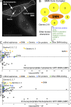
Identification of proteins that interact with SMN and the SMN complex. The GFP binder was used to immunopurify GFP-SMN from a stable HeLa cell line as compared with the nonexpressing parental cell line. Like endogenous SMN, GFP-SMN is found in both cytoplasmic and nucleoplasmic pools and accumulates in gems within nuclei (A). Bar, 15 μM. Detailed biochemical and proteomic studies have revealed that the core SMN complex is composed of SMN itself and Gemins 2–8 (B). The stoichiometry is not known and, although not depicted here, the complex can oligomerize. Also listed are several other proteins that have been shown to interact with the SMN complex by similar experimental approaches. In the study presented here, separate experiments were performed for cytoplasmic and nuclear extracts to independently assess interacting partners and compare these two pools. The log SILAC (i.e., heavy/light arginine and/or lysine) ratio calculated for each protein identified in the cytoplasmic GFP-SMN immunoprecipitation experiment is plotted versus total peptide intensity in C. The nucleoplasmic GFP-SMN immunoprecipitation data are plotted in a similar fashion (D).
Figure 5.
Figure 5.
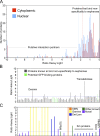
Systematic analysis of SILAC datasets. Quantitative mass spectrometric data generated by the cytoplasmic and nuclear GFP-SMN immunoprecipitation experiments were subjected to a standard analysis workflow. First, the frequency of specific SILAC (heavy/light amino acid) ratios were plotted for the entire datasets to determine the distribution of these ratios among the proteins identified (A). Environmental contaminants such as keratins have very low ratios and cluster near 0. In the cytoplasmic experiment, proteins that bind nonspecifically to the bead matrix cluster in a bell curve distribution around 1, as expected for proteins that bind equally in the light and heavy form. The threshold for detection of bona fide interaction partners was set at a conservative level above that (hashed red line). Note that in the nuclear experiment the SILAC ratios for the bead contaminants were shifted to the left, clustering in a bell curve distribution around the higher value of 1.5. In this case the threshold (hashed blue line) must also be shifted. SMN itself, all of the core SMN complex members, and several known interacting partners fell above this threshold and were identified in this first analysis step. However, less abundant or lower affinity binding partners may be found at or below these conservative threshold values. Analysis of the datasets is thus further extended by applying the Sepharose bead proteome as a filter and grouping the SILAC ratios of those proteins that have been identified as binding nonspecifically to this bead matrix, as shown here for the cytoplasmic dataset (B). Most proteins known to bind Sepharose (gray) and potential GFP-binding proteins (green) have the expected ratios near or below threshold, but a few are significantly above threshold and must be considered as potentially real interacting proteins, albeit with a lower priority for further analysis. SILAC ratios calculated for the remaining proteins in the dataset, i.e., those not known to bind nonspecifically to either the GFP tag or the bead matrix, are next plotted separately (C). Over two-thirds of the proteins have SILAC ratios significantly higher than threshold. These include both known and novel interacting partners for SMN. Some of the known SMN complex interacting partners, such as PRMT5 and Unrip, have ratios closer to threshold, and thus would be overlooked in a threshold-based analysis. As expected for such a well-characterized complex, very few novel proteins were detected. One of these, USP9X, was selected for further analysis.
Figure 6.
Figure 6.
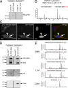
Validation of mass spectrometric results. Cytoplasmic-specific copurification of the novel protein USP9X with GFP-SMN was confirmed by Western blotting (A). Two peptides, each with a SILAC ratio >1, were found for USP9X in the SILAC analysis of a GFP-SMN pull-down from cytoplasmic extracts. The mass spectra of one of them is shown here for comparison (B). The quantifiable arginine is highlighted in red. This cytoplasmic enrichment of USP9X is consistent with immunostaining results using a monoclonal anti-USP9X antibody (C). Although predominantly cytoplasmic, there is a pool of USP9X in the nucleus (arrowhead), although it does not accumulate in gems (arrow). There is no difference in localization of USP9X in parental HeLa cells (top cell) versus HeLa cells stably expressing GFP-SMN (bottom cell). Bar, 5 μM. As a control, Western blotting was also used to confirm the enrichment of both endogenous SMN and GFP-SMN, and of the U1 snRNP protein U1A, from both cytoplasmic and nuclear extracts using the GFP binder, and the nuclear-specific enrichment of p80 coilin (D). For comparison, representative peptide spectra for these proteins from the SILAC analysis are shown (E). Quantifiable amino acids are highlighted in red, with the SILAC ratio in parentheses.

Similar articles

Cited by

References

    1. Al-Hakim, A.K., A. Zagorska, L. Chapman, M. Deak, M. Peggie, and D.R. Alessi. 2008. Control of AMPK-related kinases by USP9X and atypical Lys(29)/Lys(33)-linked polyubiquitin chains. Biochem. J. 411:249–260. - PubMed
    1. Baccon, J., L. Pellizzoni, J. Rappsilber, M. Mann, and G. Dreyfuss. 2002. Identification and characterization of Gemin7, a novel component of the survival of motor neuron complex. J. Biol. Chem. 277:31957–31962. - PubMed
    1. Blagoev, B., I. Kratchmarova, S.E. Ong, M. Nielsen, L.J. Foster, and M. Mann. 2003. A proteomics strategy to elucidate functional protein-protein interactions applied to EGF signaling. Nat. Biotechnol. 21:315–318. - PubMed
    1. Campbell, L., K.M. Hunter, P. Mohaghegh, J.M. Tinsley, M.A. Brasch, and K.E. Davies. 2000. Direct interaction of Smn with dp103, a putative RNA helicase: a role for Smn in transcription regulation? Hum. Mol. Genet. 9:1093–1100. - PubMed
    1. Carissimi, C., J. Baccon, M. Straccia, P. Chiarella, A. Maiolica, A. Sawyer, J. Rappsilber, and L. Pellizzoni. 2005. Unrip is a component of SMN complexes active in snRNP assembly. FEBS Lett. 579:2348–2354. - PubMed

Publication types

MeSH terms