. 2008 Nov 5;27(21):2873-82.
doi: 10.1038/emboj.2008.207. Epub 2008 Oct 16.

Regulated release of ERdj3 from unfolded proteins by BiP

Affiliations

Regulated release of ERdj3 from unfolded proteins by BiP

Yi Jin et al. EMBO J. .

Abstract

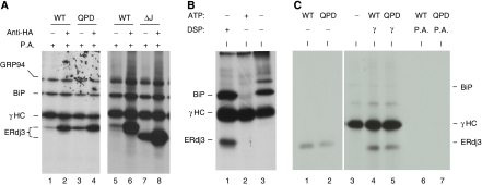
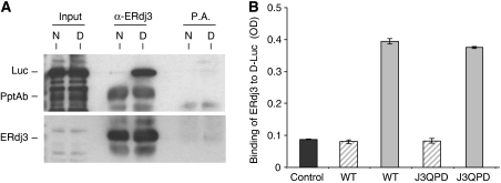
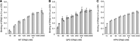
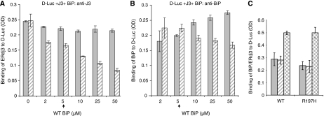
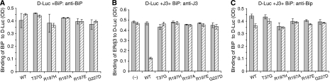
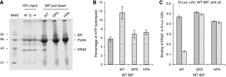
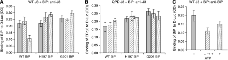
DnaJ proteins often bind to unfolded substrates and recruit their Hsp70 partners. This induces a conformational change in the Hsp70 that stabilizes its binding to substrate. By some unknown mechanism, the DnaJ protein is released. We examined the requirements for the release of ERdj3, a mammalian ER DnaJ, from substrates and found that BiP promoted the release of ERdj3 only in the presence of ATP. Mutations in ERdj3 or BiP that disrupted their interaction interrupted the release of ERdj3. BiP mutants that were defective in any step of the ATPase cycle were also unable to release ERdj3. These results demonstrate that a functional interaction between ERdj3 and BiP, including both a direct interaction and the ability to stimulate BiP's ATPase activity are required to release ERdj3 from substrate and support a model where ERdj3 must recruit BiP and stimulate its high-affinity association with the substrate through activation of ATP hydrolysis to trigger its own release from substrates. On the basis of similarities among DnaJs and Hsp70s, this is likely to be applicable to other Hsp70-DnaJ pairs.


Figures

Figure 1
Figure 1
ERdj3 binds to γHC directly. (A) COS-1 cells were co-transfected with cDNAs encoding γHC and the indicated HA-tagged ERdj3 constructs. Metabolically labelled, crosslinked cell lysates were immunoprecipitated with anti-HA or Protein A Sepharose alone. Isolated proteins were separated by reducing SDS–PAGE. (B) Ag8.8 cells were metabolically labelled for 16 h with [35S]methionine and cysteine and incubated with (lane 1) or without (lanes 2 and 3) DSP. Cell lysates were prepared with (lane 2) or without (lanes 1 and 3) ATP and immunoprecipitated with Protein A Sepharose. (C) Wild-type (WT) and QPD mutant (Mut) ERdj3 were in vitro translated and run directly (lanes 1 and 2) or incubated with the free γHC immobilized on Protein A Sepharose beads (lanes 4 and 5) prepared as in lane 2 in Figure 1B or with Protein A beads alone (lanes 6 and 7).
Figure 2
Figure 2
WT and QPD ERdj3 bind to denatured luciferase similarly in vitro. (A) Temperature denatured (D) or native (N) luciferase (Luc) was directly loaded on a gel (first two lanes) or incubated with bacterially produced recombinant wild-type ERdj3 and then immunoprecipitated with either anti-ERdj3 polyclonal antiserum or with Protein A Sepharose beads alone. Reaction cocktails were subjected to reducing SDS–PAGE and then transferred to a PVDF membrane. The membrane was blotted with either goat anti-luciferase antiserum followed by donkey anti-goat Ig conjugated to HRP or with the polyclonal anti-ERdj3 followed by goat anti-rabbit Ig conjugated to HRP. In both cases, the signal was detected by chemiluminescence. (B) Chemically denatured luciferase (solid grey bars) or binding buffer alone (hatched bars) was used to coat 96-well plates. Recombinant wild-type or the QPD mutant ERdj3 proteins (0.5 μM) were added to the wells and bound ERdj3 was detected with a polyclonal anti-ERdj3 antiserum, followed by donkey anti-rabbit Ig conjugated to alkaline phosphatase. The DNTP substrate was added and after developing, the plates were read on a spectrophotometer and the signal was expressed in OD units. A luciferase-coated well that did not receive ERdj3 protein was treated similarly and serves as a negative control for the antibody (dark grey). All samples were run in triplicate and error bars are indicated.
Figure 3
Figure 3
WT and mutant (QPD and HPN) ERdj3 bind to D-Luc similarly and ATP does not affect their binding. Chemically denatured luciferase was used to coat the wells and the indicated amounts of WT (A) QPD (B), or HPN (C) ERdj3 were added to the wells with (hatched bars) or without (solid bars) ATP. The plates were developed with anti-ERdj3 as described in Figure 2. The arrow indicates the concentration of ERdj3 that was used in the following experiments.
Figure 4
Figure 4
BiP releases ERdj3 from D-Luc in an ATP-dependent manner. (A) Chemically denatured luciferase was added to the wells followed by ERdj3 binding as described earlier. After washing, the indicated amounts of WT BiP were added to the ERdj3–luciferase complexes with (hatched bar) or without (solid bar) ATP and incubated for an additional 1 h at room temperature. After washing, the amount of ERdj3 that remained bound to luciferase was detected with an anti-ERdj3 antiserum. (B) On a parallel plate, the amount of BiP that was associated with luciferase was determined by incubating with an anti-BiP antiserum. (C) Either wild-type or mutant (R197H) BiP was allowed to bind to luciferase. After washing, ERdj3 was added to half the wells and the amount of BiP that was bound without ERdj3 (solid bars) or with ERdj3 (hatched bars) was measured with an anti-BiP antiserum. In a parallel set of wells, ERdj3 binding was measured with an anti-ERdj3 antibody (checkered bars).
Figure 5
Figure 5
Only WT BiP releases ERdj3 from D-Luc. (A) Chemically denatured luciferase was added to the wells, which were then incubated with wild-type or mutant BiP. The binding of BiP to D-Luc was performed in the absence of ERdj3 with (hatched bars) and without ATP (solid bars) and detected with anti-BiP serum. (B, C) Denatured luciferase was added to wells and wild-type ERdj3 was allowed to bind as described. After washing, either wild-type or mutant BiP was added with (hatched bars) or without (solid bars) ATP. The amount of ERdj3 remaining was detected with an anti-ERdj3 antiserum (B) and the binding of BiP was detected with an anti-BiP antiserum (C) and expressed in OD units.
Figure 6
Figure 6
Wild-type BiP can only release wild-type ERdj3 from D-Luc. (A) Recombinant wild-type BiP (5 μg) was immunoprecipitated with a polyclonal anti-BiP antiserum and Protein A Sepharose beads. After washing, 20 μg of the indicated recombinant ERdj3 proteins was added to the BiP beads in ATPase buffer containing ATP. One-tenth of the reaction was removed for direct loading and the remaining nine-tenths were incubated at 4°C for 1 h. After washing, the samples were analysed by reducing SDS–PAGE and the gel was stained with Brilliant Coomassie Blue to visualize proteins. (B) ATPase assays were performed on wild-type BiP alone or with a four-fold molar excess of wild-type, HPN, or QPD ERdj3. ATP hydrolysis was measured by quantitating ADP and expressing it as a percentage of total nucleotide. (C) An experiment similar to that described in the previous figure was performed, except that either wild-type or mutant ERdj3 was bound to luciferase first. After washing, wild-type BiP was added with (hatched bars) or without (solid bars) ATP. The amount of ERdj3 that remained bound was detected with an anti-ERdj3 antiserum and expressed in OD units.
Figure 7
Figure 7
Both BiP and ATP must be added simultaneously to induce the release of ERdj3 from substrate. Luciferase-binding assays were performed with wild-type ERdj3 or the QPD mutant. The indicated BiP proteins were added with no ATP (solid bars), with ATP as done in the previous experiments (stippled bars) or BiP was allowed to bind in the absence of ATP but after washing unbound BiP away, the D-Luc–ERdj3–BiP complexes were further incubated with ATP (hatched bars). The amount of ERdj3 remaining associated with D-Luc (A, B) and the amount of BiP in the D-Luc–wild-type ERdj3 complexes (C) were determined by ELISA.

Similar articles

Cited by

References

    1. Alder NN, Shen Y, Brodsky JL, Hendershot LM, Johnson AE (2005) The molecular mechanisms underlying BiP-mediated gating of the Sec61 translocon of the endoplasmic reticulum. J Cell Biol 168: 389–399 - PMC - PubMed
    1. Awad W, Estrada I, Shen Y, Hendershot LM (2008) BiP mutants that are unable to interact with endoplasmic reticulum DnaJ proteins provide insights into interdomain interactions in BiP. Proc Natl Acad Sci USA 105: 1164–1169 - PMC - PubMed
    1. Brightman SE, Blatch GL, Zetter BR (1995) Isolation of a mouse cDNA encoding MTJ1, a new murine member of the DnaJ family of proteins. Gene 153: 249–254 - PubMed
    1. Caplan AJ, Cyr DM, Douglas MG (1993) Eukaryotic homologues of Escherichia coli dnaJ: a diverse protein family that functions with hsp70 stress proteins. Mol Biol Cell 4: 555–563 - PMC - PubMed
    1. Cheetham ME, Caplan AJ (1998) Structure, function and evolution of DnaJ: conservation and adaptation of chaperone function. Cell Stress Chaperones 3: 28–36 - PMC - PubMed

Publication types