Comparative Study
. 2008 Oct 15;28(42):10674-86.
doi: 10.1523/JNEUROSCI.1283-08.2008.

Arx is a direct target of Dlx2 and thereby contributes to the tangential migration of GABAergic interneurons

Affiliations
Comparative Study

Arx is a direct target of Dlx2 and thereby contributes to the tangential migration of GABAergic interneurons

Gaia Colasante et al. J Neurosci. .

Abstract

The Arx transcription factor is expressed in the developing ventral telencephalon and subsets of its derivatives. Mutation of human ARX ortholog causes neurological disorders including epilepsy, lissencephaly, and mental retardation. We have isolated the mouse Arx endogenous enhancer modules that control its tightly compartmentalized forebrain expression. Interestingly, they are scattered downstream of its coding region and partially included within the introns of the downstream PolA1 gene. These enhancers are ultraconserved noncoding sequences that are highly conserved throughout the vertebrate phylum. Functional characterization of the Arx GABAergic enhancer element revealed its strict dependence on the activity of Dlx transcription factors. Dlx overexpression induces ectopic expression of endogenous Arx and its isolated enhancer, whereas loss of Dlx expression results in reduced Arx expression, suggesting that Arx is a key mediator of Dlx function. To further elucidate the mechanisms involved, a combination of gain-of-function studies in mutant Arx or Dlx tissues was pursued. This analysis provided evidence that, although Arx is necessary for the Dlx-dependent promotion of interneuron migration, it is not required for the GABAergic cell fate commitment mediated by Dlx factors. Although Arx has additional functions independent of the Dlx pathway, we have established a direct genetic relationship that controls critical steps in the development of telencephalic GABAergic neurons. These findings contribute elucidating the genetic hierarchy that likely underlies the etiology of a variety of human neurodevelopmental disorders.


Figures

Figure 1.
Figure 1.
Characterization and identification of the genomic regions acting as Arx endogenous enhancers. A, VISTA genome browser schematic representation of nucleotide homology of ∼36 kb of the Arx genomic locus in mouse, human, and zebrafish. Only three different genomic regions showed homology >50% outside the transcribed sequences, and all were placed in the 3′ region downstream to the coding sequences. In light blue is represented the sequence corresponding to the PolA1 last exon. Rectangles outline Arx coding exons, with red rectangles corresponding to homeodomain coding sequences. Yellow bars highlight the two genomic regions of 3.5 and 9.5 kb initially isolated to produce transgenic embryos. B, E10.5 and E12.5 transgenic mice carrying the 3.5 kb proximal sequence upstream to the LacZ reporter gene. β-Galactosidase activity was confined to the developing cerebral cortex (cx), eminentia thalami (et), and vt (5/5 embryos analyzed). C, The 9.5 kb sequence targets expression of the reporter in the ge, vt, floor plate (fp), and pancreas (p) in E12.5 mouse transgenic embryos (4/4 embryos analyzed). mes, Mesencephalon; sp, spinal cord.
Figure 2.
Figure 2.
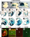
Analysis of the β-galactosidase activity profile during development in the 0.8 kb transgenic mouse line. A–C, Whole embryo at E9.5 (A) and dissected brains at E11.5 (B) and E14.5 (C) showing reporter gene activity in the ge and diencephalons (d). D–K, Coronal vibratome sections of E13.5 (D–G) and E15.5 (H–K) showing β-galactosidase activity principally localized in the medial and lateral ganglionic eminences and in cells migrating toward the cortex following tangential deep and superficial streams (arrowheads in D–K). L–O, Stainings for β-galactosidase (red) and Tbr1 (green), a molecular marker for glutamatergic neurons, do not show any colabeled cells in the cerebral cortex (L, M); on the contrary, most of the β-galactosidase cortical cells were GAD65 positive, indicating a GABAergic cell fate (N, O). cge, Caudal ganglionic eminence; cx, cerebral cortex; dmh, dorsal-medial hypothalamic nucleus; dt, dorsal thalamus; fb, forebrain; hy, hypothalamus; mes, mesencephalon; rb, rhombencephalon; sc, spinal cord; se, septum.
Figure 3.
Figure 3.
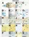
Tissue- and cell-specific localization of β-galactosidase in adult brains of 0.8 kb transgenic mice. A, Sagittal section of adult anterior brain showing reporter gene activity in the striatum (str), cortex (cx), hippocampus (hi), and olfactory bulb (ob). B, The entire rostral migratory stream (rms) from the telencephalic ventricle to the bulb resulted positive for β-galactosidase activity. C, In the cx, many cells scattered throughout all the layers expressed the reporter gene. D, Ventral forebrain structures such as the str, the septum (se), and the ot included a large cohort of β-galactosidase+ cells. E, Caudal coronal brain sections showed reporter activity in the reticular thalamic nuclei (rt). F, In the adult hypothalamic regions, β-galactosidase activity is localized in cells of the dorsal-medial hypothalamic nuclei (dmh). G, Enlargement on the telencephalic ventricular surface showing a dense β-galactosidase staining lining the ventricle corresponding probably to newly generated GABAergic neuronal precursors. H–O, Colabeling for β-galactosidase and different markers of GABAergic neurons in adult cerebral cortex. H, I, β-Galactosidase/GABA double staining shows a large amount of double-positive cells (I, arrows). Conversely, β-galactosidase+/GABA− cells were not apparently scored. J–O, Specific markers of the nonoverlapping GABAergic subsets parvalbumin (J, K), calretinin (L, M), and NPY (N, O) costained with subgroups of reporter-labeled cortical neurons (arrows in K, M, O). K, M, Higher-magnification views of the boxed areas in J and L, respectively. P–S, Colabeling for reporter gene activity and the GABAergic markers GABA (Q), NPY (R), and calretinin (S) in hippocampus. Also in this structure, as in the cortex, a virtually complete overlapping was detected between stainings of GABA or markers of specific GABAergic subsets and β-galactosidase. hy, Hypothalamus.
Figure 4.
Figure 4.
Single-cell resolution labeling of embryonic hippocampal neuronal neurons (10–14 d in vitro) from wild-type or 0.8 kb transgenic mice. A–D, Cell fate analysis of Arx-positive cells from wild-type animals. Arx colocalizes with the general neuronal marker TuJ1 (A) and with the markers of GABAergic neurons GAD65 (B, C) and VGAT (D). D, Inset, A high-magnification image highlighting Arx nuclear staining. E–H, β-Galactosidase+ cells are also labeled by antibodies against Arx (F) or GAD65 (G), but not by the Vglut1 antibody, specific for glutamatergic neurons (H). In E, the localization of β-galactosidase activity is shown in blue by differential interference contrast. Arrows in A–D point at somata of neurons. Arrowheads in F–H indicate red dots corresponding to sites of accumulation of β-galactosidase, whereas arrow in H points at the soma of a Vglut1+ neuron, where β-galactosidase is absent. The broken lines in F outline the edge of a neuron positive for β-galactosidase (identified by differential interference microscopy).
Figure 5.
Figure 5.
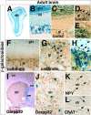
Reporter gene activity in adult olfactory bulb (ob) and striatum (str) of 0.8 kb transgenic mice. A, B, Adult olfactory bulb is strongly positive for β-galactosidase staining with many cells labeled in the gl and gcl. C–E, X-gal and GAD65 colabeling identifies many β-galactosidase+ cells as GABAergic neurons in both glomerular (D, arrows) and granular (E) layers. F, G, Periglomerular calbindin-expressing cells coexpress the reporter gene (G, arrows). H, Periglomerular dopaminergic cells express the reporter gene as scored by β-galactosidase and TH costaining. I–L, Analysis of β-galactosidase activity in the adult striatum. I, A relatively small fraction of scattered β-galactosidase+ cells are detected in adult striatum. J, DARPP32 staining reveals that transgene-expressing cells are not striatal principal medium spiny neurons. Conversely, β-galactosidase cells can express either NPY (K) or ChAT (L), suggesting their striatal GABAergic and cholinergic neuronal nature, respectively. cc, Corpus callosum; cx, cerebral cortex; ml, mitral cell layer; se, septum.
Figure 6.
Figure 6.
Identification of the mUAS enhancer core sequence and its dependence on Dlx2 activity. A, Phylogenetic comparison showing the high homology of an ∼200 nt fragment included in the UAS3 sequence. Asterisks indicate conserved nucleotide residues. Two conserved binding sites for homeodomain proteins are highlighted in yellow and named HD-1 and HD-2. B, Both the entire 0.8 kb and 200 nt sequences are strongly activated by Dlx2 in transient cotransfection assays in P19 cells, whereas a 0.8 kb sequence deprived of the 200 bp fragment is not responding to Dlx2 activity. A series of deletion constructs removing each single or both HD-1/2 binding sites (yellow and brown boxes, respectively) reveal their requirement for triggering the proper Dlx2-induced activity. Values shown represent the mean relative luciferase activity obtained by three independent experiments ± SEM. C, EMSA using the HD-1 and HD-2 sequences shows a strong binding to a Dlx2 in vitro translated protein. Preincubation with an increasing amount of nonlabeled oligonucleotides abolishes the formation of the Dlx2-HD1 or Dlx2-HD2 complexes. D, Chromatin immunoprecipitation experiments showing Dlx2 binding to the mUAS3 sequence in vivo. PCR analysis was performed on the immunoprecipitated chromatin isolated from E14.5 ventral forebrain tissue using primers corresponding to the mUAS3 sequence (Arx) and Npn2 and IL-1β as positive and negative control genes, respectively. As negative control, immunoprecipitation was performed without antibody (Mock). As input, unprecipitated chromatin was used for amplifications. bg, Basal ganglia; cx, cerebral cortex; mes, mesencephalon; rh, rhombencephalon; th, thalamus.
Figure 7.
Figure 7.
Dlx2 overexpression induces reporter gene activity and Arx protein expression in vivo. Dlx2-IRES-GFP construct was electroporated in 0.8 kb transgenic mouse brain slices at E13.5. The tissue was sectioned coronally and analyzed 30 h later. Adjacent sections of the same electroporated tissue were used for all the staining presented. A–H, Dlx2 ectopic expression induces GAD65 expression in both the cerebral cortex (A–D) and the mesencephalon (E–H). I–X, Likewise, β-galactosidase activity (I–P) and Arx protein expression (Q–X) are activated by Dlx2 forced expression in a comparable manner both in the cerebral cortex (I–L, Q–T) and mesencephalon (M–P, U–X). D, H, L, P, T, X, Merged staining between GFP and induced gene expression reveals an almost complete overlapping between the two stainings, indicating a substantially cell-autonomous effect of Dlx2 overexpression.
Figure 8.
Figure 8.
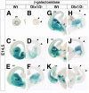
β-Galactosidase activity in E14.5 brain sections of the Arx 0.8 kb transgenic mice in wild-type (Wt) or Dlx1/2 mutant background. Sections on similar coronal levels are compared between wild-type (left side) and Dlx1/2 mutant (right side) genotypes. β-Galactosidase activity is reduced in Dlx1/2-deficient brain tissue. In these brains, reporter gene expression is marginally detectable in LGE, MGE, and vt (D, F). Conversely, β-galactosidase is still detectable, although reduced, in the mutant caudal ganglionic eminence (cge) (H, J, L). Dlx1/2 mutant tissues are devoid of any reporter staining in the cerebral cortex (cx), indicating a complete absence of GABAergic neuronal migration from the ventral telencephalon (arrowheads in B, D, F, H, J, L), by contrast to wt tissue (A, C, E, G, I, K). ob, Olfactory bulb; se, septum.
Figure 9.
Figure 9.
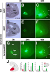
Arx overexpression in Dlx1/2 mutant MGE partially rescues tangential migration of interneurons. A–C, Representative example of GFP+ cells that migrated toward the LGE (C, arrowheads) and the cortex (cx) (C, arrows) 48 h after transplantation of the electroporated MGE in a corresponding position of the contralateral side (area highlighted with dots in A) of wild-type (WT) E14.5 forebrain slice organotypic cultures. D, E, G, Transplantation of the electroporated Dlx1/2 mutant MGE with GFP (D, E) or Arx-IRES-GFP (G) transplanted into the contralateral side of Dlx1/2 mutant forebrain brain slices. F, I, Migration of GFP+ cells after 48 h from the transplantation in Dlx1/2 mutant tissue. Whereas GFP cells failed to migrate outwards from the grafting (F), Arx-overexpressing cells show an extensive migration ability, with cells migrated distant from the transplanted MGE tissue toward the cortex (arrows) and the lateral ganglionic eminence (arrowheads) (I). J, Quantification of numbers of GFP+ cells in the cortex of WT and Dlx1/2 mutant tissues. For counting, four consecutive domains organotypic brain slices were independently analyzed (from 1 proximal to the transplanted MGE to 4, the most distal region), with the first two domains covering the LGE domain and the last two comprising the lateral and mediolateral cortices, respectively. Nine different brain slices for each experimental setup were counted, obtained in three independent experiments. The total numbers of GFP+ cells for Dlx mutant cells (red bars), Arx-overexpressing (overexp.) cells in the Dlx1/2 mutant tissue (gray bars), and control cells in WT tissue (green bars) were as follows: in domain 1, 13 ± 4, 67 ± 11, and 91 ± 1 5, respectively; in domain 2, 4 ± 3, 29 ± 10, and 48 ± 15, respectively; in domain 3, 2 ± 2, 12 ± 5, and 33 ± 7, respectively; in domain 4, 0, 2 ± 2, and 18 ± 5, respectively. KO, Knock-out.
Figure 10.
Figure 10.
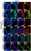
Dlx2 ability to ectopically induce GABAergic gene expression is retained in the absence of Arx activity. A–C, A single coronal section from an E14.5 Arx mutant mouse brain electroporated 30 h earlier with Dlx2iresGFP. Dlx2-targeted tissue in the lateral cerebral cortex is activating GAD65 expression. D–F, A single coronal section from an electroporated Arx mutant mouse brain. Dlx2 ectopic expression activates the LacZ gene activity, which is knocked into the Arx endogenous locus and acts as a sensor of the Arx endogenous enhancer activity. C, Merge image of A and B. F, Merge image of D and E. D, E, Enlargements of the brain sections shown as a whole in insets D′ and E′. cx, Cerebral cortex; se, septum.

Similar articles

Cited by

References

    1. Anderson SA, Qiu M, Bulfone A, Eisenstat DD, Meneses J, Pedersen R, Rubenstein JL. Mutations of the homeobox genes Dlx-1 and Dlx-2 disrupt the striatal subventricular zone and differentiation of late born striatal neurons. Neuron. 1997a;19:27–37. - PubMed
    1. Anderson SA, Eisenstat DD, Shi L, Rubenstein JL. Interneuron migration from basal forebrain to neocortex: dependence on Dlx genes. Science. 1997b;278:474–476. - PubMed
    1. Anderson SA, Marín O, Horn C, Jennings K, Rubenstein JL. Distinct cortical migrations from the medial and lateral ganglionic eminences. Development. 2001;128:353–363. - PubMed
    1. Banker GA, Cowan WM. Rat hippocampal neurons in dispersed cell culture. Brain Res. 1977;126:397–442. - PubMed
    1. Bejerano G, Pheasant M, Makunin I, Stephen S, Kent WJ, Mattick JS, Haussler D. Ultraconserved elements in the human genome. Science. 2004;304:1321–1325. - PubMed

Publication types

MeSH terms

LinkOut - more resources