Mouse hepatitis virus liver pathology is dependent on ADP-ribose-1''-phosphatase, a viral function conserved in the alpha-like supergroup
- PMID: 18922871
- PMCID: PMC2593347
- DOI: 10.1128/JVI.02082-08
Mouse hepatitis virus liver pathology is dependent on ADP-ribose-1''-phosphatase, a viral function conserved in the alpha-like supergroup
Abstract
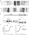
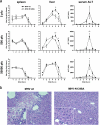
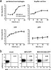
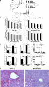
Viral infection of the liver can lead to severe tissue damage when high levels of viral replication and spread in the organ are coupled with strong induction of inflammatory responses. Here we report an unexpected correlation between the expression of a functional X domain encoded by the hepatotropic mouse hepatitis virus strain A59 (MHV-A59), the high-level production of inflammatory cytokines, and the induction of acute viral hepatitis in mice. X-domain (also called macro domain) proteins possess poly-ADP-ribose binding and/or ADP-ribose-1''-phosphatase (ADRP) activity. They are conserved in coronaviruses and in members of the "alpha-like supergroup" of phylogenetically related positive-strand RNA viruses that includes viruses of medical importance, such as rubella virus and hepatitis E virus. By using reverse genetics, we constructed a recombinant murine coronavirus MHV-A59 mutant encoding a single-amino-acid substitution of a strictly conserved residue that is essential for coronaviral ADRP activity. We found that the mutant virus replicated to slightly reduced titers in livers but, strikingly, did not induce liver disease. In vitro, the mutant virus induced only low levels of the inflammatory cytokines tumor necrosis factor alpha and interleukin-6 (IL-6). In vivo, we found that IL-6 production, in particular, was reduced in the spleens and livers of mutant virus-infected mice. Collectively, our data demonstrate that the MHV X domain exacerbates MHV-induced liver pathology, most likely through the induction of excessive inflammatory cytokine expression.
Figures

Similar articles
-
Toll-like receptor-2 exacerbates murine acute viral hepatitis.Immunology. 2016 Oct;149(2):204-24. doi: 10.1111/imm.12627. Epub 2016 Aug 10. Immunology. 2016. PMID: 27273587 Free PMC article.
-
Targeted recombination within the spike gene of murine coronavirus mouse hepatitis virus-A59: Q159 is a determinant of hepatotropism.J Virol. 1998 Dec;72(12):9628-36. doi: 10.1128/JVI.72.12.9628-9636.1998. J Virol. 1998. PMID: 9811696 Free PMC article.
-
Expression of hemagglutinin/esterase by a mouse hepatitis virus coronavirus defective-interfering RNA alters viral pathogenesis.Virology. 1998 Mar 1;242(1):170-83. doi: 10.1006/viro.1997.8993. Virology. 1998. PMID: 9501044 Free PMC article.
-
The nsp3 macrodomain promotes virulence in mice with coronavirus-induced encephalitis.J Virol. 2015 Feb;89(3):1523-36. doi: 10.1128/JVI.02596-14. Epub 2014 Nov 26. J Virol. 2015. PMID: 25428866 Free PMC article.
-
Coronavirus infection of polarized epithelial cells.Trends Microbiol. 1995 Dec;3(12):486-90. doi: 10.1016/s0966-842x(00)89018-6. Trends Microbiol. 1995. PMID: 8800844 Free PMC article. Review.
Cited by
-
Temperature Sensitivity: A Potential Method for the Generation of Vaccines against the Avian Coronavirus Infectious Bronchitis Virus.Viruses. 2020 Jul 14;12(7):754. doi: 10.3390/v12070754. Viruses. 2020. PMID: 32674326 Free PMC article.
-
Selective Pharmaceutical Inhibition of PARP14 Mitigates Allergen-Induced IgE and Mucus Overproduction in a Mouse Model of Pulmonary Allergic Response.Immunohorizons. 2022 Jul 11;6(7):432-446. doi: 10.4049/immunohorizons.2100107. Immunohorizons. 2022. PMID: 35817532 Free PMC article.
-
PARP12 is required to repress the replication of a Mac1 mutant coronavirus in a cell- and tissue-specific manner.J Virol. 2023 Sep 28;97(9):e0088523. doi: 10.1128/jvi.00885-23. Epub 2023 Sep 11. J Virol. 2023. PMID: 37695054 Free PMC article.
-
Recombination in avian gamma-coronavirus infectious bronchitis virus.Viruses. 2011 Sep;3(9):1777-99. doi: 10.3390/v3091777. Epub 2011 Sep 23. Viruses. 2011. PMID: 21994806 Free PMC article.
-
Interaction of SARS and MERS Coronaviruses with the Antiviral Interferon Response.Adv Virus Res. 2016;96:219-243. doi: 10.1016/bs.aivir.2016.08.006. Epub 2016 Sep 9. Adv Virus Res. 2016. PMID: 27712625 Free PMC article. Review.
References
-
- Asif, M., J. W. Lowenthal, M. E. Ford, K. A. Schat, W. G. Kimpton, and A. G. Bean. 2007. Interleukin-6 expression after infectious bronchitis virus infection in chickens. Viral Immunol. 20479-486. - PubMed
-
- Bertoletti, A., and M. K. Maini. 2000. Protection or damage: a dual role for the virus-specific cytotoxic T lymphocyte response in hepatitis B and C infection? Curr. Opin. Microbiol. 3387-392. - PubMed
Publication types
MeSH terms
Substances
LinkOut - more resources
Full Text Sources
Research Materials

