CD44s and CD44v6 expression in head and neck epithelia
- PMID: 18852874
- PMCID: PMC2566597
- DOI: 10.1371/journal.pone.0003360
CD44s and CD44v6 expression in head and neck epithelia
Abstract
Background: CD44 splice variants are long-known as being associated with cell transformation. Recently, the standard form of CD44 (CD44s) was shown to be part of the signature of cancer stem cells (CSCs) in colon, breast, and in head and neck squamous cell carcinomas (HNSCC). This is somewhat in contradiction to previous reports on the expression of CD44s in HNSCC. The aim of the present study was to clarify the actual pattern of CD44 expression in head and neck epithelia.
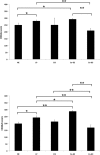
Methods: Expression of CD44s and CD44v6 was analysed by immunohistochemistry with specific antibodies in primary head and neck tissues. Scoring of all specimens followed a two-parameters system, which implemented percentages of positive cells and staining intensities from - to +++ (score = % x intensity; resulting max. score 300). In addition, cell surface expression of CD44s and CD44v6 was assessed in lymphocytes and HNSCC.
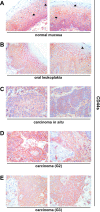
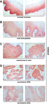
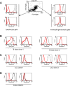
Results: In normal epithelia CD44s and CD44v6 were expressed in 60-95% and 50-80% of cells and yielded mean scores with a standard error of a mean (SEM) of 249.5+/-14.5 and 198+/-11.13, respectively. In oral leukoplakia and in moderately differentiated carcinomas CD44s and CD44v6 levels were slightly increased (278.9+/-7.16 and 242+/-11.7; 291.8+/-5.88 and 287.3+/-6.88). Carcinomas in situ displayed unchanged levels of both proteins whereas poorly differentiated carcinomas consistently expressed diminished CD44s and CD44v6 levels. Lymphocytes and HNSCC lines strongly expressed CD44s but not CD44v6.
Conclusion: CD44s and CD44v6 expression does not distinguish normal from benign or malignant epithelia of the head and neck. CD44s and CD44v6 were abundantly present in the great majority of cells in head and neck tissues, including carcinomas. Hence, the value of CD44s as a marker for the definition of a small subset of cells (i.e. less than 10%) representing head and neck cancer stem cells may need revision.
Conflict of interest statement
Figures


Similar articles
-
Expression of CD44 splice variants in squamous epithelia and squamous cell carcinomas of the head and neck.J Pathol. 1996 May;179(1):66-73. doi: 10.1002/(SICI)1096-9896(199605)179:1<66::AID-PATH544>3.0.CO;2-5. J Pathol. 1996. PMID: 8691348
-
Evaluation of soluble CD44v6 as a potential serum marker for head and neck squamous cell carcinoma.Clin Cancer Res. 1999 Nov;5(11):3534-41. Clin Cancer Res. 1999. PMID: 10589769
-
CD44 and its v6 spliced variant in lung tumors: a role in histogenesis?Cancer. 1997 Jul 1;80(1):34-41. Cancer. 1997. PMID: 9210706
-
The EGF receptor system in head and neck carcinomas and normal tissues. Immunohistochemical and quantitative studies.Dan Med Bull. 1998 Apr;45(2):121-34. Dan Med Bull. 1998. PMID: 9587699 Review.
-
Cancer stem cells in head and neck squamous cell cancer.J Clin Oncol. 2008 Jun 10;26(17):2871-5. doi: 10.1200/JCO.2007.15.1613. J Clin Oncol. 2008. PMID: 18539966 Review.
Cited by
-
The prognostic significance of CD44V6, CDH11, and β-catenin expression in patients with osteosarcoma.Biomed Res Int. 2013;2013:496193. doi: 10.1155/2013/496193. Epub 2013 Jul 18. Biomed Res Int. 2013. PMID: 23971040 Free PMC article.
-
5'-Ectonucleotidase CD73/NT5E supports EGFR-mediated invasion of HPV-negative head and neck carcinoma cells.J Biomed Sci. 2023 Aug 24;30(1):72. doi: 10.1186/s12929-023-00968-6. J Biomed Sci. 2023. PMID: 37620936 Free PMC article.
-
The biology of head and neck cancer stem cells.Oral Oncol. 2012 Jan;48(1):1-9. doi: 10.1016/j.oraloncology.2011.10.004. Epub 2011 Nov 8. Oral Oncol. 2012. PMID: 22070916 Free PMC article. Review.
-
Expression of COX-2, CD44v6 and CD147 and relationship with invasion and lymph node metastasis in hypopharyngeal squamous cell carcinoma.PLoS One. 2013 Sep 3;8(9):e71048. doi: 10.1371/journal.pone.0071048. eCollection 2013. PLoS One. 2013. PMID: 24019861 Free PMC article.
-
Activation of Matrix Hyaluronan-Mediated CD44 Signaling, Epigenetic Regulation and Chemoresistance in Head and Neck Cancer Stem Cells.Int J Mol Sci. 2017 Aug 24;18(9):1849. doi: 10.3390/ijms18091849. Int J Mol Sci. 2017. PMID: 28837080 Free PMC article. Review.
References
-
- Aruffo A, Stamenkovic I, Melnick M, Underhill CB, Seed B. CD44 is the principal cell surface receptor for hyaluronate. Cell. 1990;61:1303–1313. - PubMed
-
- Naor D, Wallach-Dayan SB, Zahalka MA, Sionov RV. Involvement of CD44, a molecule with a thousand faces, in cancer dissemination. Semin Cancer Biol. 2008;18:260–267. - PubMed
-
- Ponta H, Sherman L, Herrlich PA. CD44: from adhesion molecules to signalling regulators. Nat Rev Mol Cell Biol. 2003;4:33–45. - PubMed
-
- Trowbridge IS, Lesley J, Schulte R, Hyman R, Trotter J. Biochemical characterization and cellular distribution of a polymorphic, murine cell-surface glycoprotein expressed on lymphoid tissues. Immunogenetics. 1982;15:299–312. - PubMed
-
- Kuhn S, Koch M, Nubel T, Ladwein M, Antolovic D, et al. A complex of EpCAM, claudin-7, CD44 variant isoforms, and tetraspanins promotes colorectal cancer progression. Mol Cancer Res. 2007;5:553–567. - PubMed
MeSH terms
Substances
LinkOut - more resources
Full Text Sources
Medical
Miscellaneous

