Review
. 2008 Nov;108(11):4670-93.
doi: 10.1021/cr0782729. Epub 2008 Oct 3.

Phosphorylated proteins and control over apatite nucleation, crystal growth, and inhibition

Affiliations
Review

Phosphorylated proteins and control over apatite nucleation, crystal growth, and inhibition

Anne George et al. Chem Rev. 2008 Nov.
No abstract available


Figures

Figure 1
Figure 1
A proposed generalized scheme for matrix-regulated mineralization reactions. Modified and reprinted with permission from Reference 280 [Veis, Reviews in Mineralogy & Geochemistry, V. 54]. Copyright 2003 Mineralogical Society of America.
Figure 2
Figure 2
Three dimensional models of a putative collagen microfibril (five molecules in the quarter-stagger-overlap arrangement). Left: A unit cell, containing the equivalent of all segments from a single molecule in a single D-period. The gap region is less dense, with only four molecules, as compared to the five strands within the overlap zone. Right: A 5-stranded microfibril with the molecules traced through 5 D-periods showing the helical entwinement and reorientation of each single 4.4 D-period long molecule along the microfibril. The differences in molecular flexibility within the gap zones, as compared to the overlap zones are evident. Modified and reprinted with the permission from Figure 3 B and 3E of Reference 19 [Orgel et al. PNAS 103: 9001-9005 (2006)]. Copyright 2006, National Academy of Sciences, USA.
Figure 3
Figure 3
Correlation of collagen packing and sequence in the gap and overlap segments of 1 D period. a. Schematic showing charged residues as dark tilted stripes across all three chains, the segment correlated, in b, with the EM e-band, shown in the positively labeled 1 D period of a fibril. c. A section of type I collagen triple helix with a proline and hydroxyproline deficient region, residues 414 to 424. According to Silver et al this is a flexible region capable of binding both Ca and PO4 ions, and changing the conformation of the helix, based on the use of the SYBYL molecular modeling program and energy minimization after the addition of the mineral ions at the sites proposed. a. and b. Modified and reprinted with permission from Reference 280 [Veis, Reviews in Mineralogy & Geochemistry, V. 54]. Copyright 2003 Mineralogical Society of America. c. Modified and reprinted with permission from Reference 27 [Silver, et.al. Biomacromolecules]. Copyright 2001 American Chemical Society.
Figure 4
Figure 4
The amino acid sequence of human DSPP [PubMed ID: Q9NZW4]. Green: Signal Sequence; Red: Endopeptidase cleavage sites; Black: DSP Sequence; Blue: DPP(DMP2) sequence, including, in Dark Red the DSS Repeat Region, with a few DSS motif interruptions.
Figure 5
Figure 5
The tandem arrangement of the genes comprising the SIBLING family members on human chromosome 4q21-23 region.
Figure 6
Figure 6
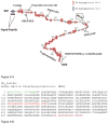
A. Schematic representation of the structural model for osteopontin adapted from Sodek et al.. The model highlights the presence of serine and threonine phosphorylation sites; region containing the poly aspartic acid residue; glycosylation sites, thrombin cleavage site and the integrin binding domain. B. The sequence of human osteopontin, taken from Pub. Med. Protein data bank, ID AAA59974. The signal peptide sequence is in green. Reprinted with permission from Reference 93. Copyright 2000, International Association of Dental Research.
Figure 7
Figure 7
A. Schematic representation of the structural model for bone sialoprotein adapted from Ganss et al.. The model highlights the highly flexible structure, the presence of an hydroxyapatite binding domain at the N-terminus, and O and N-linked glycosylation sites. The Integrin binding RGD domain is located at the carboxyl end of the molecule. B. The sequence of human BSP, taken from the Pub. Med Protein Data Bank, ID AAA60549. The signal sequence is in green, the very acidic, mineral bindng domain is in red. Reprinted with permission from Reference 185. Copyright 1999, International Association of Dental Research.
Figure 8
Figure 8
A. Schematic representation of the structural model for MEPE, depicting the highly flexible nature of the protein backbone. The model shows several PHEX cleavage sites and the hydroxyapatite inhibiting ASARM motif at the carboxyl end of the molecule. Adapted from Rowe et al.. B. The sequence of human MEPE, taken from Pub. Med. Protein Data Bank, ID NP_064588. The signal peptide sequence is in green, the cell-proliferation stimulatory sequence is in red, and the C-terminal ASARM peptide is in purple. Reprinted with permission from Reference 188. Copyright 2000, Elsevier Limited.
Figure 9
Figure 9
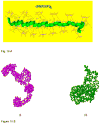
A. Structural analysis of DMP1 as deduced from small angle X-ray scattering experiments. Low resolution models of DMP1 determined using the ab initio program GASBOR depicting the structure in the absence and presence of calcium. In the absence of calcium DMP1 adopts an elongated structure. Within 1 minute of calcium binding DMP1 adopts an extended structure at one end of the molecule with a compact globular After incubation for 3 minutes DMP1 undergoes extensive dimerization. Reprinted with permission from Reference 217. Copyright 2005, American Chemical Society. B. Proposed model for the dual functional role of DMP1. Model demonstrates that DMP1 can inhibit spontaneous mineral precipitation and promote controlled mineral nucleation on a collagenous template. Reprinted with permission from Reference 217. Copyright 2005, American Chemical Society. C. The sequence of rat DMP1 taken from Pub. Med. Protein Data Bank, ID NP_987089. The signal peptide sequence is in green, the mineral binding domains are in red, and the cell attachment domains are in purple.
Figure 9
Figure 9
A. Structural analysis of DMP1 as deduced from small angle X-ray scattering experiments. Low resolution models of DMP1 determined using the ab initio program GASBOR depicting the structure in the absence and presence of calcium. In the absence of calcium DMP1 adopts an elongated structure. Within 1 minute of calcium binding DMP1 adopts an extended structure at one end of the molecule with a compact globular After incubation for 3 minutes DMP1 undergoes extensive dimerization. Reprinted with permission from Reference 217. Copyright 2005, American Chemical Society. B. Proposed model for the dual functional role of DMP1. Model demonstrates that DMP1 can inhibit spontaneous mineral precipitation and promote controlled mineral nucleation on a collagenous template. Reprinted with permission from Reference 217. Copyright 2005, American Chemical Society. C. The sequence of rat DMP1 taken from Pub. Med. Protein Data Bank, ID NP_987089. The signal peptide sequence is in green, the mineral binding domains are in red, and the cell attachment domains are in purple.
Figure 10
Figure 10
A. Molecular Modeling of the DSS domain by energy minimization. Molecular modeling shows the energy minimized lateral view of the structure showing the sommewhat extended folding of the backbone and the relative dispositions of the phosphate and the carboxyl groups relative to the backbone. A notable feature is the puckering or the mobile nature of the backbone which thus provides maximum separation between like-charged ionic groups consistent with the electrostatic repulsions and the creation of specific phosphate-carboxylate surfaces. B. Structural analysis of DPP as deduced from SAXS experiments. Low resolution structural models were determined using the GASBOR program. (i) Model generated in the absence of calcium ion. (ii) Model generated in the presence of calcium. Reprinted with permission from Reference 255. Copyright 2005 S. Krager AG, Basel.

Similar articles

Cited by

References

    1. Lowenstam HA. Science. 1981;211:1126. - PubMed
    1. Eanes ED. Monog Oral Sci. 2001;18:130. - PubMed
    1. Grynpas MD, Omelon S. Bone. 2007;41:162. - PubMed
    1. Crane NJ, Popescu V, Morris MD, Steenhuis P, Ignelzi MA., Jr Bone. 2006;39:434. - PubMed
    1. Veis A, Sabsay B. Biomineralization and Biological Metal Accumulation. D Reidel Pub Co; Dordrecht, Holland: 1982. p. 273.

Publication types