. 2008 Dec 5;283(49):34053-61.
doi: 10.1074/jbc.M805729200. Epub 2008 Sep 26.

Signal regulatory proteins (SIRPS) are secreted presynaptic organizing molecules

Affiliations

Signal regulatory proteins (SIRPS) are secreted presynaptic organizing molecules

Hisashi Umemori et al. J Biol Chem. .

Abstract

Formation of chemical synapses requires exchange of organizing signals between the synaptic partners. Using synaptic vesicle aggregation in cultured neurons as a marker of presynaptic differentiation, we purified candidate presynaptic organizers from mouse brain. A major bioactive species was the extracellular domain of signal regulatory protein alpha (SIRP-alpha), a transmembrane immunoglobulin superfamily member concentrated at synapses. The extracellular domain of SIRP-alpha is cleaved and shed in a developmentally regulated manner. The presynaptic organizing activity of SIRP-alpha is mediated in part by CD47. SIRP-alpha homologues, SIRP-beta and -gamma also have synaptic vesicle clustering activity. The effects of SIRP-alpha are distinct from those of another presynaptic organizer, FGF22: the two proteins induced vesicle clusters of different sizes, differed in their ability to promote neurite branching, and acted through different receptors and signaling pathways. SIRP family proteins may act together with other organizing molecules to pattern synapses.


Figures

FIGURE 1.
FIGURE 1.
Assay and identification of presynaptic organizing molecules. A, assay of presynaptic differentiation. Chick motoneurons were stained with anti-neurofilament antibody to visualize neurites and anti-synapsin antibody to mark synaptic vesicles. The crude brain extract induced neurite elongation, branching, and synaptic vesicle aggregation (middle). The purified fraction (after Mono P; see Table 1) induced vesicle aggregation, but not neurite branching or elongation. Bar is 20 μm for neurofilaments, 10 μm for synapsins. B, the final active fractions were separated by SDS-PAGE and stained with Colloidal Blue. The major band (∼70 kDa; arrow) was identified as SIRP-α by mass spectrometry.
FIGURE 2.
FIGURE 2.
Cell-attached SIRP-α induces aggregation of synaptic vesicle proteins in co-cultured motoneurons. A, COS cells (green cells) expressing full-length SIRP-α plus GFP (SIRPα) or GFP alone (Control) were co-cultured with motoneurons for 3 days and stained for synapsin (red). Synapsin aggregates occur in neurites at sites of contact with SIRP-α-positive COS cells (examples indicated by arrowheads). Bar is 7.5 μm. B, quantification of results from motoneuron-COS cell co-cultures. % neurites with more than one varicosity at sites of contact with COS cells. Bars show mean ± S.E. for four experiments. *, differs from control at p < 0.01. C, COS cells expressing full-length SIRP-α were co-cultured with motoneurons for 3 days and stained for SV2 (red) and SIRP-α (green). SV2 in neurites aggregated at sites of contact with SIRP-α-expressing COS cells. Bar is 6 μm.
FIGURE 3.
FIGURE 3.
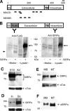
Release of SIRP-α extracellular domain from cultured cells. A, schematic illustration of the molecular structure of mouse SIRP-α and the location of peptide sequences obtained by MALDI and MS/MS. All the peptides are located in the extracellular domain of SIRP-α. SS, signal sequence; TM, transmembrane; aa, amino acids. B, the extracellular domain of SIRP-α is released from transfected COS cells. Conditioned media and cell lysates prepared from COS cells transfected with a FLAG-tagged SIRP-α construct were probed with antibodies to the FLAG tag and the cytoplasmic domain. Conditioned media contained a band (indicated by the arrowhead) that is present in the anti-FLAG blot but not in the anti-SIRP-α blot, indicating that it is a truncated and released SIRP-α. Arrows indicate bands recognized by both antibodies in cell lysates that correspond to full-length SIRP-α. C, Western blotting of muscle extracts prepared from E16 and adult mice with an antibody to the extracellular domain of SIRP-α (top). SIRP-α is more abundant in E16 when synapses are actively forming. In the E16 extract, the antibody detected two major bands of ∼85 and ∼70 kDa. D, SIRP-α in innervated and denervated muscles. The right sciatic nerve of adult mice was surgically cut, and 1 week later, right (denervated, De) and left (innervated, In) leg muscles were tested for SIRPα by Western blotting (top). Denervation significantly increased the SIRPα expression. E, SIRP-α in cell lysates of C2C12 myoblasts (MB) and differentiated myotubes (MT) (top). The level of SIRP-α expression is higher in myotubes that myoblasts. The bottom panels in CE show re-blots for α-tubulin to confirm equal loading of extracts or lysates. F, higher level of SIRP-α extracellular domain in culture media of C2C12 myotubes (MT) than myoblasts (MB), assayed by immunoprecipitation followed by Western blotting using an antibody to the extracellular domain of SIRP-α.
FIGURE 4.
FIGURE 4.
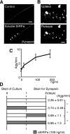
Presynaptic differentiation induced by the soluble extracellular domain of SIRP-α. A, motoneurons were incubated with or without purified soluble SIRP-α ectodomain (sSIRP-α) and stained for synapsin. sSIRP-α induced synapsin-positive aggregates (examples indicated by arrowheads). Bar is 20 μm. B, vesicles in sSIRP-α-induced varicosities are capable of depolarization-dependent recycling. Neurons were depolarized in the presence of FM1–43 dye for 90 s, then washed and photographed. Varicosities (examples indicated by arrowheads) took up dye (uptake). The dye was then released by an additional round of depolarization (release). Control motoneurons without sSIRP-α treatment did not take up FM1–43 dye (data not shown). Bar is 20 μm. C, dose dependence. Motoneuron cultures were treated with soluble SIRP-α at the indicated concentration for 48 h, then vesicle clustering was quantified (number of synapsin-positive aggregates per mm neurite). D, time course. Motoneurons were cultured for 2 days with sSIRP-α (100 ng/ml) during the times indicated in gray. The number of synapsin-positive aggregates per mm of neurite was quantified and shown (Agg/mm). sSIRP-α induced varicosities most efficiently when present on the second day in vitro.*, p < 0.05.
FIGURE 5.
FIGURE 5.
CD47 partially mediates SIRP-α-induced presynaptic differentiation. A, CD47 is expressed in motoneurons. Chick motoneurons were cultured for 2 days and stained for CD47 (top) or synapsin (bottom). B, COS cells expressing full-length SIRP-α plus GFP (SIRPα) or GFP alone were co-cultured with chick motoneurons for 3 days with or without blocking antibodies to SIRPα, CD47, or CD3, and stained for synapsin as described in Fig. 2. Graph shows % of neurites with synapsin-rich varicosities at sites of contact with GFP-positive COS cells. SIRP-α-induced vesicle aggregation is completely blocked by anti-SIRP-α antibody and partially inhibited by anti-CD47 antibodies. *, differs from SIRP-α without antibodies at p < 0.01.
FIGURE 6.
FIGURE 6.
SIRP family members have presynaptic organizing activity and are expressed by the developing muscle and brain. A, chick motoneurons were cultured for 2 days with the recombinant extracellular domain of human SIRP family members (sSIRPs) at 100 ng/ml, and stained for synapsin. All three SIRP family members induced synapsin aggregation. Bar is 20 μm. B, RT-PCR analysis showing that mouse SIRP-α and SIRP-β1 are expressed by E16 muscle and P10 brain. Glyceraldehyde-3-phosphate dehydrogenase (G3PDH) was used as a control. C, motoneurons were cultured with sSIRPs as in A in the presence or absence of blocking antibodies to CD47, and stained for synapsin. Graph shows the number of synapsin aggregates per mm of neurite. All three SIRP family members induced synapsin aggregation (differ from control at p < 0.01), with sSIRP-α and sSIRP-γ showing higher activity than sSIRP-β1(white bars). Anti-CD47 antibodies partially blocked the effect of sSIRP-α and sSIRP-γ, but had no effect on that of sSIRP-β1(black bars). Bars show mean ± S.E. for 100 neurites per condition. *, differs from corresponding controls without anti-CD47 antibodies at p < 0.01.
FIGURE 7.
FIGURE 7.
Different presynaptic organizing effects and mechanisms for SIRP-α and FGF22. A, motoneurons were cultured for 2 days with phosphate-buffered saline (control), sSIRP-α, or FGF22, and stained for synapsin. sSIRP-α induced relatively bigger synapsin-positive aggregates than FGF22, whereas FGF22 induced branching in addition to vesicle clustering. Bar is 15 μm. B, extent of neurite branching induced by FGF22 and sSIRP-α. Bars show mean ± S.E. for 100 neurites per condition. *, differs from control at p < 0.01. C, size of vesicle-rich aggregates induced by FGF22 and sSIRP-α. Bars show mean ± S.E. for 100 aggregates per condition. *, differs from control at p < 0.01. D, effect of genistein (a tyrosine kinase inhibitor; 100 nm), pertussis toxin (PTX, an inhibitor of G protein-receptor interaction; 100 ng/μl), dibutyryl-cAMP (dbcAMP; a cAMP analogue; 100 μm), and U0126 (a MAPK inhibitor; 10 μm) on FGF22- and sSIRP-α-induced vesicle aggregation. FGF22-induced formation of synapsin aggregates is completely blocked by genistein, whereas sSIRP-α-induced varicosity formation is partially inhibited by PTX, dbcAMP, and U0126. None of the inhibitors affected basal levels of aggregation. Bars show mean ± S.E. for at least 50 neurites per condition. *, differs from corresponding controls without inhibitors at p < 0.01.

Similar articles

Cited by

References

    1. Sanes, J. R., and Lichtman, J. W. (1999) Annu. Rev. Neurosci. 22 389-442 - PubMed
    1. Waites, C. L., Craig, A. M., and Garner, C. C. (2005) Annu. Rev. Neurosci. 28 251-274 - PubMed
    1. Fox, M. A., and Umemori, H. (2006) J. Neurochem. 97 1215-1231 - PubMed
    1. Scheiffele, P. (2003) Annu. Rev. Neurosci. 26 485-508 - PubMed
    1. Yamagata, M., Sanes, J. R., and Weiner, J. A. (2003) Curr. Opin. Cell Biol. 15 621-632 - PubMed