. 2008 Nov;237(11):3157-68.
doi: 10.1002/dvdy.21712.

Activation of the Wnt/beta-catenin signaling reporter in developing mouse olfactory nerve layer marks a specialized subgroup of olfactory ensheathing cells

Affiliations

Activation of the Wnt/beta-catenin signaling reporter in developing mouse olfactory nerve layer marks a specialized subgroup of olfactory ensheathing cells

Ya-Zhou Wang et al. Dev Dyn. 2008 Nov.

Abstract

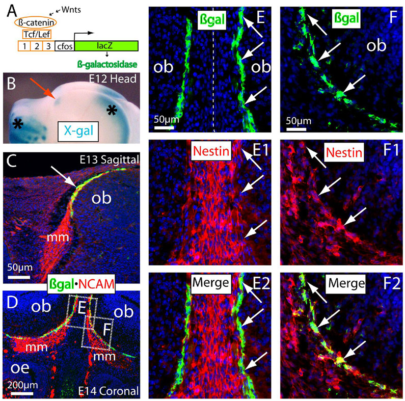
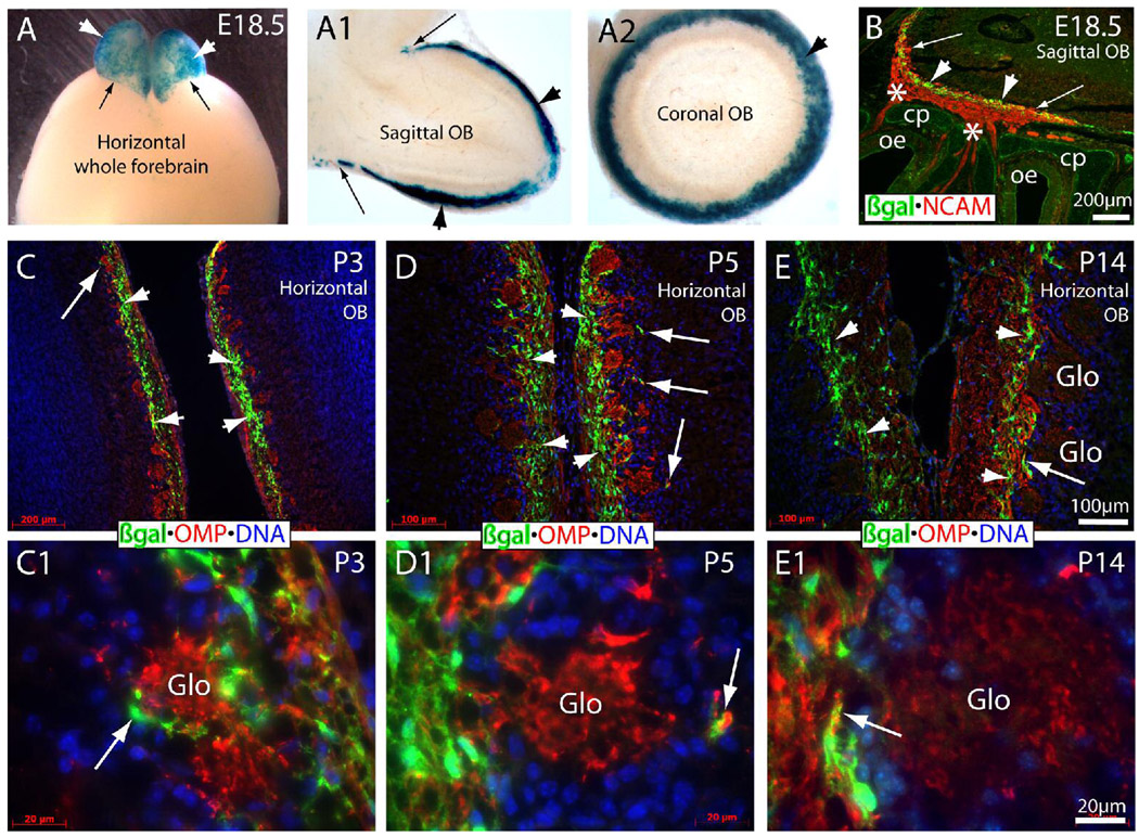
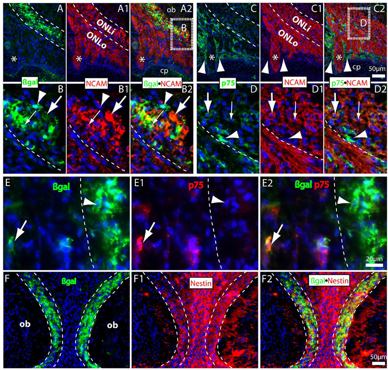
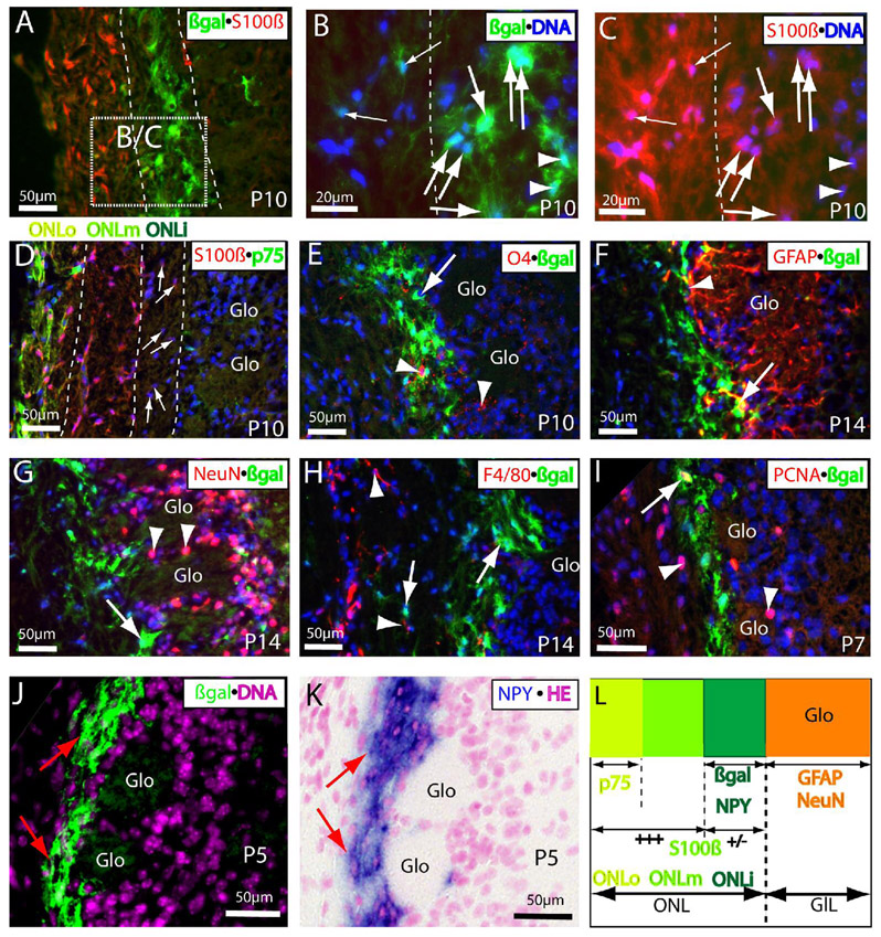
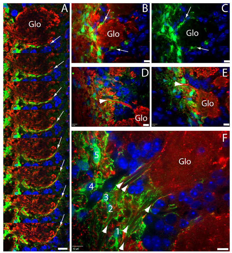
Wnt reporter TOPgal mice carry a beta-galactosidase (betagal) gene under the control of the Wnt/beta-catenin signaling responsive elements. We found that the intensely immunolabeled betagal+ cells were co-immunolabeled with Nestin and formed a tangentially oriented single-cell layer in the "connecting or docking zone" where the olfactory sensory axons attached to the brain surface during mid-gestation. During early postnatal development, betagal+ cells were located in the inner olfactory nerve layer (ONLi) and co-labeled with olfactory ensheathing cell (OEC) markers S100beta and NPY but not with lineage-specific markers for neurons, oligodendrocytes, astrocytes, and microglia, demonstrating that the TOPgal marked a subpopulation of OECs. By confocal microscopy, we found that TOPgal activated processes extended along the developing glomerulus and formed multiple tunnel-like structures that ensheathe and bridge olfactory sensory axonal bundles from ONLi to the glomerulus, which may play a key role in glomerulus formation and convergent sorting of the peripheral olfactory axons.


Figures

Figure 1
Figure 1
Localization and lineage identity of the TOPgal-labeled cells in the olfactory bulb anlage. (A) The Wnt reporter TOPgal construct. (B) Wholemount X-gal staining (blue) of E12 heads of TOPgal mice demonstrated the positively stained (blue) island in the presumably olfactory bulb anlage (red arrow). Note that the dominantly stained X-gal products in the rostral telencephalon and facial regions (asterisks) of the adjacent area faithfully reflected the known activation of Wnt signaling in these sites. (C) Double immunolabeling for β-galactosidase (green) and NCAM (red) on sagittal head sections demonstrated that the Wnt reporter activated cells were single-cell layered and tangentially localized in the connecting zone (white arrow indicates the yellow labeled cell layer with the overlapped red and green signals) where olfactory sensory neuron axons attached to the olfactory bulb anlage at E13. Note that no intense βgal immunolabeling was found in other adjacent tissues including the thick olfactory axon bundles that is a part of migratory mass (mm) on the projecting way from olfactory epithelium to the bulb. (D) Double immunolabeling for βgal and NCAM on coronal sections through olfactory bulb and epithelium at E14. The dash-enclosed areas indicate relevant regions of different tissue sections with a higher magnification in panels E–F2 (single optical sections by confocal microscopy). (E–E2) Double immunolabeling of βgal and Nestin in the middle region between two olfactory bulbs at E14. Most intensely immunolabeled βgal+ cells are co-labeled with weak Nestin-immunoreactivities in the connecting zone (arrows). (F–F2) βgal+/nestin+ cells (arrows) in the connecting zone of the migratory mass. ob, olfactory bulb; oe, olfactory epithelium. The cell nuclei were counterstained by DAPI (blue in C–F2).
Figure 2
Figure 2
Wnt reporter activated “connecting zone” cells in developing olfactory nerve layer (ONL) during glomerulus formation from E18.5 to P14 of mice. (A–A2) X-gal staining of whole forebrain (A) and 150-µm-thick sections of sagittal (A1) and coronal (A2) olfactory bulb at E18.5. Arrowheads indicate the intensely stained X-gal products on the bulb surface/olfactory nerve layer. Arrows indicate the sharp border between the stained bulb and non-stained forebrain surface. Note that no positive staining was present inside the bulb in the thick sections (A1,A2). (B) Double immunolabeling of βgal (green) and NCAM (red) on the sagittal section of E18.5 heads. Intensely immunolabeled βgal+ cells formed a multiple-cell layer (arrowheads) in the connecting zone/developing inner ONL where some NCAM+ olfactory axons crossed the connecting zone and entered into the forming glomerular layer (arrows). Asterisks indicate the thick bundles of NCAM+ olfactory axons crossed through the cribriform plate (cp). (C,C1) The βgal+ cells (green) in the ONL of P3 horizontal bulb sections. Arrow (in C,C1) indicates the same region with different magnifications for the βgal+ process as a capsule-like structure surrounding the forming glomerulus immunolabeled by OMP (red) on the bulb side. Arrowheads (in C) indicate the area of significantly expanded βgal+ multiple-cell layer in ONL. (D,D1) The thickness and density of the βgal+ multiple-cell layer was increased further (arrowheads) in P5 ONL. βgal+ processes were still found in the periglomerulus (arrows) on the bulb side. (E,E1) The thickness and density of βgal+ cells were significantly declined and restricted to the inner most ONL at P14 (arrowheads) when most glomeruli are formed. The βgal+ process mixed with OMP+ olfactory axons were found in the olfactory axonal entrance to the glomerulus (arrows). OB, olfactory bulb; Glo, glomerulus (visualized by OMP+ olfactory axons); OMP, olfactory marker protein. The cell nuclei were counterstained by DAPI (blue in C–E1).
Figure 3
Figure 3
Double immunolabeling for the TOPgal-labeled “connecting zone” cells with OEC markers on coronal sections of E18.5 olfactory bulb. (A–B2) βgal+ cells (green) consist a multiple-cell layer intermingled with NCAM+ olfactory axons (red) in the inner ONL (dashed lines in parallel). Some of them were co-immunolabeled with NCAM in both perinuclear cytoplasm (small arrows in B–B2) and processes (large arrows in B–B2). Arrowheads (in B–B2) indicate a βgal+/NCAM cell located in the front line of the connecting zone. (Panels B–B2 were magnified from the dash-lined area indicated in A2). (C–D2) Weakly immunolabeled p75+ OECs (green) were also found in the inner ONL that most of them were co-immunolabeled with NCAM in the perinuclear cytoplasm (arrows). Note that p75+ OECs were dominantly distributed in outer ONL by surrounding the large olfactory axonal bundles (asterisks) but no overlapping (arrowheads) with NCAM immunolabeling. (Panels D–D2 were magnified from the dash-lined area indicated in C2; Arrowheads in D–D2 indicate an intense p75+ cells that was not stained with NCAM in boundary between ONLi and ONLo). (E–E2) A few βgal+ (green) and p75+ (red) double-immunolabeled OEC-like cells were found in the E18.5 ONLo (arrows). Arrowheads indicate an intensely immunolabeled βgal+ cell without p75-immunolabeling in the inner ONL (that divided by a dashed line). (F–F2) Double immunolabeling of βgal and Nestin on a coronal section with left and right bulbs shown at E18.5. Most βgal+ cells (green) in the ONLi were co-immunolabeled with Nestin (red). Note that Nestin immunoreactivities were also found in the intermediate zone between two bulbs and in the developing glomerular layer. cp, cribriform plate. Asterisks (in A–C2) indicate a large olfactory axonal bundle crossed the cribriform plate. The cell nuclei were counterstained by DAPI (blue).
Figure 4
Figure 4
The Wnt reporter TOPgal marked a distinct subpopulation of OECs in the postnatal ONL. (A–C) βgalintense+/S100βweak+ cells (indicated by large arrows in the same orientations) were detected in the inner ONL at P10. Panels B and C were magnified from the dashed square in panel A. Note that some intense βgal+ cells in the inner ONLi were extremely low immunoreactivities with S100β (arrowheads), and some double immunolabeled βgal+/S100β+ cells (small arrows) were found in the middle or outer ONL. Dashed lines in B and C indicate the border between inner and middle ONL. (D) Three subpopulations of OECs exist in the P10 ONL: S100β+/p75+ in the outer (ONLo), S100β+/p75 in the middle (ONLm), and the weakly S100β immunolabeled OECs (arrows) in the inner (ONLi). (E) O4+ Schwann lineage cells (red indicated by arrowheads) were scattered in the inner ONL where with intense βgal-immunolabeling, but no co-localization in the same cells was found at P10. (F) GFAP+ astrocyte lineage cells and processes (red) were stained intensely in the glomeruli at P14 with a clear border next to the βgal+ inner ONL, but no cells with double immunolabeling were found except that a few processes could be running together tightly in the periglomerular region (arrow). (G) No double-immunolabeling of βgal (green indicated by arrow) with neuronal marker NeuN (red indicated by arrowheads) was found in the periglomerular zone or entire ONL at P14. (H) F4/80+ microglia were found in the outer ONL, and no co-immunolabeling with βgal was found at P14. (I) A small portion of βgal+ cells in the ONL was found co-immunolabeled with PCNA+ (proliferating cell nuclear antigen) at P7 (arrow). Note that PCNA+ cells were found more frequently in adjacent layers, the middle ONL and glomerular layer (arrowheads). (J,K) The distribution pattern of βgal immunolabeling in the ONLi (green indicated by arrows in J) mirrors the NPY expression detected by in situ hybridization (blue indicated by arrows in K) in the same region on an immediately adjacent neighboring section at P5. (L) Schematic summary for the distribution, antigenic characters, and relationship of the Wnt reporter activated or inactivated OECs in the ONL around P5 to P14. The Wnt reporter activated cells in the early postnatal olfactory bulb are a specialized subgroup of OECs with intense βgal and weak S100β immunolabeling as well as NPY expression that are restricted in the ONLi immediately adjacent to the glomerular layer (GlL; In which the cells are CNS origins with characteristic immunolabeling for astrocyte (GFAP) and neuronal (NeuN) markers). Glo, glomerulus. A–J, the cell nuclei were counterstained by DAPI (blue in A–I, pink in J); K, counterstained by hematoxylin and eosin (HE).
Figure 5
Figure 5
Confocal microscopy of TOPgal+ OECs/processes. (A) A long extension (arrows) of TOPgal+ processes along the edge of a glomerulus at P3 in serial adjacent scanning images. (B,C) Capsule-like TOPgal+ processes (arrows) surrounding a glomerulus at P5. (D,E) Two examples of tunnel-like structures (arrowheads) of TOPgal+ processes wrapping axonal bundles towards the glomerulus at P5. (F) Multiple tunnel-like structures (arrowheads) formed by TOPgal+ processes bridging a single glomerulus and ONL at P10. Five TOPgal+ OECs (no. 1~5) on this panel are lining up in the inner most ONL, and some of them (particularly the no. 5 cell) with net-like processes ensheath multiple axonal bundles in the transverse orientation. B–F, single optical sections. Scale bars, 10 µm. Nuclei were counterstained by DAPI (blue).

Similar articles

Cited by

References

    1. Aoki K, Osumi-Yamashita N, Ninomiya Y, Eto K. Differential expression of NCAM, vimentin and MAP1B during initial pathfinding of olfactory receptor neurons in the mouse embryo. Anat Embryol (Berl) 1995;192:211–220. - PubMed
    1. Au E, Roskams AJ. Olfactory ensheathing cells of the lamina propria in vivo and in vitro. Glia. 2003;41:224–236. - PubMed
    1. Au WW, Treloar HB, Greer CA. Sublaminar organization of the mouse olfactory bulb nerve layer. J Comp Neurol. 2002;446:68–80. - PubMed
    1. Bailey MS, Puche AC, Shipley MT. Development of the olfactory bulb: evidence for glia-neuron interactions in glomerular formation. J Comp Neurol. 1999;415:423–448. - PubMed
    1. Balmer CW, LaMantia AS. Noses and neurons: induction, morphogenesis, and neuronal differentiation in the peripheral olfactory pathway. Dev Dyn. 2005;234:464–481. - PubMed

Publication types

MeSH terms

LinkOut - more resources