A role for sphingosine kinase 1 in dextran sulfate sodium-induced colitis
- PMID: 18815359
- PMCID: PMC2626622
- DOI: 10.1096/fj.08-118109
A role for sphingosine kinase 1 in dextran sulfate sodium-induced colitis
Abstract
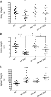
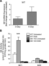
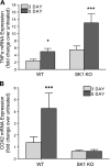
The bioactive lipid sphingosine-1-phosphate (S1P) is emerging as an important mediator of immune and inflammatory responses. S1P formation is catalyzed by sphingosine kinase (SK), of which the SK1 isoenzyme is activated by tumor necrosis alpha (TNF-alpha). SK1 has been shown to be required for mediating TNF-alpha inflammatory responses in cells, including induction of cyclooxygenase 2 (COX-2). Because TNF-alpha and COX-2 are increased in patients with inflammatory bowel disease (IBD), we investigated the role of SK1 in a murine model of colitis. SK1(-/-) mice treated with dextran sulfate sodium (DSS) had significantly less blood loss, weight loss, colon shortening, colon histological damage, and splenomegaly than did wild-type (WT) mice. In addition, SK1(-/-) mice had no systemic inflammatory response. Moreover, WT but not SK1(-/-) mice treated with dextran sulfate sodium had significant increases in blood S1P levels, colon SK1 message and activity, and colon neutrophilic infiltrate. Unlike WT mice, SK1(-/-) mice failed to show colonic COX-2 induction despite an exaggerated TNF-alpha response; thus implicating for the first time SK1 in TNF-alpha-mediated COX-2 induction in vivo. Inhibition of SK1 may prove to be a valuable therapeutic target by inhibiting systemic and local inflammation in IBD.
Figures


Similar articles
-
Distinct roles for hematopoietic and extra-hematopoietic sphingosine kinase-1 in inflammatory bowel disease.PLoS One. 2014 Dec 2;9(12):e113998. doi: 10.1371/journal.pone.0113998. eCollection 2014. PLoS One. 2014. PMID: 25460165 Free PMC article.
-
The sphingosine kinase 1/sphingosine-1-phosphate pathway mediates COX-2 induction and PGE2 production in response to TNF-alpha.FASEB J. 2003 Aug;17(11):1411-21. doi: 10.1096/fj.02-1038com. FASEB J. 2003. PMID: 12890694
-
Loss of neutral ceramidase increases inflammation in a mouse model of inflammatory bowel disease.Prostaglandins Other Lipid Mediat. 2012 Dec;99(3-4):124-30. doi: 10.1016/j.prostaglandins.2012.08.003. Epub 2012 Aug 31. Prostaglandins Other Lipid Mediat. 2012. PMID: 22940715 Free PMC article.
-
Role of sphingosine 1-phosphate receptors, sphingosine kinases and sphingosine in cancer and inflammation.Adv Biol Regul. 2016 Jan;60:151-159. doi: 10.1016/j.jbior.2015.09.001. Epub 2015 Sep 25. Adv Biol Regul. 2016. PMID: 26429117 Review.
-
Sphingosine kinase: Role in regulation of bioactive sphingolipid mediators in inflammation.Biochimie. 2010 Jun;92(6):707-15. doi: 10.1016/j.biochi.2010.02.008. Epub 2010 Feb 13. Biochimie. 2010. PMID: 20156522 Free PMC article. Review.
Cited by
-
Host sphingosine kinase 1 worsens pancreatic cancer peritoneal carcinomatosis.J Surg Res. 2016 Oct;205(2):510-517. doi: 10.1016/j.jss.2016.05.034. Epub 2016 Jul 9. J Surg Res. 2016. PMID: 27664902 Free PMC article.
-
Isoflurane activates intestinal sphingosine kinase to protect against bilateral nephrectomy-induced liver and intestine dysfunction.Am J Physiol Renal Physiol. 2011 Jan;300(1):F167-76. doi: 10.1152/ajprenal.00467.2010. Epub 2010 Oct 20. Am J Physiol Renal Physiol. 2011. PMID: 20962114 Free PMC article.
-
STAT3 in epithelial cells regulates inflammation and tumor progression to malignant state in colon.Neoplasia. 2013 Sep;15(9):998-1008. doi: 10.1593/neo.13952. Neoplasia. 2013. PMID: 24027425 Free PMC article.
-
Counter-regulation of opioid analgesia by glial-derived bioactive sphingolipids.J Neurosci. 2010 Nov 17;30(46):15400-8. doi: 10.1523/JNEUROSCI.2391-10.2010. J Neurosci. 2010. PMID: 21084596 Free PMC article.
-
Sphingosine-1-Phosphate Signaling and Metabolism Gene Signature in Pediatric Inflammatory Bowel Disease: A Matched-case Control Pilot Study.Inflamm Bowel Dis. 2018 May 18;24(6):1321-1334. doi: 10.1093/ibd/izy007. Inflamm Bowel Dis. 2018. PMID: 29788359 Free PMC article.
References
-
- Obeid L M, Linardic C M, Karolak L A, Hannun Y A. Programmed cell death induced by ceramide. Science (New York) 1993;259:1769–1771. - PubMed
-
- Venable M E, Lee J Y, Smyth M J, Bielawska A, Obeid L M. Role of ceramide in cellular senescence. J Biol Chem. 1995;270:30701–30708. - PubMed
-
- Seufferlein T, Rozengurt E. Sphingosine induces p125FAK and paxillin tyrosine phosphorylation, actin stress fiber formation, and focal contact assembly in Swiss 3T3 cells. J Biol Chem. 1994;269:27610–27617. - PubMed
-
- Lee S C, Kuan C Y, Wen Z D, Yang S D. The naturally occurring PKC inhibitor sphingosine and tumor promoter phorbol ester potentially induce tyrosine phosphorylation/activation of oncogenic proline-directed protein kinase FA/GSK-3alpha in a common signalling pathway. J Protein Chem. 1998;17:15–27. - PubMed
-
- Kimura T, Watanabe T, Sato K, Kon J, Tomura H, Tamama K, Kuwabara A, Kanda T, Kobayashi I, Ohta H, Ui M, Okajima F. Sphingosine 1-phosphate stimulates proliferation and migration of human endothelial cells possibly through the lipid receptors, Edg-1 and Edg-3. Biochem J. 2000;348:71–76. - PMC - PubMed
Publication types
MeSH terms
Substances
Grants and funding
LinkOut - more resources
Full Text Sources
Other Literature Sources
Molecular Biology Databases
Research Materials

