

A duplication at chromosome 11q12.2-11q12.3 is associated with spinocerebellar ataxia type 20
- PMID: 18801880
- PMCID: PMC2588641
- DOI: 10.1093/hmg/ddn283
A duplication at chromosome 11q12.2-11q12.3 is associated with spinocerebellar ataxia type 20
Abstract
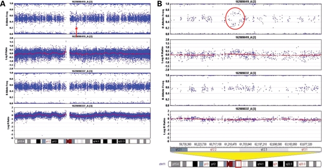
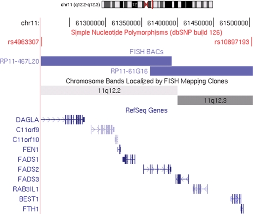
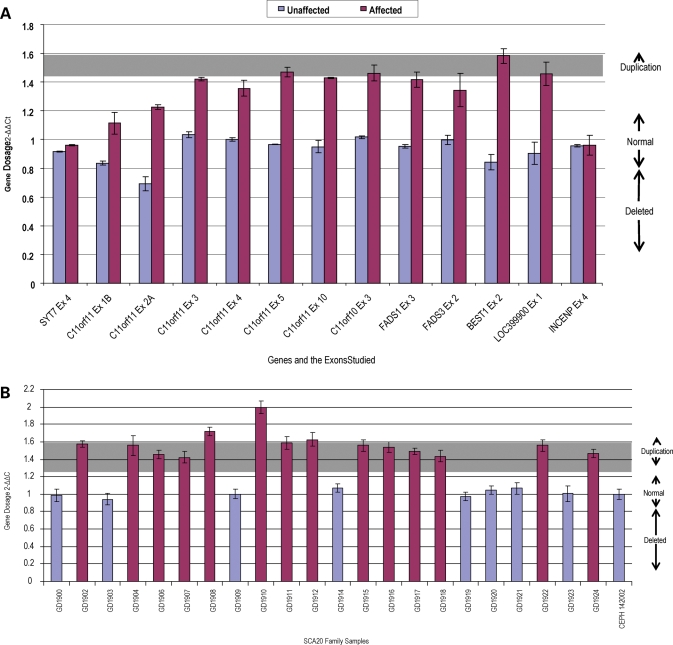
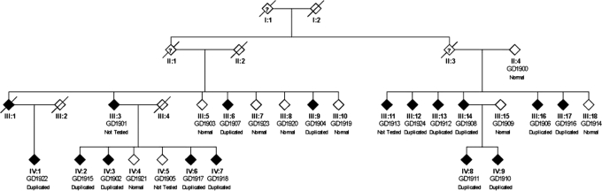
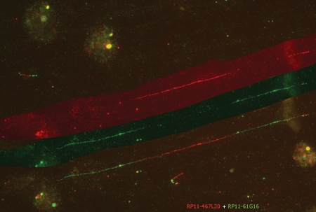
Spinocerebellar ataxia type 20 (SCA20) has been linked to chromosome 11q12, but the underlying genetic defect has yet to be identified. We applied single-nucleotide polymorphism genotyping to detect structural alterations in the genomic DNA of patients with SCA20. We found a 260 kb duplication within the previously linked SCA20 region, which was confirmed by quantitative polymerase chain reaction and fiber fluorescence in situ hybridization, the latter also showing its direct orientation. The duplication spans 10 known and 2 unknown genes, and is present in all affected individuals in the single reported SCA20 pedigree. While the mechanism whereby this duplication may be pathogenic remains to be established, we speculate that the critical gene within the duplicated segment may be DAGLA, the product of which is normally present at the base of Purkinje cell dendritic spines and contributes to the modulation of parallel fiber-Purkinje cell synapses.
Figures
Similar articles
-
A 7.5-Mb duplication at chromosome 11q21-11q22.3 is associated with a novel spastic ataxia syndrome.Mov Disord. 2015 Feb;30(2):262-6. doi: 10.1002/mds.26059. Epub 2014 Dec 27. Mov Disord. 2015. PMID: 25545641 Free PMC article.
-
Spinocerebellar ataxia type 20.Cerebellum. 2005;4(1):55-7. doi: 10.1080/14734220410019048. Cerebellum. 2005. PMID: 15895561
-
Spinocerebellar ataxia with mental retardation (SCA13).Cerebellum. 2005;4(1):43-6. doi: 10.1080/14734220510007923. Cerebellum. 2005. PMID: 15895558 Review.
-
Spinocerebellar ataxia type 26 maps to chromosome 19p13.3 adjacent to SCA6.Ann Neurol. 2005 Mar;57(3):349-54. doi: 10.1002/ana.20371. Ann Neurol. 2005. PMID: 15732118
-
Spinocerebellar ataxia type 20.Handb Clin Neurol. 2012;103:567-73. doi: 10.1016/B978-0-444-51892-7.00038-3. Handb Clin Neurol. 2012. PMID: 21827916 Review.
Cited by
-
Towards a complete resolution of the genetic architecture of disease.Trends Genet. 2010 Oct;26(10):438-42. doi: 10.1016/j.tig.2010.07.004. Trends Genet. 2010. PMID: 20813421 Free PMC article.
-
Endocannabinoid dysfunction in neurological disease: neuro-ocular DAGLA-related syndrome.Brain. 2022 Oct 21;145(10):3383-3390. doi: 10.1093/brain/awac223. Brain. 2022. PMID: 35737950 Free PMC article.
-
Emerging pathogenic pathways in the spinocerebellar ataxias.Curr Opin Genet Dev. 2009 Jun;19(3):247-53. doi: 10.1016/j.gde.2009.02.009. Epub 2009 Apr 1. Curr Opin Genet Dev. 2009. PMID: 19345087 Free PMC article. Review.
-
Autosomal dominant cerebellar ataxia type I: a review of the phenotypic and genotypic characteristics.Orphanet J Rare Dis. 2011 May 28;6:33. doi: 10.1186/1750-1172-6-33. Orphanet J Rare Dis. 2011. PMID: 21619691 Free PMC article. Review.
-
Identification of Extremely Rare Pathogenic CNVs by Array CGH in Saudi Children with Developmental Delay, Congenital Malformations, and Intellectual Disability.Children (Basel). 2023 Mar 31;10(4):662. doi: 10.3390/children10040662. Children (Basel). 2023. PMID: 37189911 Free PMC article.
References
-
- Orr H.T., Zoghbi H.Y. Trinucleotide repeat disorders. Annu. Rev. Neurosci. 2007;30:575–621. - PubMed
-
- Ranum L.P., Day J.W. Dominantly inherited, non-coding microsatellite expansion disorders. Curr. Opin. Genet. Dev. 2002;12:266–271. - PubMed
-
- Schöls L., Bauer P., Schmidt T., Schulte T., Riess O. Autosomal dominant cerebellar ataxias: clinical features, genetics, and pathogenesis. Lancet Neurol. 2004;3:291–304. - PubMed
-
- Houlden H., Johnson J., Gardner-Thorpe C., Lashley T., Hernandez D., Worth P., Singleton A.B., Hilton D.A., Holton J., Revesz T., et al. Mutations in TTBK2, encoding a kinase implicated in tau phosphorylation, segregate with spinocerebellar ataxia type 11. Nat. Genet. 2007;39:1434–1436. - PubMed
-
- Waters M.F., Minassian N.A., Stevanin G., Figueroa K.P., Bannister J.P., Nolte D., Mock A.F., Evidente V.G., Fee D.B., Muller U., et al. Mutations in voltage-gated potassium channel KCNC3 cause degenerative and developmental central nervous system phenotypes. Nat. Genet. 2006;38:447–451. - PubMed
Publication types
MeSH terms
Grants and funding
LinkOut - more resources
Full Text Sources
Other Literature Sources
Molecular Biology Databases
Research Materials
