. 2008 Dec;82(23):11495-502.
doi: 10.1128/JVI.01548-08. Epub 2008 Sep 17.

The TRIM5alpha B-box 2 domain promotes cooperative binding to the retroviral capsid by mediating higher-order self-association

Affiliations

The TRIM5alpha B-box 2 domain promotes cooperative binding to the retroviral capsid by mediating higher-order self-association

Xing Li et al. J Virol. 2008 Dec.

Abstract

The retroviral restriction factor, TRIM5alpha, blocks infection of a spectrum of retroviruses soon after virus entry into the cell. TRIM5alpha consists of RING, B-box 2, coiled-coil, and B30.2(SPRY) domains. The B-box 2 domain is essential for retrovirus restriction by TRIM5alpha, but its specific function is unknown. We show here that the B-box 2 domain mediates higher-order self-association of TRIM5alpha(rh) oligomers. This self-association increases the efficiency of TRIM5alpha binding to the retroviral capsid, thus potentiating restriction of retroviral infection. The contribution of the B-box 2 domain to cooperative TRIM5alpha association with the retroviral capsid explains the conditional nature of the restriction phenotype exhibited by some B-box 2 TRIM5alpha mutants; the potentiation of capsid binding that results from B-box 2-mediated self-association is essential for restriction when B30.2(SPRY) domain-mediated interactions with the retroviral capsid are weak. Thus, B-box 2-dependent higher-order self-association and B30.2(SPRY)-dependent capsid binding represent complementary mechanisms whereby sufficiently dense arrays of capsid-bound TRIM5alpha proteins can be achieved.


Figures
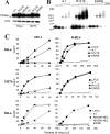
FIG. 1.
FIG. 1.
Expression and antiretroviral activity of TRIM5α variants. (A) The expression level of wild-type (w.t.) TRIM5αrh, R121E, and ER/RE with C-terminal HA tags was determined by Western blotting lysates from HeLa and Cf2Th cells stably expressing these proteins. Cell lysates with equal amount of total proteins were resolved by SDS-PAGE, and the Western blot was probed with an anti-HA antibody and an anti-β-actin antibody to control for loading. (B) Oligomerization of TRIM5α variants was examined. Cell lysates from 293T cells transiently expressing the indicated TRIM5α proteins were cross-linked with increasing concentrations of glutaraldehyde (0, 1, 2, 4, and 8 mM). The cross-linked products were resolved by SDS-PAGE and visualized by Western blotting with an anti-HA antibody. (C) The effects of the TRIM5α B-box 2 variants on retroviral infection were assessed. HeLa or Cf2Th cells stably expressing the wild-type TRIM5αrh protein (w.t.) and mutant TRIM proteins, or control cells transduced with the empty LPCX vector, were incubated with various amounts of HIV-1-GFP or N-MLV-GFP. Infected GFP-positive cells were counted by FACS. The experiments were repeated with comparable results.
FIG. 2.
FIG. 2.
Turnover and subcellular localization of TRIM5αrh B-box 2 variants. (A) HeLa cells expressing wild-type TRIM5αrh and TRIM5αrh ER/RE were treated with cycloheximide to block protein synthesis for a 5-h period in the absence or presence of the proteasome inhibitor MG115. Cells were harvested and lysed at 1-h intervals. Cell lysates containing equal amounts of total protein were analyzed by Western blotting with anti-HA and anti-actin antibodies. (B) HeLa or Cf2Th cells stably expressing the HA-tagged TRIM5αrh variants were stained with an anti-HA antibody, followed by an anti-rat secondary antibody conjugated to Alexa 488. The TRIM5αrh proteins are shown in green, and the nuclei are stained blue with DAPI.
FIG. 3.
FIG. 3.
Contribution of the B-box 2 domain to higher-order self-association of TRIM5αrh. (A) The coprecipitation of HA-tagged TRIM5αrh variants with FLAG-tagged TRIM5αrh was examined. 293T cells were transfected transiently with pLPCX plasmids expressing C-terminally HA-tagged wild-type (w.t.) TRIM5αrh, R121E, or ER/RE or N-terminally FLAG-tagged wild-type (w.t.) TRIM5αrh. Cytosolic lysates containing the TRIM5αrh variants were prepared, and the concentration of TRIM5 protein in the lysates was adjusted to account for differences in the levels of expression, as described in Materials and Methods. The adjusted lysates were then mixed in a 1:1 ratio and used for precipitation with an anti-FLAG antibody. The amounts of HA- and FLAG-tagged proteins in the lysates and immunoprecipitates (IPs) were analyzed by Western blotting (WB) with HRP-conjugated anti-HA and anti-FLAG antibodies. (B) The ability of TRIM5αrh ER/RE and R121E to associate with themselves in the coimmunoprecipitation assay was examined. The assay was carried out as described above using N-terminally FLAG-tagged TRIM5αrh ER/RE and R121E in addition to the FLAG-tagged wild-type TRIM5αrh protein (FLAG-w.t.). (C) The requirement for the B30.2(SPRY) domain for the higher-order self-association of TRIM5αrh was examined. An N-terminally FLAG-tagged truncation mutant of TRIM5rh (RBCC-L2), which lacks a B30.2(SPRY) domain (16), was used in the coimmunoprecipitation assay along with HA-tagged wild-type TRIM5αrh and TRIM5αrh ER/RE.
FIG. 4.
FIG. 4.
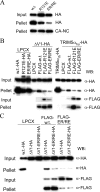
Contribution of the B-box 2 domain to TRIM5α recruitment to the capsid. (A) The binding of the wild-type TRIM5αrh protein (w.t.) and the R121E and ER/RE TRIM5αrh mutants to the HIV-1 CA-NC complexes was examined. Cell lysates of 293T cells transiently expressing the C-terminally HA-tagged TRIM5αrh variants were used in the HIV-1 CA-NC binding assay. To compare the binding efficiency quantitatively, the lysates containing the highly expressed R121E and ER/RE proteins were diluted with lysates from 293T cells transiently transfected with the empty pLPCX plasmid to achieve input levels comparable to that of w.t. TRIM5αrh. The top and middle panels show Western blots with an anti-HA antibody to detect the amount of TRIM5αrh proteins in the input and pellet, respectively; the bottom panel shows the amount of HIV-1 CA-NC protein that was pelleted through the 70% sucrose cushion. (B) The ability of wild-type (w.t.) TRIM5αrh to recruit the ΔV1 TRIM5αrh mutant and TRIM5αhu to HIV-1 CA-NC complexes was examined. Cell lysates made from 293T cells transiently transfected with the empty pLPCX plasmid were mixed with HA-tagged wild-type TRIM5αrh, R121E, or ER/RE in a 1:1 ratio. The mixed lysates were then used in the in vitro HIV-1 CA-NC binding assay as controls to demonstrate that they bind the assembled capsid complexes. HA-tagged ΔV1 and TRIM5αhu, which do not bind the capsid well when present alone (6, 21, 36, 39), were mixed with lysates from cells transfected with pLPCX or cells expressing FLAG-tagged wild-type TRIM5αrh, R121E, or ER/RE and subjected to the binding assay. The input amounts of lysates containing FLAG-tagged wild-type TRIM5αrh, R121E, and ER/RE were adjusted so that the amounts of FLAG-R121E and FLAG-ER/RE bound to the HIV-1 CA-NC complexes were at least as great as that of the FLAG-tagged wild-type TRIM5αrh protein. Cell lysates containing HA-tagged TRIM5αrh mixed with lysates from pLPCX-transfected cells were included as a positive control in the TRIM5αhu recruitment experiment. The amounts of HA-tagged and FLAG-tagged proteins present in the input and the pellet were detected by Western blotting with an anti-HA antibody (Roche) and an anti-FLAG antibody (Sigma), respectively. To compensate for the high level of R121E expression, all HA-tagged and FLAG-tagged R121E input and pellet samples were loaded onto the SDS-polyacrylamide gel at half the volume of the other protein samples, except in the pellet sample of the TRIM5αhu recruitment experiment. Due to the different input levels of the FLAG-tagged wild-type TRIM5αrh, R121E, and ER/RE, in some cases the FLAG-w.t. protein is not visible on the Western blot at the exposure shown. (C) The ability of the ER/RE mutant to recruit itself to HIV-1 capsid complexes was examined. A C-terminally HA-tagged mutant containing both the ΔV1 B30.2(SPRY) domain deletion and the ER/RE B-box 2 changes was used in the recruitment assay, as described above. Cell lysates containing HA-tagged wild-type TRIM5αrh, ΔV1, and ΔV1-ER/RE were mixed with pLPCX-transfected cell lysates or lysates containing FLAG-tagged wild-type TRIM5αrh or ER/RE in a 1:1 ratio and used in the capsid binding assay.
FIG. 5.
FIG. 5.
HIV-1-restricting ability of TRIM5-Cyp A fusion proteins with changes in the B-box 2 domain. HeLa cells stably expressing wild-type TRIM5αrh or rh5RBCC-Cyp B-box 2 mutants, or cells transduced with the empty LPCX vector, were exposed to increasing amounts of HIV-1-GFP. Infected, GFP-positive cells were counted by FACS. The results of a single experiment are shown; similar results were obtained in a repeat experiment.

Similar articles

Cited by

References

    1. Anderson, J. L., E. M. Campbell, X. Wu, N. Vandegraaff, A. Engelman, and T. J. Hope. 2006. Proteasome inhibition reveals that a functional preintegration complex intermediate can be generated during restriction by diverse TRIM5 proteins. J. Virol. 809754-9760. - PMC - PubMed
    1. Beenders, B., P. L. Jones, and M. Bellini. 2007. The tripartite motif of nuclear factor 7 is required for its association with transcriptional units. Mol. Cell. Biol. 272615-2624. - PMC - PubMed
    1. Borden, K. L., M. N. Boddy, J. Lally, N. J. O'Reilly, S. Martin, K. Howe, E. Solomon, and P. S. Freemont. 1995. The solution structure of the RING finger domain from the acute promyelocytic leukaemia proto-oncoprotein PML. EMBO J. 141532-1541. - PMC - PubMed
    1. Campbell, E. M., O. Perez, J. L. Anderson, and T. J. Hope. 2008. Visualization of a proteasome-independent intermediate during restriction of HIV-1 by rhesus TRIM5α. J. Cell Biol. 180549-561. - PMC - PubMed
    1. Cao, T., K. L. Borden, P. S. Freemont, and L. D. Etkin. 1997. Involvement of the rfp tripartite motif in protein-protein interactions and subcellular distribution. J. Cell Sci. 110(Pt. 14)1563-1571. - PubMed

Publication types

LinkOut - more resources