

TDP-43 accumulation in inclusion body myopathy muscle suggests a common pathogenic mechanism with frontotemporal dementia
- PMID: 18796596
- PMCID: PMC2586594
- DOI: 10.1136/jnnp.2007.131334
TDP-43 accumulation in inclusion body myopathy muscle suggests a common pathogenic mechanism with frontotemporal dementia
Abstract
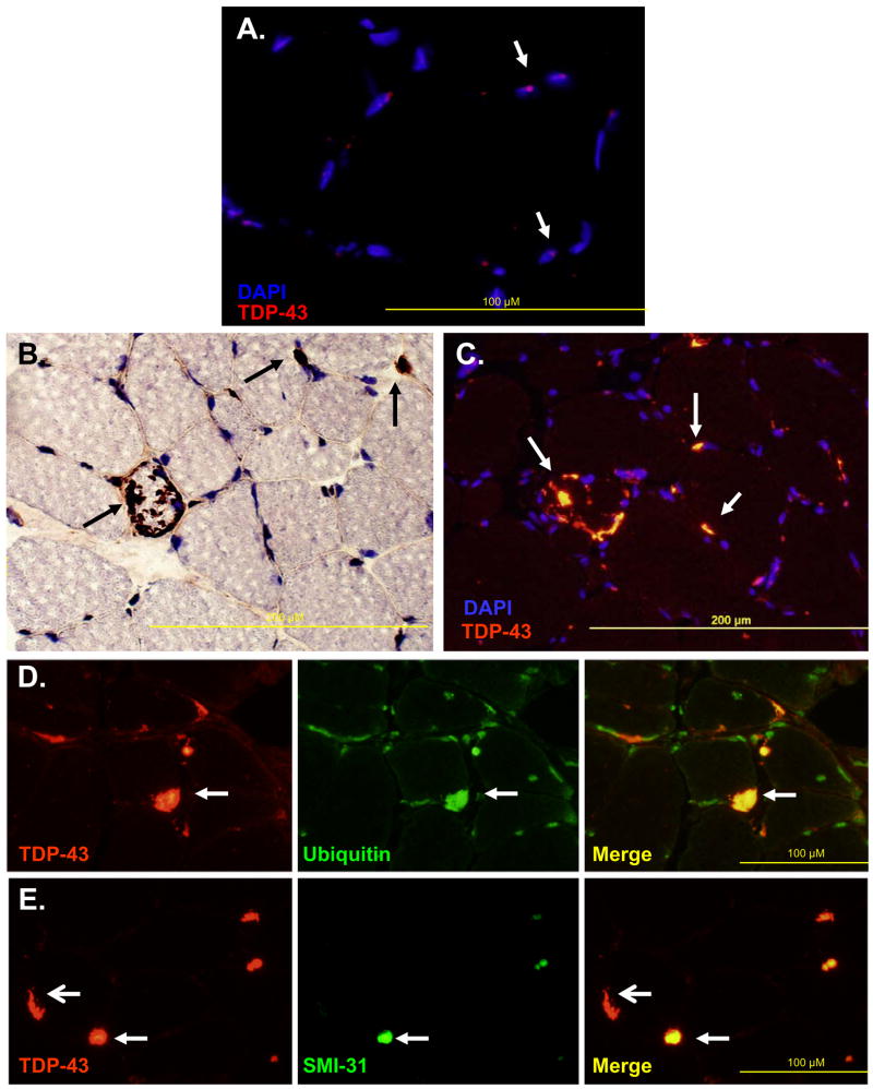
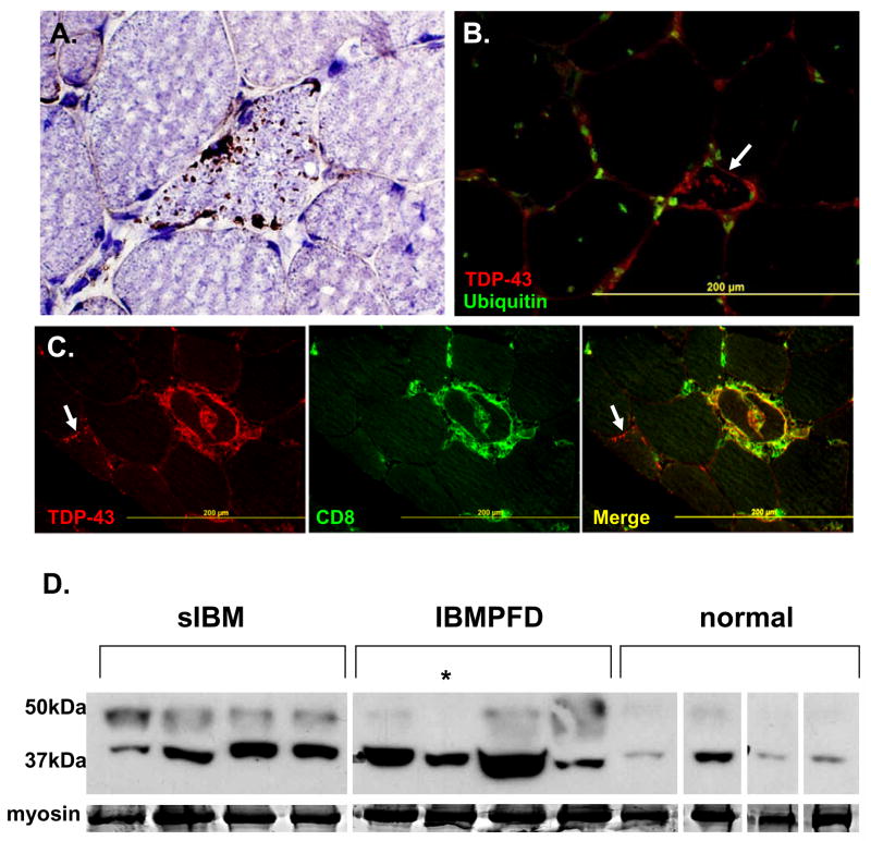
TAR DNA binding protein-43 (TDP-43) is found in ubiquitinated inclusions (UBIs) in some frontotemporal dementias (FTD-U). One form of FTD-U, due to mutations in the valosin containing protein (VCP) gene, occurs with an inclusion body myopathy (IBMPFD). Since IBMPFD brain has TDP-43 in UBIs, we looked for TDP-43 inclusions in IBMPFD muscle. In normal muscle, TDP-43 is present in nuclei. In IBMPFD muscle, TDP-43 is additionally present as large inclusions within UBIs in muscle cytoplasm. TDP-43 inclusions were also found in 78% of sporadic inclusion body myositis (sIBM) muscles. In IBMPFD and sIBM muscle, TDP-43 migrated with an additional band on immunoblot similar to that reported in FTD-U brains. This study adds sIBM and hereditary inclusion body myopathies to the growing list of TDP-43 positive inclusion diseases.
Figures
Similar articles
-
Valosin-containing protein disease: inclusion body myopathy with Paget's disease of the bone and fronto-temporal dementia.Neuromuscul Disord. 2009 May;19(5):308-15. doi: 10.1016/j.nmd.2009.01.009. Epub 2009 Apr 19. Neuromuscul Disord. 2009. PMID: 19380227 Free PMC article. Review.
-
The multiple faces of valosin-containing protein-associated diseases: inclusion body myopathy with Paget's disease of bone, frontotemporal dementia, and amyotrophic lateral sclerosis.J Mol Neurosci. 2011 Nov;45(3):522-31. doi: 10.1007/s12031-011-9627-y. Epub 2011 Sep 3. J Mol Neurosci. 2011. PMID: 21892620 Review.
-
Valosin containing protein associated fronto-temporal lobar degeneration: clinical presentation, pathologic features and pathogenesis.Curr Alzheimer Res. 2011 May;8(3):252-60. doi: 10.2174/156720511795563773. Curr Alzheimer Res. 2011. PMID: 21222596 Free PMC article. Review.
-
TAR DNA-Binding protein 43 accumulation in protein aggregate myopathies.J Neuropathol Exp Neurol. 2009 Mar;68(3):262-73. doi: 10.1097/NEN.0b013e3181996d8f. J Neuropathol Exp Neurol. 2009. PMID: 19225410
-
TDP-43 in the ubiquitin pathology of frontotemporal dementia with VCP gene mutations.J Neuropathol Exp Neurol. 2007 Feb;66(2):152-7. doi: 10.1097/nen.0b013e31803020b9. J Neuropathol Exp Neurol. 2007. PMID: 17279000
Cited by
-
Drosophila Answers to TDP-43 Proteinopathies.J Amino Acids. 2012;2012:356081. doi: 10.1155/2012/356081. Epub 2012 Apr 18. J Amino Acids. 2012. PMID: 22577517 Free PMC article.
-
The new missense G376V-TDP-43 variant induces late-onset distal myopathy but not amyotrophic lateral sclerosis.Brain. 2024 May 3;147(5):1768-1783. doi: 10.1093/brain/awad410. Brain. 2024. PMID: 38079474 Free PMC article.
-
Heteromeric amyloid filaments of ANXA11 and TDP-43 in FTLD-TDP type C.Nature. 2024 Oct;634(8034):662-668. doi: 10.1038/s41586-024-08024-5. Epub 2024 Sep 11. Nature. 2024. PMID: 39260416 Free PMC article.
-
Sporadic inclusion body myositis: possible pathogenesis inferred from biomarkers.Curr Opin Neurol. 2010 Oct;23(5):482-8. doi: 10.1097/WCO.0b013e32833d3897. Curr Opin Neurol. 2010. PMID: 20664349 Free PMC article. Review.
-
A cross-sectional analysis of clinical evaluation in 35 individuals with mutations of the valosin-containing protein gene.Neuromuscul Disord. 2018 Sep;28(9):778-786. doi: 10.1016/j.nmd.2018.06.007. Epub 2018 Jun 27. Neuromuscul Disord. 2018. PMID: 30097247 Free PMC article.
References
-
- Neumann M, Sampathu DM, Kwong LK, Truax AC, Micsenyi MC, Chou TT, et al. Ubiquitinated TDP-43 in frontotemporal lobar degeneration and amyotrophic lateral sclerosis. Science. 2006;314(5796):130–3. - PubMed
-
- Forman MS, Mackenzie IR, Cairns NJ, Swanson E, Boyer PJ, Drachman DA, et al. Novel ubiquitin neuropathology in frontotemporal dementia with valosin-containing protein gene mutations. J Neuropathol Exp Neurol. 2006;65(6):571–81. - PubMed
-
- Neumann M, Mackenzie IR, Cairns NJ, Boyer PJ, Markesbery WR, Smith CD, et al. TDP-43 in the ubiquitin pathology of frontotemporal dementia with VCP gene mutations. J Neuropathol Exp Neurol. 2007;66(2):152–7. - PubMed
-
- Hubbers CU, Clemen CS, Kesper K, Boddrich A, Hofmann A, Kamarainen O, et al. Pathological consequences of VCP mutations on human striated muscle. Brain. 2006 - PubMed
-
- Askanas V, Engel WK. Inclusion-body myositis: a myodegenerative conformational disorder associated with Abeta, protein misfolding, and proteasome inhibition. Neurology. 2006;66(2 Suppl 1):S39–48. - PubMed
MeSH terms
Substances
Grants and funding
LinkOut - more resources
Full Text Sources
Other Literature Sources
Medical
Miscellaneous