. 2008 Oct;118(10):3491-502.
doi: 10.1172/JCI35740.

Neutrophil primary granule proteins HBP and HNP1-3 boost bacterial phagocytosis by human and murine macrophages

Affiliations

Neutrophil primary granule proteins HBP and HNP1-3 boost bacterial phagocytosis by human and murine macrophages

Oliver Soehnlein et al. J Clin Invest. 2008 Oct.

Abstract

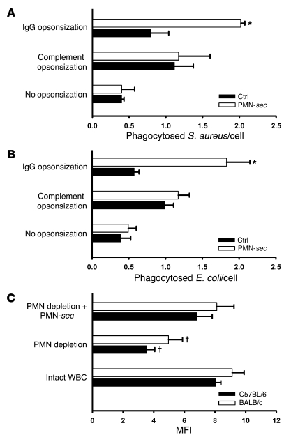
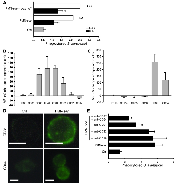
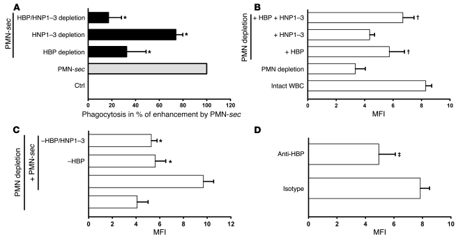
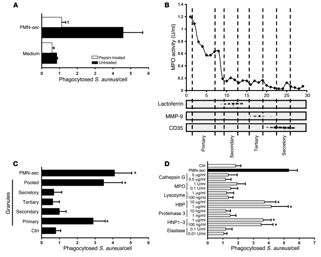
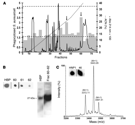
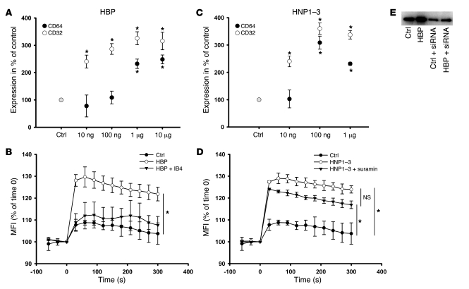
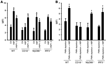
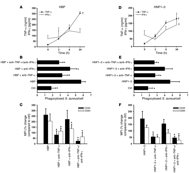
In acute inflammation, infiltrating polymorphonuclear leukocytes (also known as PMNs) release preformed granule proteins having multitudinous effects on the surrounding environment. Here we present what we believe to be a novel role for PMN-derived proteins in bacterial phagocytosis by both human and murine macrophages. Exposure of macrophages to PMN secretion markedly enhanced phagocytosis of IgG-opsonized Staphylococcus aureus both in vitro and in murine models in vivo. PMN secretion activated macrophages, resulting in upregulation of the Fcgamma receptors CD32 and CD64, which then mediated the enhanced phagocytosis of IgG-opsonized bacteria. The phagocytosis-stimulating activity within the PMN secretion was found to be due to proteins released from PMN primary granules; thorough investigation revealed heparin-binding protein (HBP) and human neutrophil peptides 1-3 (HNP1-3) as the mediators of the macrophage response to PMN secretion. The use of blocking antibodies and knockout mice revealed that HBP acts via beta2 integrins, but the receptor for HNP1-3 remained unclear. Mechanistically, HBP and HNP1-3 triggered macrophage release of TNF-alpha and IFN-gamma, which acted in an autocrine loop to enhance expression of CD32 and CD64 and thereby enhance phagocytosis. Thus, we attribute what may be a novel role for PMN granule proteins in regulating the immune response to bacterial infections.


Figures

Figure 1
Figure 1. PMN-sec products enhance phagocytosis in macrophages.
(A and B) Human macrophages were treated with PMN-sec or medium alone (ctrl) for 24 hours. After stimulation, fluorescent S. aureus (A) or E. coli (B) were injected into the medium. Bacteria were either opsonized with IgG or with complement or left nonopsonized. The number of incorporated bacteria per cell was quantified by fluorescence microscopy. For each analysis, 3–6 independent experiments were performed. *P < 0.05 versus respective control. (C) Comparison of phagocytic activity of peritoneal macrophages obtained from BALB/c or C57BL/6 mice. Macrophages were isolated from mice with intact WBC, neutropenic mice, and from neutropenic mice treated with PMN-sec. MFI as a measure of phagocytosed IgG-opsonized bacteria was read in a plate reader. For each analysis, 6 independent experiments were performed. P < 0.05 versus intact WBC and PMN depletion plus PMN-sec of the respective strain.
Figure 2
Figure 2. PMN-sec enhances activation of macrophages and expression of FcγRs.
(A) Human macrophages were incubated with PMN-sec for either 2 or 24 hours or with medium. In some wells, the PMN-sec was washed off after the 2- or 24-hour incubation period and replaced by medium. Subsequently the phagocytic activity was quantified. For each analysis, 4 independent experiments were performed. *P < 0.05 versus control; **P < 0.05 versus control and the respective 2-hour treatment group. (B and C) Expression of activation markers (B) and phagocytic receptors (C) in macrophages in response to treatment with PMN-sec for 24 hours. Expression is given as percentage change compared with basal expression. All values are isotype corrected. For each analysis, 6–8 independent experiments were performed. (D) Representative images of antibody staining for CD32 and CD64 in macrophages after treatment with PMN-sec or medium. Scale bar: 10 μm. (E) Human macrophages were incubated with PMN-sec or medium for 24 hours. Blocking antibodies toward CD64, CD32, or CD16 were added 30 minutes before incubation with IgG-opsonized S. aureus. The number of phagocytosed bacteria after 1 hour was quantified. For each analysis, 6 independent experiments were performed. P < 0.05 versus group treated with PMN-sec.
Figure 5
Figure 5. Contribution of HBP and HNP1–3 to enhanced phagocytosis.
(A) Human macrophages were treated with medium, PMN-sec, or secretion depleted of HBP, HNP1–3, or both. Phagocytic activity is expressed in percentage of enhanced phagocytosis above control in response to PMN-sec, which was set to 100%. For each analysis, 4 independent experiments were performed. *P < 0.05 versus group treated with PMN-sec. (B) Comparison of phagocytic activity of peritoneal macrophages from mice with intact WBC or neutropenic mice. Neutropenic mice were also injected i.p. with HBP (10 μg/mouse), HNP1–3 (2 μg/mouse), or both. MFI, as a measure of phagocytosed IgG-opsonized bacteria, was read in a plate reader. For each analysis, 5 independent experiments were performed. P < 0.05 versus fluorescence intensity in macrophages obtained from PMN-depleted mice. (C) Thioglycollate-treated neutropenic mice received PMN-sec, PMN-sec depleted of HBP, or PMN-sec depleted of both HBP and HNP1–3. *P < 0.05 versus group treated with PMN-sec. For each analysis, 5 independent experiments were performed. (D) A polyclonal antibody to HBP or control IgG were injected i.p. into thioglycollate-treated mice. The phagocytic capacity of peritoneal macrophages was tested ex vivo 4 days after initiating peritonitis. P < 0.05 versus isotype control. For each analysis, 4 independent experiments were performed.
Figure 3
Figure 3. Identification of active PMN granule components.
(A) PMN-sec was digested with pepsin and the remaining activity was tested. For each analysis, 4 independent experiments were performed. P < 0.05 versus group treated with active PMN-sec. (B) Localization of neutrophil granules in fractions (x axis) obtained by subcellular fractionation of PMNs is shown by marker analysis. MPO activity was measured as a marker for primary granules. Western blots of the fractions probed with antibodies to lactoferrin, MMP-9, and CD35 indicate the localization of secondary and tertiary granules and of secretory vesicles, respectively. (C) Human macrophages were treated with PMN-sec, fractions of the indicated PMN granules, a mixture of the 4 different granule fractions (pooled), or medium and phagocytosis assay was performed. For each analysis, 6 independent experiments were performed. *P < 0.05 versus control. (D) Human macrophages were treated with proteins and peptides stored in human primary granules. Phagocytosis was compared with that obtained from treatment with PMN-sec or medium. For each analysis, 6 independent experiments were performed. *P < 0.05 versus control.
Figure 4
Figure 4. Identification of HBP and HNP1–3 as enhancers of bacterial phagocytosis in macrophages.
(A) Fractionation of PMN-sec by reversed-phase HPLC. PMN-sec was loaded onto a C18 column. Proteins (right y axis, solid curve) were eluted with a gradient of ACN with 0.1% TFA (right y axis, dotted line). Gray bars indicate the phagocytic activity resulting from stimulation with material of 3 consecutive fractions (left y axis). Basal phagocytosis and phagocytosis in response to PMN-sec are indicated by the lower and upper dashed line, respectively. Arrows indicate active fractions (45–47 and 60–62). For each analysis, 4 independent experiments were performed. (B) Immunological detection of HBP in fraction 60–62 using dot blot (left panel). These fractions were pooled and further analyzed with Western blot analysis (right panel). As positive control for both Western and dot blot, 40 ng recombinant HBP was used. (C) HPLC fractions were screened for the presence of HNP1–3 by dot blot analysis. Positive staining was detected in fractions 46 (insert). Inserts of dot blots in B and C were run on the same gel but were noncontiguous. As positive control 20 ng HNP1 was used. Determination of the mass values of material in fraction 46 with MALDI-MS gave molecular weights close to the theoretical values of HNP1–3.
Figure 6
Figure 6. Characterization of the enhanced phagocytosis by HBP and HNP1–3.
(A and C) Human macrophages were treated with increasing concentrations of (A) HBP or (C) HNP1–3 for 24 hours, and the expression of CD32 and CD64 was assessed by immunofluorescence. Data are expressed as a percentage of basal expression, which was set to 100%. For each analysis, 5 independent experiments were performed. *P < 0.05 versus control. (B and D) Intracellular Ca2+-mobilization in macrophages labeled with the Ca2+-sensitive dye fluo4/AM in response to stimulation with (B) HBP (1 μg/ml) and (D) HNP1–3 (1 μg/ml). After labeling and washing, fluorescence was read in a plate reader every 30 seconds, from 60 seconds before stimulation until 300 seconds after stimulation. Data are expressed as percent of fluorescence intensity at time 0. IB4 (10 μg/ml) or suramin (100 μM) were added to analyze the involvement of β2 integrins and P2Y6 receptors in the respective activation. For each analysis, 4 independent experiments were performed. *P < 0.05 versus control treatment. (E) Western blot for CD64 of whole cell lysate of human macrophages treated with HBP (1 μg/ml, 24 hours) in the presence or absence of siRNA to CD64.
Figure 7
Figure 7. Enhanced phagocytosis mediated by HBP depends on CD18 but not TLR signaling, while enhanced phagocytosis induced by HNP1–3 is independent of CD18 and TLR signaling.
(A) Peritoneal macrophages were isolated from neutropenic mice of various strains and treated ex vivo with medium, HBP (1 μg/ml), or HNP1–3 (1 μg/ml) for 24 hours. Thereafter, the phagocytic capacity was assessed using IgG-opsonized S. aureus. For each analysis, 4 independent experiments were performed. *P < 0.05 versus respective control. (B) Comparison of phagocytic capacity of peritoneal macrophages from different strains. Mice were rendered neutropenic and then injected i.p. with either PBS or HBP. For each analysis, 3–5 independent experiments were performed. P < 0.05 versus neutropenic mice of the respective strain.
Figure 8
Figure 8. Involvement of TNF-α and IFN-γ in the HBP- and HNP1–3–mediated enhanced phagocytosis.
(A and D) Human macrophages were treated with (A) HBP or (D) HNP1–3 and the concentration of TNF-α and IFN-γ was determined by ELISA at different time points. For each analysis, 4 independent experiments were performed. P < 0.05 indicates significant effect of treatment. (B and E) The contribution of TNF-α and IFN-γ was assessed in the phagocytosis assay after stimulation of human macrophages with (B) HBP or (E) HNP1–3 by addition of neutralizing antibodies to TNF-α (5 μg/ml) or IFN-γ (10 μg/ml). For each analysis, 4 independent experiments were performed. *P < 0.05 versus HBP or HNP1–3 treatment groups. (C and F) Importance of TNF-α and IFN-γ for the upregulation of CD32 and CD64 on human macrophages in response to (C) HBP or (F) HNP1–3 evaluated through use of neutralizing antibodies. Receptor expression is assessed by immunofluorescence staining and displayed as percent change compared with control. For each analysis, 5 independent experiments were performed. *P < 0.05 versus HBP or HNP1–3 treatment groups.

Similar articles

Cited by

References

    1. Witko-Sarsat V., Rieu P., Descamps-Latscha B., Lesavre P., Halbwachs-Mecarelli L. Neutrophils: molecules, functions and pathophysiological aspects. Lab. Invest. . 2000;80:617–653. - PubMed
    1. Chertov O., Yang D., Howard O.M., Oppenheim J.J. Leukocyte granule proteins mobilize innate host defenses and adaptive immune responses. Immunol. Rev. . 2000;177:68–78. doi: 10.1034/j.1600-065X.2000.17702.x. - DOI - PubMed
    1. Borregaard N., Cowland J.B. Granules of the human neutrophilic polymorphonuclear leukocyte. Blood. 1997;89:3503–3521. - PubMed
    1. Shiohara M., et al. Phenotypic and functional alterations of peripheral blood monocytes in neutrophil-specific granule deficiency. J. Leukoc. Biol. 2004;75:190–197. - PubMed
    1. Tavor S., et al. Macrophage functional maturation and cytokine production are impaired in C/EBP epsilon-deficient mice. Blood. 2002;99:1794–1801. doi: 10.1182/blood.V99.5.1794. - DOI - PubMed

Publication types

MeSH terms