. 2008 Nov;82(22):11167-80.
doi: 10.1128/JVI.01218-08. Epub 2008 Sep 10.

Temporal dynamics of cytomegalovirus chromatin assembly in productively infected human cells

Affiliations

Temporal dynamics of cytomegalovirus chromatin assembly in productively infected human cells

Alexandra Nitzsche et al. J Virol. 2008 Nov.

Abstract

The genomes of herpesviruses, including human cytomegalovirus (CMV), are double-stranded DNA molecules maintained as episomes during infection. The viral DNA lacks histones when encapsidated in the virion. However, it has been found histone associated inside infected cells, implying unidentified chromatin assembly mechanisms. Our results indicate that components of the host cell nucleosome deposition machinery target intranuclear CMV DNA, resulting in stepwise viral-chromatin assembly. CMV genomes undergo limited histone association and nucleosome assembly as early as 30 min after infection via DNA replication-independent mechanisms. Low average viral-genome chromatinization is maintained throughout the early stages of infection. The late phase of infection is characterized by a striking increase in average histone occupancy coupled with the process of viral-DNA replication. While the initial chromatinization affected all analyzed parts of the CMV chromosome, a subset of viral genomic regions, including the major immediate-early promoter, proved to be largely resistant to replication-dependent histone deposition. Finally, our results predict the likely requirement for an unanticipated chromatin disassembly process that enables packaging of histone-free DNA into progeny capsids.


Figures

FIG. 1.
FIG. 1.
Nucleosomal arrangement of intracellular CMV DNA during productive infection. (A) MRC-5 cells were mock infected or infected with CMV. At 2 or 48 h postinfection, permeabilized cells were reacted with increasing amounts of MNase for the indicated incubation times at room temperature (24 and 48 U) or 37°C (0 and 96 U). DNA was prepared and separated in ethidium bromide-stained 1.2% agarose gels (top). The same samples were transferred to nylon membranes and hybridized to a 32P-labeled probe derived from a bacterial artificial chromosome clone of the complete CMV (Towne) genome (32) (bottom). To ensure comparable signal intensities despite varying amounts of viral DNA, a shorter film exposure was chosen for the 48-h than for the 2-h samples. Marker, 100-bp DNA ladder (New England Biolabs). (B) At the indicated postinfection time points, permeabilized cells were incubated for 30 min with 96 U MNase at 37°C, and samples were treated as described for panel A. For the 48- and 96-h samples, only 1/10 or 1/50 of the DNA amounts used in the mock and 0.5- and 16-h lanes were loaded, respectively.
FIG. 2.
FIG. 2.
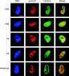
Subnuclear distribution of core histones in relation to late viral replication compartments. MRC-5 cells were infected with CMV for 48 h, fixed with paraformaldehyde/methanol, and stained with a mouse monoclonal antibody specifically detecting the CMV ppUL44 DNA polymerase accessory protein and rabbit polyclonal antibodies directed against the C-terminal histone fold domain of H2A, H2B, H3, or H4. A rabbit antiserum specific for histone H3 dimethylated at lysine 9 (H3K9me2) was used as a negative control. Samples were subsequently stained with DAPI, a mouse-specific Alexa Fluor 594, and a rabbit-specific Alexa Fluor 488 conjugate. Representative nuclei showing DAPI, ppUL44, and the respective histone staining are shown. Additionally, merge images of ppUL44 and histone signals are presented. Scale bar, 10 μm.
FIG. 3.
FIG. 3.
Differential chromatinization of the CMV genome at early versus late times postinfection as analyzed by FAIRE. MRC-5 cells were infected with CMV and collected at 2, 16, or 48 h postinfection. Enrichment for nucleosome-depleted chromatin by FAIRE extraction was performed, and DNA from the aqueous phase was quantified by real-time PCR using primer pairs specific for seven selected viral indicator loci and human GAPDH. The results were normalized to GAPDH and are representative of triplicate experiments with standard deviations. They are presented as the ratio of viral DNA recovered from non-formaldehyde-fixed cells divided by the amounts of the same DNA in the corresponding cross-linked samples. The data therefore reflect the degrees of nucleosome assembly in the respective viral genomic regions.
FIG. 4.
FIG. 4.
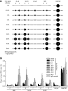
Temporal patterns of histone H3 occupancy in selected regions of the CMV genome. MRC-5 cells were infected with CMV, and cell extracts were subjected to ChIP using an antibody specifically directed against the C-terminal domain of H3 at the indicated times (0.5 to 96 h) postinfection. Normal rabbit IgG was used to control for nonspecific precipitation. Quantitative PCR was performed on input and coprecipitated (output) DNAs with primers specific for the indicated viral genomic regions and human GAPDH. (A) The circular areas represent the mean output-to-input DNA ratios determined from at least two independent ChIP assays, each quantified in duplicate. Where white circles are missing, no specific PCR products were detected. (B) The data set used for the schematic representation in panel A is shown as bars (mean values) with standard deviations. For the IgG controls, average values from all 10 time points are shown.
FIG. 5.
FIG. 5.
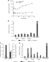
Consecutive stages of replication-independent and -dependent histone H3 deposition on the CMV genome. (A) MRC-5 cells were infected with CMV for 0.5 to 72 h, relative amounts of viral and cellular DNAs were determined at the UL32-T and GAPDH loci by quantitative PCR, and the results were normalized to GAPDH at 0.5 h (set to 1). The data present the mean amounts of DNA from three independent experiments with standard deviations. (B) MRC-5 cells were pretreated with PAA (200 μg/ml) or aphidicolin (2 μM) for 1 h or left untreated prior to infection with CMV. Infection continued for 2 h in the presence of the inhibitors. ChIP was performed using an antibody against the C-terminal domain of histone H3, and the amounts of input and coprecipitated DNA were determined by quantitative PCR with eight specific primer pairs, as indicated. PCR results from two independent experiments, each quantified in duplicate, are presented as mean output-to-input DNA fractions normalized to the output-to-input ratio of GAPDH without drug treatment (set to 1). The error bars indicate standard deviations. (C) MRC-5 cells were infected with CMV and either left untreated or treated with PAA (200 μg/ml) immediately following infection. After 24 h, the culture medium was changed, and fresh drug was added where applicable. Samples were collected at 2 and 48 h postinfection. Relative amounts of viral (means of all seven viral indicator loci) and cellular (GAPDH) DNAs were determined by quantitative PCR and normalized to GAPDH at 2 h postinfection (set to 1). The bars represent mean values with standard deviations from three experiments. (D) The samples from panel C were subjected to ChIP as described for panel B. PCR results from two independent experiments, each quantified in duplicate, are presented as mean output-to-input DNA fractions normalized to the output-to-input ratio of GAPDH at 2 h (set to 1). The error bars indicate standard deviations.
FIG. 6.
FIG. 6.
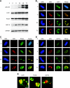
Increased accumulation of human nucleosome assembly proteins and association with intranuclear viral replication compartments during CMV infection. (A) MRC-5 cells were mock infected (0 h) or infected with CMV for 8 to 72 h, as indicated, and proteins from whole-cell extracts were separated in 10% polyacrylamide-SDS gels. After Western blot transfer, the proteins were reacted with the respective antibodies as shown in Table 2. (B to D) Mock- or CMV-infected MRC-5 cells were fixed and permeabilized with paraformaldehyde/methanol (B and C) or paraformaldehyde/Triton X-100 (D) at the indicated time points and incubated with primary antibodies specifically detecting the CMV IE2 (0 and 8 h) or ppUL44 (24 and 72 h) proteins, together with antibodies directed against CAF1 p48, PCNA, or ASF1A, as indicated. Samples were subsequently stained with an Alexa Fluor 594 conjugate, an Alexa Fluor 488 conjugate, and DRAQ5. Single- and dual-color merge confocal images of representative nuclei are shown. Scale bars, 10 μm. (E) Three-dimensional projections of z stacks showing details of the spatial relationship between CMV replication compartments (ppUL44) (red) and complexes containing the indicated cellular chromatin assembly proteins (green) at 24 h postinfection. The frames were acquired with a step width of 0.38 μM and rendered with the Zeiss LSM510 software. Scale bar, 1 μm.
FIG. 7.
FIG. 7.
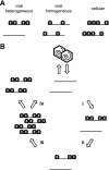
Models of CMV chromatin assembly and disassembly. (A) Schematic of two possible chromatin distributions in early-phase CMV genome populations. By ChIP or FAIRE analysis, both the heterogeneous and the rather homogeneous distributions would be interpreted as reduced histone occupancy relative to a cellular control locus. (B) Hypothetical four-step cycle of CMV chromatin assembly and disassembly (assuming heterogeneous chromatin distributions). Step I, limited initial DNA replication-independent nucleosome assembly on “naked” input viral genomes. Step II, maintenance of low chromatinization levels and partial chromatin disassembly. Step III, DNA replication-dependent nucleosome assembly resulting in chromatinization of most or all newly synthesized viral genomes (note that a subset of viral genomic regions is largely resistant to this step). Step IV, complete chromatin disassembly in a fraction of replicated viral genomes before or during encapsidation.

Similar articles

Cited by

References

    1. Ahmad, K., and S. Henikoff. 2002. The histone variant H3.3 marks active chromatin by replication-independent nucleosome assembly. Mol. Cell 91191-1200. - PubMed
    1. Atalay, R., A. Zimmermann, M. Wagner, E. Borst, C. Benz, M. Messerle, and H. Hengel. 2002. Identification and expression of human cytomegalovirus transcription units coding for two distinct Fcγ receptor homologs. J. Virol. 768596-8608. - PMC - PubMed
    1. Booy, F. P., W. W. Newcomb, B. L. Trus, J. C. Brown, T. S. Baker, and A. C. Steven. 1991. Liquid-crystalline, phage-like packing of encapsidated DNA in herpes simplex virus. Cell 641007-1015. - PMC - PubMed
    1. Cohen, G. H., M. Ponce de Leon, H. Diggelmann, W. C. Lawrence, S. K. Vernon, and R. J. Eisenberg. 1980. Structural analysis of the capsid polypeptides of herpes simplex virus types 1 and 2. J. Virol. 34521-531. - PMC - PubMed
    1. Deshmane, S. L., and N. W. Fraser. 1989. During latency, herpes simplex virus type 1 DNA is associated with nucleosomes in a chromatin structure. J. Virol. 63943-947. - PMC - PubMed

Publication types

LinkOut - more resources