Suppression of autophagy in skeletal muscle uncovers the accumulation of ubiquitinated proteins and their potential role in muscle damage in Pompe disease
- PMID: 18782848
- PMCID: PMC2638578
- DOI: 10.1093/hmg/ddn292
Suppression of autophagy in skeletal muscle uncovers the accumulation of ubiquitinated proteins and their potential role in muscle damage in Pompe disease
Abstract
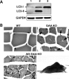
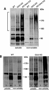
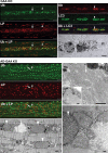
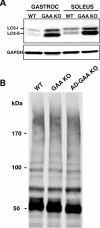
The role of autophagy, a catabolic lysosome-dependent pathway, has recently been recognized in a variety of disorders, including Pompe disease, the genetic deficiency of the glycogen-degrading lysosomal enzyme acid-alpha glucosidase. Accumulation of lysosomal glycogen, presumably transported from the cytoplasm by the autophagic pathway, occurs in multiple tissues, but pathology is most severe in skeletal and cardiac muscle. Skeletal muscle pathology also involves massive autophagic buildup in the core of myofibers. To determine if glycogen reaches the lysosome via autophagy and to ascertain whether autophagic buildup in Pompe disease is a consequence of induction of autophagy and/or reduced turnover due to defective fusion with lysosomes, we generated muscle-specific autophagy-deficient Pompe mice. We have demonstrated that autophagy is not required for glycogen transport to lysosomes in skeletal muscle. We have also found that Pompe disease involves induction of autophagy but manifests as a functional deficiency of autophagy because of impaired autophagosomal-lysosomal fusion. As a result, autophagic substrates, including potentially toxic aggregate-prone ubiquitinated proteins, accumulate in Pompe myofibers and may cause profound muscle damage.
Figures


Similar articles
-
When more is less: excess and deficiency of autophagy coexist in skeletal muscle in Pompe disease.Autophagy. 2009 Jan;5(1):111-3. doi: 10.4161/auto.5.1.7293. Epub 2009 Jan 30. Autophagy. 2009. PMID: 19001870 Free PMC article.
-
Murine muscle cell models for Pompe disease and their use in studying therapeutic approaches.Mol Genet Metab. 2009 Apr;96(4):208-17. doi: 10.1016/j.ymgme.2008.12.012. Epub 2009 Jan 22. Mol Genet Metab. 2009. PMID: 19167256 Free PMC article.
-
Autophagy in skeletal muscle: implications for Pompe disease.Int J Clin Pharmacol Ther. 2009;47 Suppl 1(Suppl 1):S42-7. doi: 10.5414/cpp47042. Int J Clin Pharmacol Ther. 2009. PMID: 20040311 Free PMC article. Review.
-
Suppression of mTORC1 activation in acid-α-glucosidase-deficient cells and mice is ameliorated by leucine supplementation.Am J Physiol Regul Integr Comp Physiol. 2014 Nov 15;307(10):R1251-9. doi: 10.1152/ajpregu.00212.2014. Epub 2014 Sep 17. Am J Physiol Regul Integr Comp Physiol. 2014. PMID: 25231351 Free PMC article.
-
Failure of Autophagy in Pompe Disease.Biomolecules. 2024 May 13;14(5):573. doi: 10.3390/biom14050573. Biomolecules. 2024. PMID: 38785980 Free PMC article. Review.
Cited by
-
Age-Related Maintenance of the Autophagy-Lysosomal System Is Dependent on Skeletal Muscle Type.Oxid Med Cell Longev. 2020 Jul 24;2020:4908162. doi: 10.1155/2020/4908162. eCollection 2020. Oxid Med Cell Longev. 2020. PMID: 32774673 Free PMC article.
-
Sporadic inclusion body myositis: possible pathogenesis inferred from biomarkers.Curr Opin Neurol. 2010 Oct;23(5):482-8. doi: 10.1097/WCO.0b013e32833d3897. Curr Opin Neurol. 2010. PMID: 20664349 Free PMC article. Review.
-
Exercise ameliorates the detrimental effect of chloroquine on skeletal muscles in mice via restoring autophagy flux.Acta Pharmacol Sin. 2014 Jan;35(1):135-42. doi: 10.1038/aps.2013.144. Epub 2013 Dec 16. Acta Pharmacol Sin. 2014. PMID: 24335841 Free PMC article.
-
Low survival rate and muscle fiber-dependent aging effects in the McArdle disease mouse model.Sci Rep. 2019 Mar 26;9(1):5116. doi: 10.1038/s41598-019-41414-8. Sci Rep. 2019. PMID: 30914683 Free PMC article.
-
Mechanisms of skeletal muscle aging: insights from Drosophila and mammalian models.Dis Model Mech. 2013 Nov;6(6):1339-52. doi: 10.1242/dmm.012559. Epub 2013 Oct 2. Dis Model Mech. 2013. PMID: 24092876 Free PMC article. Review.
References
-
- Levine B., Klionsky D.J. Development by self-digestion: molecular mechanisms and biological functions of autophagy. Dev. Cell. 2004;6:463–477. - PubMed
-
- Klionsky D.J. Autophagy: from phenomenology to molecular understanding in less than a decade. Nat. Rev. Mol. Cell Biol. 2007;8:931–937. - PubMed
-
- Settembre C., Fraldi A., Jahreiss L., Spampanato C., Venturi C., Medina D., de Pablo R., Tacchetti C., Rubinsztein D.C., Ballabio A. A block of autophagy in lysosomal storage disorders. Hum. Mol. Genet. 2008;17:119–129. - PubMed
Publication types
MeSH terms
Substances
Grants and funding
LinkOut - more resources
Full Text Sources
Other Literature Sources
Medical
Molecular Biology Databases
Research Materials

