. 2008 Oct 8;27(19):2484-94.
doi: 10.1038/emboj.2008.177. Epub 2008 Sep 4.

Ubiquitin binding and conjugation regulate the recruitment of Rabex-5 to early endosomes

Affiliations

Ubiquitin binding and conjugation regulate the recruitment of Rabex-5 to early endosomes

Rafael Mattera et al. EMBO J. .

Abstract

Rab GTPases and ubiquitination are critical regulators of transmembrane cargo sorting in endocytic and lysosomal targeting pathways. The endosomal protein Rabex-5 intersects these two layers of regulation by being both a guanine nucleotide exchange factor (GEF) for Rab5 and a substrate for ubiquitin (Ub) binding and conjugation. The ability of trafficking machinery components to bind ubiquitinated proteins is known to have a function in cargo sorting. Here, we demonstrate that Ub binding is essential for the recruitment of Rabex-5 from the cytosol to endosomes, independently of its GEF activity and of Rab5. We also show that monoubiquitinated Rabex-5 is enriched in the cytosol. These observations are consistent with a model whereby a cycle of Ub binding and monoubiquitination regulates the association of Rabex-5 with endosomes.


Figures
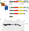
Figure 1
Figure 1
Domain organization, design and expression of Rabex-5 constructs used in this study. (A) Rabex-5 domains include the A20-like Cys2/Cys2 zinc-finger (ZnF), which displays Ub protein ligase (E3) activity and binds Ub, and the motif interacting with ubiquitin (MIU) (Lee et al, 2006; Mattera et al, 2006; Penengo et al, 2006), the Vps9 module and its associated helical bundle (HB) harbouring the Rab5/Rab21 guanine nucleotide exchange factor (GEF) catalytic core (Delprato et al, 2004), the predicted amphipatic helix (CC) that binds Rabaptin-5 and acts as an autoinhibitory element of the HB-Vps9 tandem GEF activity (Mattera et al, 2006; Delprato and Lambright, 2007; Kalesnikoff et al, 2007; Zhu et al, 2007), and a C-terminal proline-rich region (PR). Residues substituted in the full-length construct (Y25 and Y26 in the ZnF, A58 in the MIU, D314, P318 and Y355 in the Vps9 domain) are indicated in the top scheme. Deletion constructs are shown underneath. The structure of the Rabex-5 ZnF and MIU domains (magenta and green ribbons, respectively) bound to two Ub molecules (blue and gold surfaces; adapted from Lee et al, 2006) is shown on the left. Tyr25/Tyr26 and Ala58 represent residues critical for Ub binding to the ZnF and MIU domains, respectively. Numbering corresponds to the bovine Rabex-5 used in this study. (B) Expression of myc-tagged Rabex-5 constructs in transiently transfected HeLa cells was assessed by SDS–PAGE of extracts and immunoblotting (IB) with anti-myc antiserum. The light bands with slower mobility (e.g. ∼73 kDa for myc–Rabex-5 WT, left lane) represent the monoubiquitinated recombinant proteins (see also Figure 4); the main bands correspond to the unmodified myc-tagged proteins, whereas the light bands with higher mobility are most likely degradation products. Notice that constructs with MIU substitutions (A58D and Y25A/A58D) do not undergo monoubiquitination. Monoubiquitination of myc–Rabex-5 Y25A/Y26A and Δ13–49 is also reduced or abolished when compared with WT. Numbers on the left indicate the position of molecular mass markers (in kilodaltons).
Figure 2
Figure 2
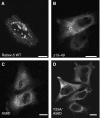
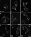
Mutations impairing Ub binding alter the localization of Rabex-5. HeLa cells were transiently transfected and fixed with 4% formaldehyde ∼24 h after transfection. Fixed cells were incubated with mouse anti-myc and Alexa 568-conjugated anti-mouse antisera. The substitutions in the ZnF and MIU domains interfere with Ub binding to Rabex-5 (Lee et al, 2006; Mattera et al, 2006; Penengo et al, 2006). myc–Rabex-5 wild type (WT) (A) is predominantly recruited to large (∼2 μm) vesicles that are positive for the early endosomal marker EEA1 (see Figure 3); in contrast, expression of comparable levels of myc–Rabex-5 Ub-binding mutants, such as Δ13–49 (deletion of the ZnF) (B), A58D (C) and Y25A/A58D (D), results in predominantly cytosolic accumulation (some threads and membrane ruffles are also visible). Enlarged vesicles containing the recombinant proteins were observed in 99, 2, 1 and 1% of cells expressing intermediate to high levels of myc–Rabex-5 WT or the Δ13–49, A58D and Y25A/A58D mutants, respectively (n=396, 643, 534 and 372, respectively). Approximately 8–12% of cells transfected with either Rabex-5 A58D or Rabex-5 Y25A/A58D also exhibited large artificial aggregates containing the recombinant proteins that were clearly discernable from the vesicles containing myc–Rabex-5 WT; this aggregation was less frequent (0–3%) in cells transfected with the ΔZnF or Y25A/Y26A mutants. Scale bars=10 μm.
Figure 3
Figure 3
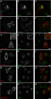
Recruitment of myc–Rabex-5 WT but not Ub-binding mutants to early endosomes. HeLa cells were transfected and fixed as described in the legend to Figure 2. Fixed cells were incubated with rabbit anti-myc and mouse anti-EEA1 antisera, followed by incubation with Alexa 568-conjugated anti-rabbit and Alexa 488-conjugated anti-mouse antisera. Rabex-5 WT (AC) is recruited to early endosomes, whereas the Rabex-5 Ub-binding mutants (DR) exhibit a predominantly cytosolic distribution. Although expression of all constructs results in enlargement of early endosomes as compared with untransfected cells, only myc–Rabex-5 WT is recruited to these structures. The enlargement of early endosomes in cells transfected with myc–Rabex-5 WT was greater than in cells expressing the Ub-binding mutants. In some instances, particularly for Rabex-5 Y25A/Y26A (G–I, insets), it was possible to observe decoration of enlarged early endosomes by the Ub-binding mutants amidst their predominantly cytosolic distribution. Insets in (G–I) represent a × 3 magnification of areas in the dotted squares. (C, F, I, L, O, R) Merged images of the panels at their left. Scale bars=10 μm.
Figure 4
Figure 4
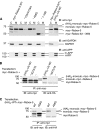
Subcellular fractionation of myc–Rabex-5 constructs and ubiquitination assays. (A) HeLa cells were transfected with the indicated constructs and subjected to nitrogen cavitation ∼24 h after transfection. Postnuclear (900 g) supernatants were centrifuged at 300 000 g to yield cytosolic (C) and crude membrane (M) fractions that were analysed by SDS–PAGE and immunoblotting (IB). Cytosol and membrane samples loaded onto gels represent 0.75% of the total of each fraction. The mobilities of (HA)3-monoubiquitinated myc–Rabex-5, monoubiquitinated myc–Rabex-5 (see legend of B, C) and myc–Rabex-5 are shown on the right of the upper blots. Note that both (HA)3-monoubiquitinated myc–Rabex-5 (cells co-transfected with myc–Rabex-5 and (HA)3–Ub in blot shown on right) and monoubiquitinated myc–Rabex-5 (blots on left and right) are enriched in the cytosolic fraction. GAPDH and amyloid precursor protein (APP) were used as markers of the cytosolic and membrane fractions, respectively (middle and lower blots). The two bands detected with anti-APP correspond to the mature N- plus O-glycosylated (m-APP) and immature N-glycosylated (i-APP) forms, respectively (Chyung and Selkoe, 2003). (B, C) HeLa cells were transfected with myc–Rabex-5 with or without the indicated (HA)3–Ub constructs. Cells were lysed ∼24 h after transfection and the lysates were subjected to immunoprecipitation (IP) with mouse anti-myc followed by SDS–PAGE and IB with rabbit anti-myc (left) or mouse anti-HA (right) antisera. The mobilities of (HA)3-monoubiquitinated myc–Rabex-5, monoubiquitinated myc–Rabex-5 and myc–Rabex-5 are shown on the right of the blot. Identification of the second band from top (∼73 kDa) as myc–Rabex-5 monoubiquitinated with endogenous Ub is based on (a) its immunoreactivity with anti-myc but not anti-HA (B, C); (b) the presence of this band in immunoprecipitates of cells transfected only with myc–Rabex-5 (no co-transfection with (HA)3–Ub) or co-transfected with unconjugatable (HA)3–UbΔG75/76 (B) (demonstrating it does not originate from proteolysis of (HA)3-mono-ubiquitinated-Rabex-5); (c) the absence of this band, along with that corresponding to (HA)3-monoubiquitinated-Rabex-5 (∼76 kDa) in immunoprecipitated extracts of cells transfected with Rabex-5 A58D, a mutant that does not undergo monoubiquitination (Mattera et al, 2006; Penengo et al, 2006) (C) and (d) the similar enrichment of both the 76- and 73-kDa species in the cytosolic fraction (A, blot on right). Monoubiquitination of myc–Rabex-5 by endogenous ubiquitin is not inhibited by the co-expression of (HA)3–UbΔG75/G76 Ub (anti-myc blot in B), consistent with the requirement for Ub glycine 76 in the formation of an Ub adenylate during the first step of Ub protein conjugation (Pickart et al, 1994). The lower band above the 50 kDa standard in the anti-myc blot is a myc–Rabex-5 cleavage product, whereas the anti-HA immunoreactivity at ∼50 kDa corresponds to the IgG heavy chain of the mouse anti-myc used for IP.
Figure 5
Figure 5
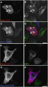
Colocalization of myc–Rabex-5 and (HA)3–Ub on early endosomes; interference of Rabex-5 recruitment to early endosomes by (HA)3–UbΔG75/76. Cells were co-transfected with myc–Rabex-5 and (HA)3–Ub or (HA)3–UbΔG75/76 and fixed 24 h after transfection. Fixed cells were incubated with chicken anti-myc, mouse anti-EEA1 and rabbit anti-HA, followed by incubation with Alexa 594-conjugated anti-chicken, Alexa 488-conjugated anti-mouse and Alexa 647-conjugated anti-rabbit antisera. Arrows in AD show colocalization of myc–Rabex-5 with (HA)3–Ub WT on early endosomes. We observed this phenotype in 90 and 28% of cells co-transfected with (HA)3–Ub or (HA)3–UbΔG75/76, respectively (n=104 and 421 cells, respectively, exhibiting medium–high expression levels of myc–Rabex-5). The phenotype in >70% of cells co-transfected with (HA)3–UbΔG75/76 is shown in (EH). (D, H) Merged images of the preceding panels. Scale bars=10 μm.
Figure 6
Figure 6
Roles of the Vps9 domain (residues 230–375), C-terminal coiled-coil (residues 405–456) and GEF activity in the recruitment of Rabex-5 to early endosomes. HeLa cells were transiently transfected with the indicated constructs (AD) (see Figure 1A for schematic representation of the constructs). Cells were fixed and stained as described in the legend to Figure 2. The substitutions in Vps9 residues (i.e. D314, P318 and Y355; EI) impair the Rab5/Rab21 GEF activity of Rabex-5 (Delprato et al, 2004). All substitutions in the Vps9 module were introduced in the context of the Rabex-5 FL construct. Scale bars=10 μm.
Figure 7
Figure 7
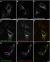
Co-expression of Rab5a S34N does not inhibit the recruitment of myc–Rabex-5 to early endosomes. (AC) HeLa cells were transfected with GFP–Rab5a WT, GFP–Rab5a Q79L (constitutively active, ‘GTP-locked') or GFP–Rab5a S34N (dominant negative, displaying higher affinity for GDP than for GTP) (Li and Stahl, 1993; Stenmark et al, 1994). Transfected cells were fixed and incubated with rabbit anti-GFP followed by Alexa 488-conjugated anti-rabbit antiserum. (DI) HeLa cells were co-transfected with GFP–Rab5a S34N and myc–Rabex-5 WT (D–F) or myc–Rabex-5 Y25A/A58D (G–I), fixed and incubated with rabbit anti-GFP and mouse anti-myc followed by incubation with Alexa 488-conjugated anti-rabbit and Alexa 568-conjugated anti-mouse antisera. (F, I) Merged images of the panels at their left. Scale bars=10 μm.
Figure 8
Figure 8
siRNA-mediated depletion of Rab5a, Rab5b, Rab5c or Rabaptin-5 does not affect the recruitment of Rabex-5 to early endosomes. HeLa cells were transfected with the indicated siRNA (AF). Approximately 36 h after start of treatment, cells were trypsinized, plated on glass coverslips and transfected (∼12 h after plating) with myc–Rabex-5. Cells were fixed ∼24 after transfection with myc–Rabex-5 (72 h cumulative treatment with siRNAs) and stained for myc–Rabex-5 as indicated in the legend to Figure 2. myc–Rabex-5 recruitment to early endosomes was observed in 98, 96, 96, 85, 98 and 97% of cells treated with siRNAs for Rab5a, Rab5b, Rab5c, Rab5a+b+c, Rabaptin-5 or TOM1 (negative control), respectively, (n=221, 354, 334, 155, 302 and 352, respectively; cells with intermediate–high levels of myc–Rabex-5 were counted). A relatively lower transfection efficiency with myc–Rabex-5 (∼50% of the average in the other experimental groups) was observed in cells treated with Rab5a+b+c siRNA. Scale bars=10 μm.
Figure 9
Figure 9
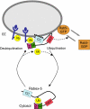
Proposed model for regulation of Rabex-5 association to early endosomes by Ub binding and monoubiquitination. The scheme depicts the proposed recruitment of Rabex-5 to early endosomes by virtue of its ability to bind ubiquitinated cargo through the ZnF (Z) and MIU (M) domains, and by the interaction of the C-terminal coiled-coil (CC) with as yet unidentified factors. The activation of Rab5 catalysed by the GEF activity of the HB-Vps9 tandem catalytic core (denoted as GEF) is also shown. We hypothesize that cargo conveyance followed by UBD-dependent monoubiquitination results in dissociation of Rabex-5 from endosomes to the cytosol, where the covalently linked Ub moiety in cis (position shown is arbitrary) may bind intramolecularly to the ZnF/MIU domains. Intermolecular interactions in Rabex-5 oligomers where the Ub binds to ZnF/MIU domains in trans are also possible. Deubiquitination of Rabex-5, presumably by endosomal deubiquitinating enzymes, releases this interaction allowing resumption of the cycle. The model is consistent with the enrichment of non-ubiquitinated Rabex-5 and monoubiquitinated Rabex-5 in the membrane and cytosolic fractions, respectively, and is thought to apply to Rabex-5 in dynamic equilibrium with the endosomal compartment.

Similar articles

Cited by

References

    1. Amit I, Yakir L, Katz M, Zwang Y, Marmor MD, Citri A, Shtiegman K, Alroy I, Tuvia S, Reiss Y, Roubini E, Cohen M, Wides R, Bacharach E, Schubert U, Yarden Y (2004) Tal, a Tsg101-specific E3 ubiquitin ligase, regulates receptor endocytosis and retrovirus budding. Genes Dev 18: 1737–1752 - PMC - PubMed
    1. Brett TJ, Traub LM, Fremont DH (2002) Accessory protein recruitment motifs in clathrin-mediated endocytosis. Structure 10: 797–809 - PubMed
    1. Burkhard P, Stetefeld J, Strelkov SV (2001) Coiled coils: a highly versatile protein folding motif. Trends Cell Biol 11: 82–88 - PubMed
    1. Chyung JH, Selkoe DJ (2003) Inhibition of receptor-mediated endocytosis demonstrates generation of amyloid beta-protein at the cell surface. J Biol Chem 278: 51035–51043 - PubMed
    1. Clague MJ, Urbé S (2006) Endocytosis: the DUB version. Trends Cell Biol 16: 551–559 - PubMed

Publication types

MeSH terms

Substances