. 2008 Oct;118(10):3411-9.
doi: 10.1172/JCI35365.

Patients with relapsing-remitting multiple sclerosis have normal Treg function when cells expressing IL-7 receptor alpha-chain are excluded from the analysis

Affiliations

Patients with relapsing-remitting multiple sclerosis have normal Treg function when cells expressing IL-7 receptor alpha-chain are excluded from the analysis

Laure Michel et al. J Clin Invest. 2008 Oct.

Abstract

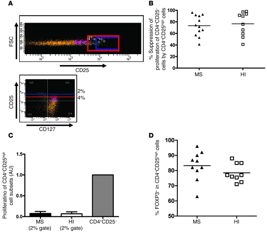
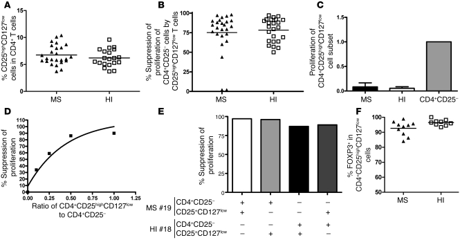
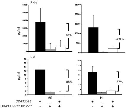
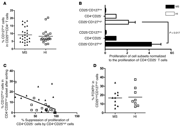
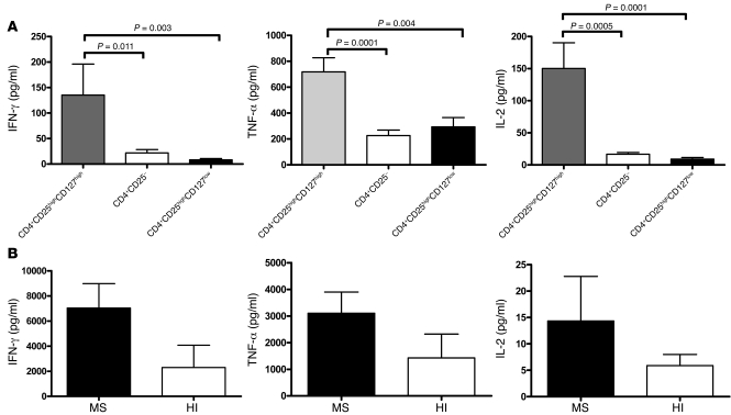
Multiple sclerosis (MS) is a chronic inflammatory disease that results in demyelination in the central nervous system, and a defect in the regulatory function of CD4+CD25high T cells has been implicated in the pathogenesis of the disease. Here, we reanalyzed the function of this T cell subset in patients with MS, but we depleted cells expressing IL-7 receptor alpha-chain (CD127), a marker recently described as present on activated T cells but not Tregs. Similar to other studies, we observed a marked defect in the suppressive function of unseparated CD4+CD25high T cells isolated from MS patients. However, when CD127(high) cells were removed from the CD4+CD25high population, patient and control cells inhibited T cell proliferation and cytokine production equally. Likewise, when the CD25 gate used to sort the cells was stringent enough to eliminate CD127high cells, CD4+CD25high T cells from patients with MS and healthy individuals had similar regulatory function. Additional analysis indicated that the CD127high cells within the CD4+CD25high T cell population from patients with MS appeared more proliferative and secreted more IFN-gamma and IL-2 than the same cells from healthy individuals. Taken together, we conclude that CD4+CD25highCD127low Tregs from MS patients and healthy individuals exhibit similar suppressive functions. The decreased inhibitory function of unfractioned CD4+CD25high cells previously observed might be due to abnormal activation of CD127high T cells in patients with MS.


Figures

Figure 1
Figure 1. Frequency, proliferation, and suppressive activity of the top 4% of sorted CD4+CD25high T cells from MS patients and HIs.
(A) Comparison of the percentage of CD4+CD25high T cells from MS patients and controls. The cut-off for high-staining CD25 was placed at 6 × 10³ of mean fluorescence intensity. No statistical difference can be shown between the groups (patients, n = 21; HI, n = 17). (B) Regulatory properties of CD4+CD25high cells were examined in 11 untreated patients with RR-MS and 11 healthy controls. Cocultures of CD4+CD25 and CD4+CD25high cells were performed at a 1:1 ratio and under anti-CD3 stimulation. Proliferation was measured by incorporation of 3H-thymidine after 5 days of incubation. The percentage of suppression of responding cell proliferation (CD4+CD25) by CD4+CD25high cells was determined as 1 – (proliferation of coculture / proliferation of responder population alone) × 100, where proliferation is expressed as cpm. CD4+CD25high T cells from MS patients exhibited less suppressive activity when the gate for sorting was positioned as shown in Supplemental Figure 1 (P < 0.05, Mann-Whitney U test). Mean values in A and B are indicated by horizontal lines. (C) Comparison of the proliferation of CD4+CD25high T cells in MS patients and HIs relative to the proliferation of the CD4+CD25 T cell subset. Proliferation of CD4+CD25high cells was not significantly different between patients (n = 11) and HIs (n = 11). When the top 4% of CD4+CD25high T cells were sorted, the cells were not fully anergic, suggesting the presence of activated T cells. Data represent mean ± SD.
Figure 2
Figure 2. Suppressive activity and proliferation of the top 2% of sorted CD4+CD25high cells from MS patients and healthy controls.
(A) CD4+ lymphocytes obtained from the peripheral blood of MS patients and healthy controls were stained with Pe-Cy7–conjugated anti-CD3, FITC-conjugated anti-CD8, Alexa Fluor 647–conjugated anti-CD25, and PE-conjugated anti-CD127. Sorting was performed on the CD4+CD25highCD127low and CD4+CD25highCD127high populations. Sorting was also performed on the CD4+CD25high population with 2 different gates. In the example provided for 1 MS patient, CD4+CD25+ T cells appear in orange, CD4+CD25+CD127low T cells appear in green, and CD127high cells appear in violet. The presence of CD127high activated cells can be observed in the CD4+CD25high sorted T cells when the gate is not stringent enough (4% gating stringency, area above the red line), while in the case of a 2% gating stringency (area above the blue line), only a few CD127high cells remain within the CD4+CD25high T cell subset. FSC, forward scatter. (B) CD4+CD25 responder cells were stimulated with anti-CD3 antibody (0.1 μg/ml) in coculture with the top 2% of CD4+CD25high sorted cells. Data are the mean of duplicate wells. Regulatory properties of CD4+CD25high cells are comparable in both HIs (n = 10) and patients (n = 12). (C) Comparison of the proliferation of the top 2% of sorted CD4+CD25high T cells in MS patients and HIs relative to the proliferation of the CD4+CD25 T cell subset. The proliferation of CD4+CD25high T cells was minimal and almost null in both patients and controls. Data are mean ± SD. (D) Intracellular FOXP3 staining was performed on the top 2% of sorted CD4+CD25high T cells in 10 patients and 9 HIs. In B and D, the mean values for each group are indicated by horizontal lines.
Figure 3
Figure 3. Frequency, proliferation, and suppressive activity of CD4+CD25highCD127low cells from MS patients and healthy controls.
(A) Percentages of CD25highCD127low cells within the total CD4+ T cell population were determined by flow cytometry analysis of PBMCs. No statistical difference was found between the groups (25 patients and 20 HIs). (B) Regulatory properties of CD4+CD25highCD127low cells were comparable in both HIs (n = 24) and patients (n = 25). (C) Comparison of the proliferation of CD4+CD25highCD127low T cells in MS patients and HIs normalized against the proliferation of the CD4+CD25 T cell subset. The proliferation of CD4+CD25highCD127low T cells was minimal and approaching 0 in both patients and controls. (D) Example in 1 patient of variations of the suppression of proliferation by CD4+CD25highCD127low cells in the cocultures at varying ratios of CD4+CD25highCD127low to CD4+CD25. Decreasing the number of CD4+CD25highCD127low T cells resulted in less suppressor activity. (E) CD4+CD25highCD127low T cells from 1 patient with MS inhibited proliferation of responder T cells isolated from either the autologous individual or the healthy control. Conversely, Tregs from 1 HI were cocultured with responder T cells from the same subject or those from the MS patient. MS#19, MS patient 19; HI#18, HI subject 18. (F) Intracellular FOXP3 staining was performed on CD4+CD25highCD127low T cells in 10 patients and 9 HIs. In A, B, and F, mean values are indicated by horizontal lines.
Figure 4
Figure 4. Suppression of cytokine production by CD4+CD25highCD127low T cells in patients and controls.
Cytokines (IL-2 and IFN-γ) were measured in supernatants taken from each well 3 days after the initiation of coculture (CD4+CD25 T cells/CD4+CD25highCD127low T cells at a 1:1 ratio) using a multiplex fluorescent bead immunoassay. Three days after initiation of the coculture, the same percentage suppression of IFN-γ (top panels) and IL-2 (bottom panels) production by CD4+CD25highCD127low was observed in the cocultures from 4 patients and 4 HIs. Data are mean ± SD.
Figure 5
Figure 5. Comparison of the proliferation of CD4+CD25highCD127low T cells and CD4+CD25highCD127high T cells.
(A) CD4+ lymphocytes obtained from the peripheral blood of MS patients and healthy controls were stained with Pe-Cy7–conjugated anti-CD3, FITC-conjugated anti-CD8, Alexa Fluor 647–conjugated anti-CD25, and PE-conjugated anti-CD127. No statistically significant difference was observed in the frequency of CD4+CD25highCD127high T cells between MS patients (n = 25) and healthy controls (n = 19). Mean value is indicated for each group. (B) CD4+CD25highCD127low T cells or CD4+CD25highCD127high T cells were cocultured with irradiated autologous PBMCs and stimulated with anti-CD3 antibody. CD25highCD127low cells were isolated from 25 patients and 23 HIs. CD25highCD127high cells were isolated from 20 patients and 20 HIs. A significant difference was observed in the proliferation of CD25highCD127high T cells between MS patients and HIs (P = 0.017, Mann-Whitney U test). Bar graphs indicate the mean ± SD. (C) Suppression of proliferation of CD4+CD25 cells by CD4+CD25high cells was calculated in 13 patients and 15 HIs. The percentage of CD127high cells present in the sorted CD4+CD25high T cells was estimated in the same manner. A correlation was found between this percentage and the regulatory properties of CD4+CD25high cells with a Pearson coefficient of r = –0.50 (P = 0.006, linear regression test). Black triangles represent data obtained from MS patients, and white squares represent data from HIs. (D) Intracellular FOXP3 staining was performed on CD4+CD25highCD127high T cells in 10 patients and 9 HIs. In B and D, horizontal lines indicate the mean.
Figure 6
Figure 6. Cytokines secreted by the CD4+CD25highCD127high, CD4+CD25, and CD4+CD25highCD127low populations in MS patients and HIs.
(A) The supernatants from each well of the proliferation assays were removed 24 hours after the beginning of the incubation. The cytokines TNF-α, IFN-γ, and IL-2 were measured from 10 MS patients in the following 3 cell subsets: CD4+CD25highCD127high, CD4+CD25, and CD4+CD25highCD127low. Mann-Whitney U tests were performed to compare the 3 cell subsets. (B) Comparison of cytokine production by CD4+CD25highCD127high T cells under CD3 polyclonal stimulation between MS patients (n = 4) and HIs (n = 4). Supernatants were removed 3 days after the beginning of the incubation. The results are the mean ± SD from 4 patients and 4 healthy controls.
Figure 7
Figure 7. Comparison of FOXP3 expression in CD4+CD25high T cells and CD4+CD25highCD127low cells.
Intracellular FOXP3 staining was performed in 9 HIs. A significant difference was observed between the top 2% of sorted CD4+CD25high and CD4+CD25highCD127low T cells (P < 0.0001, Mann-Whitney U test). The horizontal lines indicate the mean values for each group.

Similar articles

Cited by

References

    1. Baecher-Allan C., Hafler D.A. Human regulatory T cells and their role in autoimmune disease. Immunol. Rev. 2006;212:203–216. doi: 10.1111/j.0105-2896.2006.00417.x. - DOI - PubMed
    1. Tang Q., Bluestone J.A. Regulatory T-cell physiology and application to treat autoimmunity. Immunol. Rev. 2006;212:217–237. doi: 10.1111/j.0105-2896.2006.00421.x. - DOI - PubMed
    1. Asano M., Toda M., Sakaguchi N., Sakaguchi S. Autoimmune disease as a consequence of developmental abnormality of a T cell subpopulation. J. Exp. Med. 1996;184:387–396. doi: 10.1084/jem.184.2.387. - DOI - PMC - PubMed
    1. Sakaguchi S., Sakaguchi N., Asano M., Itoh M., Toda M. Immunologic self-tolerance maintained by activated T cells expressing IL-2 receptor alpha-chains (CD25). Breakdown of a single mechanism of self-tolerance causes various autoimmune diseases. J. Immunol. 1995;155:1151–1164. - PubMed
    1. Fontenot J.D., Gavin M.A., Rudensky A.Y. Foxp3 programs the development and function of CD4+CD25+ regulatory T cells. Nat. Immunol. 2003;4:330–336. doi: 10.1038/ni904. - DOI - PubMed

Publication types

MeSH terms

Substances