Comparative Study
. 2008 Sep;14(9):931-8.
doi: 10.1038/nm.1857.

The actin cytoskeleton of kidney podocytes is a direct target of the antiproteinuric effect of cyclosporine A

Affiliations
Comparative Study

The actin cytoskeleton of kidney podocytes is a direct target of the antiproteinuric effect of cyclosporine A

Christian Faul et al. Nat Med. 2008 Sep.

Abstract

The immunosuppressive action of the calcineurin inhibitor cyclosporine A (CsA) stems from the inhibition of nuclear factor of activated T cells (NFAT) signaling in T cells. CsA is also used for the treatment of proteinuric kidney diseases. As it stands, the antiproteinuric effect of CsA is attributed to its immunosuppressive action. Here we show that the beneficial effect of CsA on proteinuria is not dependent on NFAT inhibition in T cells, but rather results from the stabilization of the actin cytoskeleton in kidney podocytes. CsA blocks the calcineurin-mediated dephosphorylation of synaptopodin, a regulator of Rho GTPases in podocytes, thereby preserving the phosphorylation-dependent synaptopodin-14-3-3 beta interaction. Preservation of this interaction, in turn, protects synaptopodin from cathepsin L-mediated degradation. These results represent a new view of calcineurin signaling and shed further light on the treatment of proteinuric kidney diseases. Novel calcineurin substrates such as synaptopodin may provide promising starting points for antiproteinuric drugs that avoid the serious side effects of long-term CsA treatment.


Figures

Figure 1
Figure 1
Synaptopodin specifically interacts with 14-3-3. (a) Schematic of synaptopodin isoforms. The white box shows the first 676 amino acids that are shared between Synpo-long and Synpo-short. This fragment contains two 14-3-3 binding motifs (black ovals) and two CatL cleavage sites (arrows). (b) In the adult mouse kidney, 14-3-3β colocalizes with synaptopodin in podocytes. Scale bar, 30 μm. (c) In differentiated cultured podocytes, 14-3-3β colocalizes with synaptopodin along stress fibers (top), and both proteins remain associated after disruption of actin filaments with latrunculin A (bottom). Scale, 25 μm. (d) Synaptopodin from isolated mouse glomerular extracts (input) specifically binds GST–14-3-3β but not GST alone. (e) Coimmunoprecipitation experiments show that endogenous synaptopodin interacts with endogenous 14-3-3β in isolated mouse glomeruli. An antibody to GFP serves as negative control. IP, immunoprecipitation. (f) GFP–14-3-3β precipitates with wild-type (WT) Flag-synaptopodin and phosphomimetic Flag–Synpo-ED but not with phosphoresistant Flag–Synpo-AA or Flag-raver (control).
Figure 2
Figure 2
Identification of synaptopodin as calcineurin binding protein. (a) GFP–Synpo-short and GFP–Synpo-alt precipitate with Flag-tagged aCnA from co-transfected HEK293 cells. GFP–Synpo-alt can also interact with WT calcineurin. No binding is found with Flag-raver (control). (b) Endogenous coimmunoprecipitation experiments show that synaptopodin interacts with calcineurin in isolated mouse glomeruli. An antibody specific for GFP serves as negative control. (c) In the adult mouse kidney (top) and differentiated cultured podocytes (bottom), calcineurin partially colocalizes with synaptopodin. Scale bars, 30 μm (top panels) and 25 μm (bottom panels).
Figure 3
Figure 3
The synaptopodin–14-3-3 interaction is antagonistically regulated by PKA, CaMKII and calcineurin. (a) SDS-PAGE (top) and western blot (bottom) analyses show a reduced molecular weight of purified Flag-synaptopodin (Flag-Synpo) after dephosphorylation with λ-PPase. (b) Time-dependent phosphorylation of purified Flag–Synpo-short by PKA (top panels) and CaMKII (bottom panels). Alanine substitution of Thr216 and Ser619 (AA) strongly reduces 32P labeling when compared to WT synaptopodin. Flag blots show equal protein loading. (c) Phosphorylation of synaptopodin by PKA and CaMKII in HEK293 cells cultured in the absence of inhibitors (Con), KN62 and H89. (d) Time-dependent dephosphorylation of purified 32P-labeled Flag–Synpo-short by calcineurin (left) or in its absence (right). (e) Dephosphorylation of 32P–Flag–Synpo-short in HEK293 cells by calcineurin. (f) Dephosphorylation of purified Flag–Synpo-short by λ-PPase or calcineurin abrogates the binding of synaptopodin to immobilized GST–14-3-3β. Flag–Synpo-alt does not bind 14-3-3 and serves as negative control.
Figure 4
Figure 4
14-3-3β, E64 and CsA block the CatL-mediated degradation of synaptopodin. (a) Dose-dependent degradation of purified Flag–Synpo-short by CatL at pH 4.5, 6.5 or 7.0 leads to the generation of a 44-kDa fragment. (b) H89-mediated reduction of stress fibers and synaptopodin abundance are prevented by the cathepsin inhibitor E64 (right, top). Similarly, E64 prevents the loss of synaptopodin expression and stress fibers caused by simultaneous inhibition of PKA and CaMKII (right, bottom). Scale bar, 25 μm. (c) Western blot analysis of synaptopodin steady-state abundance in differentiated WT podocytes. In control cells (Con) the 110-kDa full-length protein is visible. Inhibition of PKA and CaMKII (H89 + KN62) causes a partial degradation of full-length synaptopodin. Treatment with CsA, E64 or E64D blocks the H89- and KN62-mediated degradation of synaptopodin. Immunoblotting for glyceraldehyde-3-phosphate dehydrogenase (GAPDH) shows equal protein loading. (d) Binding of 14-3-3β but not of α-actinin-4 partially protects synaptopodin against proteolytic processing by CatL (left). 14-3-3β (middle) or α-actinin-4 (right) are not cleaved by CatL. (e) Synaptopodin contains two evolutionarily conserved CatL cleavage sites. The first motif (ALGAE) is conserved from fish to humans (top). The second motif (ALPRPS) is preserved from mice to humans (bottom). (f) Site-directed mutagenesis of CatL cleavage sites separately (CM1 or CM2) or together (CM1+2) increases resistance of synaptopodin against CatL-mediated proteolytic processing. Flag-synaptopodin and its mutant forms were purified from HEK293 cells (left) and incubated with CatL at pH 7.0 (right).
Figure 5
Figure 5
CsA and E64 ameliorate LPS-induced proteinuria by blocking the CatL-mediated degradation of synaptopodin. (a) SDS-PAGE analysis of urines from control and LPS-injected mice (top). Quantitative analysis of albuminuria (bottom). (b) Western blot analysis of isolated glomeruli showing that LPS causes the degradation of endogenous synaptopodin and RhoA. CsA or E64 block the LPS-induced degradation of synaptopodin and RhoA. GAPDH shows equal protein loading. (c) Immunoblot analysis of Flag-synaptopodin proteins from isolated glomeruli (left) and liver extracts (right) 48 h after in vivo gene delivery. Glomerular RhoA levels are positively correlated with Flag-synaptopodin abundance. (d) Expression of Flag–Synpo-ED in podocytes after in vivo gene delivery as detected by double-labeling deconvolution microscopy with antibodies to Flag and the podocyte marker podocin. No Flag signal is seen in podocytes of delivery solution–injected mice (control). Scale bar, 50 μm. (e) Gene transfer of Synpo-CM1+2 or Synpo-ED but not WT synaptopodin, Synpo-AA or delivery solution (control) protects against LPS-induced proteinuria. NS, not significant. (f) Immunoblot analysis of synaptopodin protein expression after LPS injection and gene delivery. LPS causes degradation and decrease of synaptopodin (LPS + Con) when compared to PBS-injected mice (Con). High levels of synaptopodin can be found in LPS-injected mice after gene transfer of Synpo-CM1+2 or Synpo-ED but not of WT synaptopodin or Synpo-AA (left). Synaptopodin protein in the liver is not affected by LPS (right). Mice that did not receive synaptopodin cDNA (Con, Con + LPS) do not express synaptopodin in the liver. *P < 0.05; **P < 0.001; ***P < 0.0005.
Figure 6
Figure 6
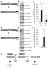
Expression of Synpo-CM1+2 in podocytes protects against proteinuria, whereas activation of calcineurin in podocytes causes proteinuria. (a) Schematic of constructs used for the generation of Synpo-CM1+2–transgenic mice. rTetR, reverse Tet repressor; VP16, herpes simplex VP16 protein; tetO, tet operator sequences; pCMV, cytomegalovirus promoter. (b) Immunoblot analysis of isolated glomeruli from doxycycline (Dox)- or vehicle (Veh)-treated double-transgenic mice. After LPS injection, synaptopodin protein abundance remains stable in Dox-treated but not in Veh-treated mice. Protein abundance of RhoA, dynamin and ZO-1 are positively correlated with synaptopodin abundance. GAPDH shows equal protein loading. (c) GFP–Synpo-CM1+2 (Dox +LPS) protects against LPS-induced proteinuria. (d) Schematic of constructs used for the generation of aCnA-transgenic mice. (e) Immunoblot analysis of isolated glomeruli showing reduced steady-state protein levels of synaptopodin, RhoA, dynamin and ZO-1 but not of α-actinin-4 in GFP-aCnA–expressing (Dox) mice. (f) Detection of proteinuria in GFP-aCnA–expressing (Dox) but not in control (Veh) mice. (g) Model for the regulation of podocyte function by calcineurin. Phosphorylation of synaptopodin by PKA or CaMKII promotes 14-3-3 binding, which protects synaptopodin against CatL-mediated cleavage, thereby contributing to the intact glomerular filtration barrier. Dephosphorylation of synaptopodin by calcineurin (CaN) abrogates the interaction with 14-3-3. This renders the CatL cleavage sites of synaptopodin accessible and promotes the degradation of synaptopodin. CsA and E64 safeguard against proteinuria by stabilizing synaptopodin steady-state protein levels in podocytes. *P < 0.005.

Comment in

Similar articles

Cited by

References

    1. Somlo S, Mundel P. Getting a foothold in nephrotic syndrome. Nat Genet. 2000;24:333–335. - PubMed
    1. Tryggvason K, Patrakka J, Wartiovaara J. Hereditary proteinuria syndromes and mechanisms of proteinuria. N Engl J Med. 2006;354:1387–1401. - PubMed
    1. Tryggvason K, Pikkarainen T, Patrakka J. Nck links nephrin to actin in kidney podocytes. Cell. 2006;125:221–224. - PubMed
    1. Faul C, Asanuma K, Yanagida-Asanuma E, Kim K, Mundel P. Actin up: regulation of podocyte structure and function by components of the actin cytoskeleton. Trends Cell Biol. 2007;17:428–437. - PubMed
    1. Mundel P, et al. Synaptopodin: an actin-associated protein in telencephalic dendrites and renal podocytes. J Cell Biol. 1997;139:193–204. - PMC - PubMed

Publication types