. 2008 Oct;4(10):609-16.
doi: 10.1038/nchembio.109. Epub 2008 Aug 24.

Identification of a copper-binding metallothionein in pathogenic mycobacteria

Affiliations

Identification of a copper-binding metallothionein in pathogenic mycobacteria

Ben Gold et al. Nat Chem Biol. 2008 Oct.

Abstract

A screen of a genomic library from Mycobacterium tuberculosis (Mtb) identified a small, unannotated open reading frame (MT0196) that encodes a 4.9-kDa, cysteine-rich protein. Despite extensive nucleotide divergence, the amino acid sequence is highly conserved among mycobacteria that are pathogenic in vertebrate hosts. We synthesized the protein and found that it preferentially binds up to six Cu(I) ions in a solvent-shielded core. Copper, cadmium and compounds that generate nitric oxide or superoxide induced the gene's expression in Mtb up to 1,000-fold above normal expression. The native protein bound copper within Mtb and partially protected Mtb from copper toxicity. We propose that the product of the MT0196 gene be named mycobacterial metallothionein (MymT). To our knowledge, MymT is the first metallothionein of a Gram-positive bacterium with a demonstrated function.


Figures
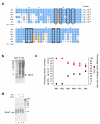
Figure 1
Figure 1
Identification of MymT as a polypeptide with MT-like metal-binding motifs. (a) Alignment of homologs of Mtb MymT identified by tBLASTn homology searches in M. bovis BCG (Mbo), M. avium paratuberculosis and M.avium 104 (Mav; they have identical sequences), M. buruli (Mbu), M. leprae (Mle), and M. marinum (Mma). Conserved residues comprising the Cys-X-His-X-X-Cys-X-Cys motif observed in SmtA from Synechococcus are noted in the line marked Syn. Conserved cysteines and histidines are boxed. The star above Mtb Met6 indicates the predicted N-terminal Met of the mature peptide. Amino acids identical to the Mtb MymT sequence are coded blue, and amino acids with similar properties, tan. (b) 15% SDS-PAGE of lysates (20 μg) stained with Coomassie from M. smegmatis transformed with a plasmid designed to over-express MymT (MT) or the empty plasmid pMV261 as a control (C). (c) Differing codon usage despite >67% amino acid identity among mycobacterial MymT homologs for the 48 amino acids beginning with Mtb MymT Met6. Amino acid similarity was determined by BLAST analysis. Amino acids identical to the Mtb sequence were used to perform the analysis “% different codons”. (d) 24 μM of recombinant Zn(II)-MymT (lane 1) was mixed with 100 mM DTT (lane 2), 1 mM CuSO4 (lane 3), or 100 mM DTT and 1 mM CuSO4 (lane 4) and separated by 20% SDS-PAGE and stained with Coomassie. CuSO4 was present at a 6-fold molar ratio to MymT cysteine content. Higher molecular weight bands correspond to the molecular weights of a minor contaminating species of uncleaved MymT / Mxe-intein fusion (~33 kDa) and the Mxe-intein (~27 kDa).
Figure 2
Figure 2
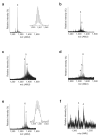
Mass spectra of MymT complexed with Cu(I) or Zn(II). The masses of solid-phase synthesized MymT and its Cu(I)n- and Zn(II)n-MymT complexes were determined from the isotope distribution of charged ions. 100 μM of apo-MymT was reconstituted with various molar ratios of the Cu(I) donor, Cu(CH3CN)4[PF6], or ZnSO4, in 0.01 N HCl (~ pH 2) and the generated complexes analyzed by nano-ESI-mass spectrometry. The mass spectrum of apo-MymT at the 4+ charge state (a), or after the addition of 3 (b), 6 (c), 7 (d) or 10 (e) equivalents of Cu(I), and (f) 5 equivalents of ZnSO4 in 50 mM ammonium acetate pH 7. The charge state was determined based on the observed isotope distribution of a specific complex ion. The number above each peak corresponds to the number of metal ions bound to MymT. Only species in the 4+ charge state are illustrated. The inset figures in (a) and (e) represent the observed isotope distribution of apo- and Cu(I)6-MymT, respectively. Similar isotope distributions were observed for all MymT-metal complexes. Full spectra (600-1500 m/z) of MymT-metal complexes in multiple charge states are shown in Supplementary Figure 3 online. Peak m/z and mass determinations are summarized in Supplementary Table 1 online.
Figure 3
Figure 3
MymT binds Cu(I) in a solvent-shielded, luminescent core. Synthetic apo-MymT (10 μM) was incubated with 0-12 molar equivalents of Cu(CH3CN)4[PF6], a donor of Cu(I) (Supplementary Fig. 2a online), in 0.001 N HCl (~ pH 3). Reactions were incubated 20 minutes at RT. (a) After excitation at A280, the emission spectrum was recorded and shown as normalized emission intensity. (b) The emission maxima at 600 nm, and (c) the data in (b) plotted as emission intensity per molar equivalent of Cu(I) to MymT. One experiment representative of three is shown.
Figure 4
Figure 4
Regulation of mymT transcript abundance. Wild-type Mtb was treated with indicated stimuli at 50 and 500 μM (unless otherwise indicated) for 2 hours. RNA was extracted and subjected to quantitative RT-PCR using molecular beacons. Data were normalized against results for sigA, a housekeeping sigma factor whose mRNA levels did not significantly vary under most experimental conditions. Data for exposure to plumbagin were normalized by RNA concentration due to significant sigA mRNA degradation. mymT mRNA expression was expressed as fold induction compared to unstimulated cultures in response to (a) divalent heavy metals: cadmium, cobalt, copper, manganese, nickel, zinc, and the metal chelator ethylenediamine tetraacetic acid (EDTA); and (b) RNI, ROI and cell-wall perturbing agents: NO released from 50 or 500 μM DETA/NO; superoxide-generators menadione and plumbagin; and 0.05% sodium dodecyl sulfate (SDS). Error bars denote standard deviations of triplicate samples from 1 of at least 2 experiments with similar results.
Figure 5
Figure 5
Generation of a ΔmymT mutant in Mtb. (a) Cloning strategy to replace mymT with a hygromycin antibiotic resistance cassette by specialized transduction. (b) Southern blot analysis of genomic DNA digested with NcoI-XhoI using a probe of upstream DNA sequence (depicted in (a)) confirmed the presence of a 1284 bp fragment in the wild-type strain and 824 bp in two ΔmymT mutants (1 and 2). (c) Wild type Mtb and the ΔmymT mutant were exposed overnight to 500 μM CuSO4 and their lysates were separated by 20% SDS-PAGE, transferred to a 0.2 μ PVDF membrane and probed with a 1:1000 dilution of α-MymT antiserum. Unlike wild type Mtb, the Mtb ΔmymT mutant did not produce MymT protein in response to a 24 hour exposure to 500 μM CuSO4. Reconstitution of the mymT gene under control of its native promoter on pMV361 restored copper-responsive MymT expression (complementing strains 1 (C1) and 2 (C2)) to the ΔmymT mutant. (d) The Mtb ΔmymT mutant exhibited less luminescence that the wild-type and complemented strains. After overnight exposure to increasing concentrations of CuSO4 in 7H9 medium, the Mtb wild-type, ΔmymT mutant, and complemented strains were washed and resuspended in ~1/10 of the original volume of PBS-0.05% Tween80. Cells were irradiated at A280 nm and their emission spectra read at A450-750 nm. OD580 measurements of copper-exposed cultures indicated less than 10% differences in cell number between strains. Data shown are at the emission peak of A595.
Figure 6
Figure 6
Sensitivity of the Mtb ΔmymT mutant to cuprous ion. WT Mtb, the ΔmymT mutant and the ΔmymT mutant reconstituted with a WT mymT allele were grown to mid-log phase and spotted (from left to right: 5 μL of cultures at an OD580 of 0.10, 0.01, and 0.001) on 7H11 agar with (a) no addition; (b) 1 mM bathocuproine disulphonate (BCS); (c) 150 μM CuSO4; (d) 150 μM CuSO4 + 1 mM BCS. The Cu(I)-specific chelating agent BCS was added in a ~10-fold molar excess prior to spotting Mtb dilutions. The orange color was apparent immediately after the addition of BCS. One representative experiment of 3 similar experiments is shown. Scale bar: 13 mm.
Figure 7
Figure 7
Generation of Cu(I) by NO. (a) Reactive nitrogen intermediates can liberate Cu(I) from Cu(I)4-MymT. Cu(I)4-MymT was exposed in the dark to 10 mM NaNO2 or 10 mM NaNO3 for indicated times and luminescence determined (pH 5.5 + NaNO3, open red circles; pH 5.5 + NaNO2, closed red circles; pH 7.0 + NaNO3, open blue squares; pH 7.0 + NaNO2, solid blue squares). (b) Reactive nitrogen intermediates disrupt luminescent Cu(I)-thiolate cores in live Mtb. The Mtb WT, the ΔmymT mutant, and the ΔmymT mutant reconstituted with a WT mymT allele were grown to late-log phase, exposed to 0, 40 or 100 μM CuSO4 for 24 hours, washed and resuspended in ~1/10 volume 50 mM KPi pH 5.5 buffer. Cells were then untreated (light gray bars) or treated 15 minutes at room temperature with water (dark gray bars), 10 mM NaNO2 (red bars), or 10 mM NaNO3 (blue bars). Error bars are SD of triplicates. (c) NO released from DETA/NO reduced Cu(II) in a dose- and time-dependent manner (closed blue circles, 1 hr; open blue circles, 24 hours). The NO-scavenger carboxy-PTIO (CPTIO) prevents Cu(I) formation (closed red circles, 1 hr; open red circles, 24 hrs). (d) After 72 hours decomposition, DETA/NO no longer supported Cu(I) formation. Error bars are SD of triplicates. Experiments were performed at least twice with similar results.

Similar articles

Cited by

References

    1. Cole ST, et al. Deciphering the biology of Mycobacterium tuberculosis from the complete genome sequence. Nature. 1998;393:537–44. - PubMed
    1. Ehrt S, et al. A novel antioxidant gene from Mycobacterium tuberculosis. J Exp Med. 1997;186:1885–96. - PMC - PubMed
    1. Ruan J, John G, Ehrt S, Riley L, Nathan C. noxR3, a novel gene from Mycobacterium tuberculosis, protects Salmonella typhimurium from nitrosative and oxidative stress. Infect Immun. 1999;67:3276–83. - PMC - PubMed
    1. Gold B, Rodriguez GM, Marras SA, Pentecost M, Smith I. The Mycobacterium tuberculosis IdeR is a dual functional regulator that controls transcription of genes involved in iron acquisition, iron storage and survival in macrophages. Mol Microbiol. 2001;42:851–65. - PubMed
    1. Rodriguez GM, Smith I. Identification of an ABC transporter required for iron acquisition and virulence in Mycobacterium tuberculosis. J Bacteriol. 2006;188:424–30. - PMC - PubMed

Publication types

MeSH terms

LinkOut - more resources