. 2008 Nov 1;323(1):19-30.
doi: 10.1016/j.ydbio.2008.07.034. Epub 2008 Aug 6.

A dosage-dependent requirement for Sox9 in pancreatic endocrine cell formation

Affiliations

A dosage-dependent requirement for Sox9 in pancreatic endocrine cell formation

Philip A Seymour et al. Dev Biol. .

Abstract

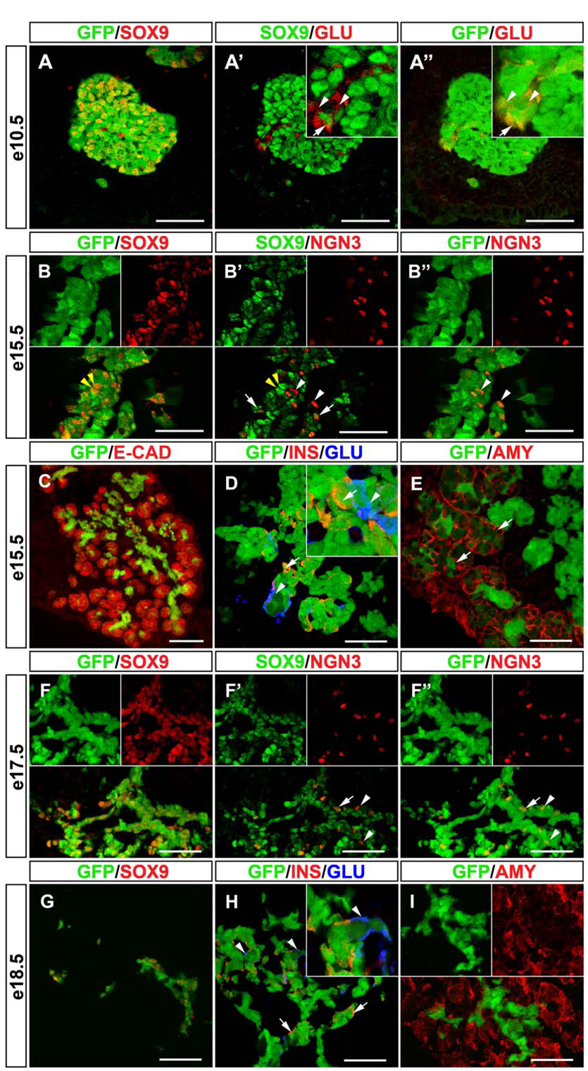
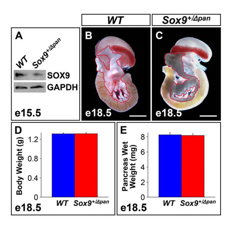
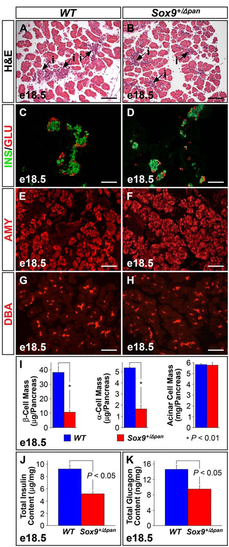
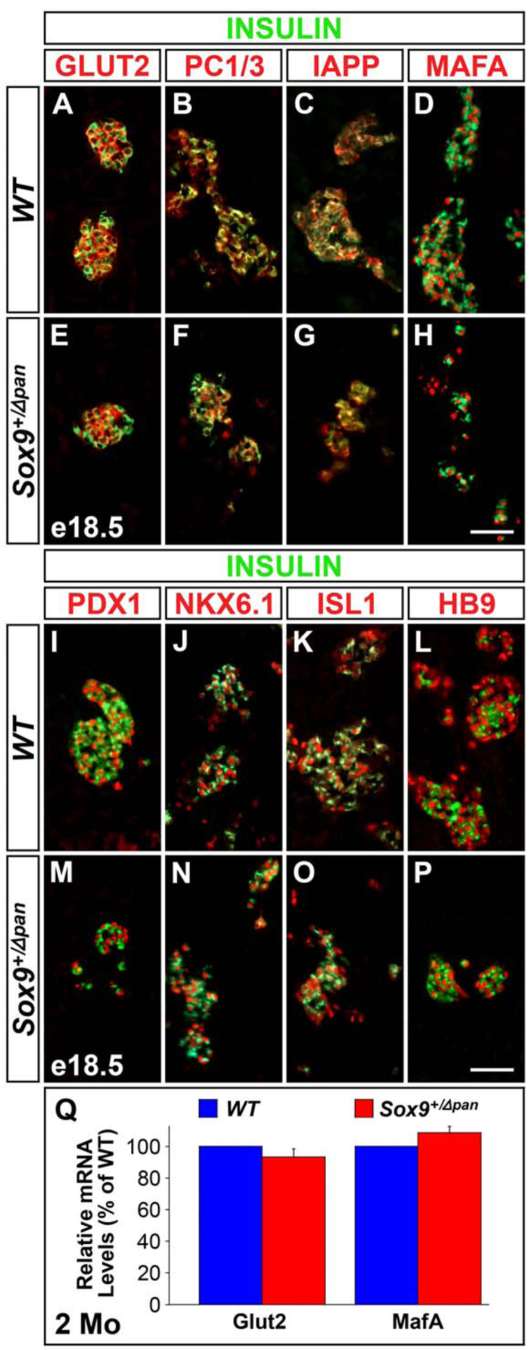
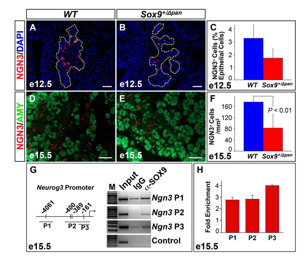
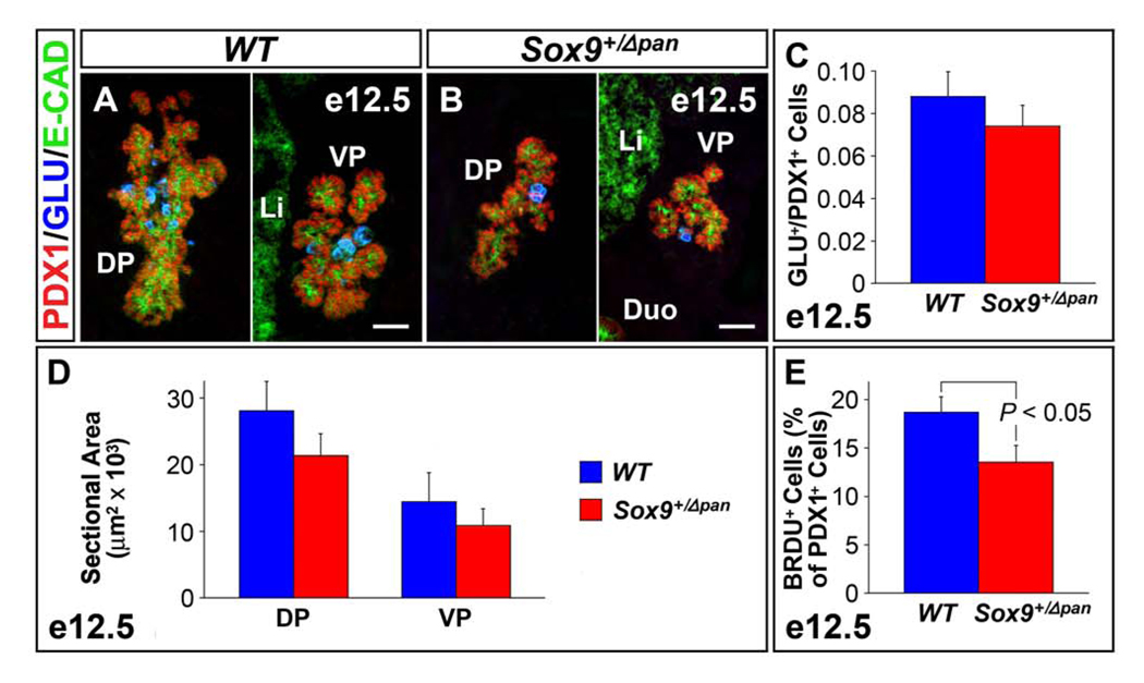
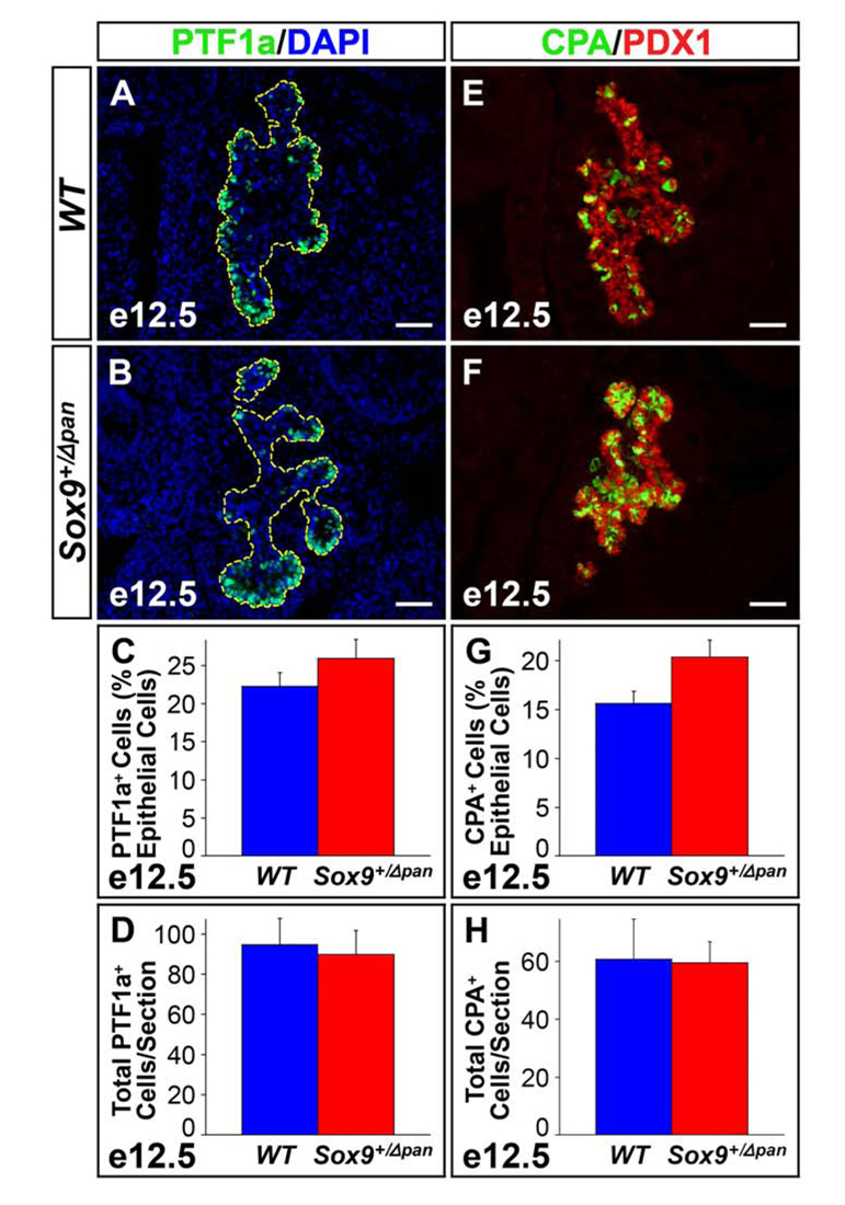
We have previously shown the transcription factor SOX9 to be required for the maintenance of multipotential pancreatic progenitor cells in the early embryonic pancreas. However, the association of pancreatic endocrine defects with the Sox9-haploinsufficiency syndrome campomelic dysplasia (CD) implies additional later roles for Sox9 in endocrine development. Using short-term lineage tracing in mice, we demonstrate here that SOX9 marks a pool of multipotential pancreatic progenitors throughout the window of major cell differentiation. During mid-pancreogenesis, both endocrine and exocrine cells simultaneously arise from the SOX9(+) epithelial cords. Our analysis of mice with 50%-reduced Sox9 gene dosage in pancreatic progenitors reveals endocrine-specific defects phenocopying CD. By birth, these mice display a specific reduction in endocrine cell mass, while their exocrine compartment and total organ size is normal. The decrease in endocrine cells is caused by reduced generation of endocrine progenitors from the SOX9(+) epithelium. Conversely, formation of exocrine progenitors is insensitive to reduced Sox9 gene dosage, thus explaining the normal organ size at birth. Our results show that not only is SOX9 required for the maintenance of early pancreatic progenitors, but also governs their adoption of an endocrine fate. Our findings therefore suggest that defective endocrine specification might underlie the pancreatic phenotype of individuals with CD.


Figures

Figure 1
Figure 1. Endocrine and Exocrine Cells Originate from the SOX9+ Epithelial Cords
(A-A”) Simultaneous detection of SOX9, glucagon and GFP in Sox9-eGFP mice and subsequent confocal analysis shows almost complete overlap of GFP and SOX9 expression in the dorsal pancreatic bud at e10.5. While most glucagon+ cells do not express SOX9 (A’, arrowheads in inset), occasional SOX9/glucagon coexpressing cells are detected (A’, arrow in inset). All glucagon+ cells express GFP (A”, arrow and arrowheads). Staining of sections from Sox9-eGFP mice for E-cadherin reveals that the GFP signal is restricted to the epithelial progenitor cords at e15.5 (C). (B-B”) Simultaneous detection of SOX9, NGN3 and GFP at e15.5 shows that the majority of the SOX9+/GFP+ cells do not express NGN3 (B,B’, yellow arrowheads), while a small proportion of the SOX9+ cells also express NGN3 (B’, white arrows). Notably, a subset of NGN3+ cells shows no SOX9 expression but retains GFP label (B’,B”, white arrowheads), suggesting that NGN3+ cells arise from the SOX9+ domain. Also, at e15.5, GFP co-localizes with insulin (D, arrows) and glucagon (D, arrowheads) as well as amylase (D, arrows). Since SOX9 protein is absent from endocrine and exocrine cells at e15.5, this indicates that both lineages originate from SOX9+ cells. (F-F”) Simultaneous detection of SOX9, NGN3 and GFP at e17.5 shows that NGN3+ cells continue to arise from SOX9+ progenitors late in embryogenesis. Note the NGN3+/SOX9 cells that are GFP+ (F’,F”, arrowheads). The arrow (F’,F”) points to a cell that expresses SOX9, NGN3 and GFP. At e18.5, GFP co-localizes with SOX9 in the pancreatic ducts (G). GFP label continues to be seen in a proportion of insulin+ (H, arrows) and glucagon+ cells (H, arrowheads). However, amylase+ cells no longer retain GFP at e18.5 (I), suggesting that endocrine, but not exocrine cells, continue to arise from SOX9+ progenitors until birth. Note that the SOX9 signal was digitally color-converted in A-A’, B-B” and F-F”. INS, insulin; GLU, glucagon; AMY, amylase; E-CAD, E-cadherin. Scale bar = 50µm in A–B and D–I; 100µm in C.
Figure 2
Figure 2. A Two-Fold Reduction in Sox9 Gene Dosage Has No Effect on Organ Size
(A) Western blot of protein extracts from e15.5 pancreata (n=3) shows a 46% decrease in SOX9 in Sox9 heterozygous pancreatic mutants (Sox9+/Δpan) vs. wild-type (WT) littermates. At e18.5, gross anatomy of the pancreas (demarcated by a red dashed line) in Sox9+/Δpan mice (C) is undistinguishable from that of wild-type littermates (B). In concordance, body weight (D) and pancreas wet weight (E) are equivalent in Sox9+/Δpan (n=19) and wild-type (n=31) embryos at e18.5. (D,E) The values shown represent mean values ± SEM (standard error of the mean). (B,C) Scale bar = 200µm.
Figure 3
Figure 3. Sox9-Haploinsufficiency Leads to Islet-Specific Hypoplasia
Hematoxylin and eosin (H&E) staining reveals significant islet hypoplasia in Sox9 heterozygous mutant (Sox9+/Δpan) (B) compared to wild-type (WT) pancreas (A). Numbers of insulin+ β-cells and glucagon+ α-cells are equally reduced in Sox9+/Δpan islets (C,D), while the amylase+ acinar (E,F) and DBA+ ductal compartments (G,H) of the exocrine pancreas, are indistinguishable between Sox9+/Δpan and wild-type pancreas. (I) Morphometry confirms significant reductions in both α- and β-cell mass in Sox9+/Δpan vs. control pancreata at e18.5, while the acinar cell mass, as assessed by amylase+ tissue area, is unchanged (n=3). ELISAs verify significant decreases in both total pancreatic insulin (J) and glucagon (K) in Sox9+/Δpan compared to wild-type pancreas (n=5). The values shown represent mean values ± SEM (standard error of the mean). INS, insulin; GLU, glucagon; AMY, amylase; DBA, Dolichos biflorus agglutinin, i, islet. (A–H) Scale bar = 100µm.
Figure 4
Figure 4. Sox9-Haploinsufficiency Does Not Result in Impaired Terminal Differentiation of Endocrine Cells
Immunofluorescence staining for markers of differentiated endocrine cells, including GLUT2, PC1/3, IAPP, MAFA, PDX1, NKX6.1, ISL1 and HB9 reveals all markers to be equally expressed in insulin+ cells of Sox9 heterozygous mutant (Sox9+/Δpan) (E–H; M–P) and wild-type (WT) (A–D; I–L) pancreas at e18.5. qRT-PCRs performed on pooled mRNA from 200 isolated islets per genotype, confirm that islet Glut2 and MafA mRNA levels are similar in two 2-month-old Sox9+/Δpan and WT mice. The values shown represent mean values ± SEM (standard error of the mean) (n=3) (Q). Scale bar = 50µm.
Figure 5
Figure 5. Endocrine Progenitors are Two-Fold Reduced in Sox9 Heterozygous Mutant Pancreata
Numbers of NGN3+ endocrine progenitor cells are 50% reduced in the Sox9 heterozygous mutant (Sox9+/Δpan) compared to wild-type (WT) pancreas (n=3) both as a proportion of total (DAPI+) pancreatic epithelial cells at e12.5 (A–C; pancreatic epithelium highlighted by yellow dashed line in A,B) and per square millimeter of pancreas tissue at e15.5 (D–F). Chromatin immunoprecipitation studies of cross-linked chromatin from dissected pancreata at e15.5 with anti-SOX9 antiserum show that SOX9 occupies sequences in the Neurog3 promoter. Three fragments, as previously described by Lynn et al. 2007, were amplified by PCR from either input DNA, DNA precipitated with IgG or anti-SOX9 antiserum. Sequences from the GABA-A receptor intron 5 were amplified in the control reaction (G). qRT-PCR analysis of DNA precipitated with anti-SOX9 antiserum shows a 3–4-fold enrichment over DNA precipitated with IgG (n=4) (H). The values shown represent mean values ± SEM (standard error of the mean). AMY, amylase. (A,B,D,E) Scale bar = 50µm.
Figure 6
Figure 6. Sox9-Haploinsufficiency Causes a Mild Proliferation Defect of Pancreatic Progenitors
The ratio of glucagon+ endocrine cells to PDX1+ pancreatic progenitors is not significantly different between Sox9 heterozygous mutant (Sox9+/Δpan) and wild-type (WT) pancreas (n=3) at e12.5 (A–C). By contrast, the proliferation of PDX1+ progenitors (as measured by percentage BrdU incorporation of PDX1+ cells) is 25% reduced in Sox9+/Δpan pancreas (n=3) at e12.5 (E). Consequently, mean sectional area of both dorsal and ventral Sox9+/Δpan pancreas is 25% reduced compared with those of wild-type littermates at e12.5 (D). The values shown represent mean values ± SEM (standard error of the mean). E-CAD, E-cadherin; GLU, glucagon; DP, dorsal pancreas; VP, ventral pancreas; Duo, duodenum; Li, liver. (A,B) Scale bar = 50µm.
Figure 7
Figure 7. Reduced Sox9 Gene Dosage Does Not Affect the Formation of Exocrine-Fated Cells
Relative numbers of “proto-acinar” cells, as marked by expression of PTF1a (A–D) or carboxypeptidase A (CPA) (E–H), are mildly increased in e12.5 Sox9 heterozygous mutant (Sox9+/Δpan) pancreas compared to wild-type (WT) littermates (n=3). To take into account the epithelial hypoplasia of Sox9+/Δpan pancreata, the PTF1a+ and CPA+ cell numbers were normalized to the total DAPI+ pancreatic epithelial cell area (C,G). However, absolute numbers of PTF1a+ (D) or CPA+ (H) cells per section are unchanged between Sox9+/Δpan and wild-type pancreas. (A,B) Pancreatic epithelium is highlighted by yellow dashed line. The values shown represent mean values ± SEM (standard error of the mean). (A,B,E,F) Scale bar = 50µm.

Similar articles

Cited by

References

    1. Ahlgren U, et al. beta-cell-specific inactivation of the mouse Ipf1/Pdx1 gene results in loss of the beta-cell phenotype and maturity onset diabetes. Genes Dev. 1998;12:1763–1768. - PMC - PubMed
    1. Akiyama H, et al. The transcription factor Sox9 is degraded by the ubiquitin-proteasome system and stabilized by a mutation in a ubiquitintarget site. Matrix Biol. 2005a;23:499–505. - PubMed
    1. Akiyama H, et al. Osteo-chondroprogenitor cells are derived from Sox9 expressing precursors. Proc Natl Acad Sci U S A. 2005b;102:14665–14670. - PMC - PubMed
    1. Barrionuevo F, et al. Homozygous inactivation of Sox9 causes complete XY sex reversal in mice. Biol Reprod. 2006;74:195–201. - PubMed
    1. Bernard P, et al. Dimerization of SOX9 is required for chondrogenesis, but not for sex determination. Hum Mol Genet. 2003;12:1755–1765. - PubMed

Publication types

MeSH terms