. 2008 Nov;19(11):4738-49.
doi: 10.1091/mbc.e07-10-1078. Epub 2008 Aug 20.

Erk5 controls Slug expression and keratinocyte activation during wound healing

Affiliations

Erk5 controls Slug expression and keratinocyte activation during wound healing

Valerie Arnoux et al. Mol Biol Cell. 2008 Nov.

Abstract

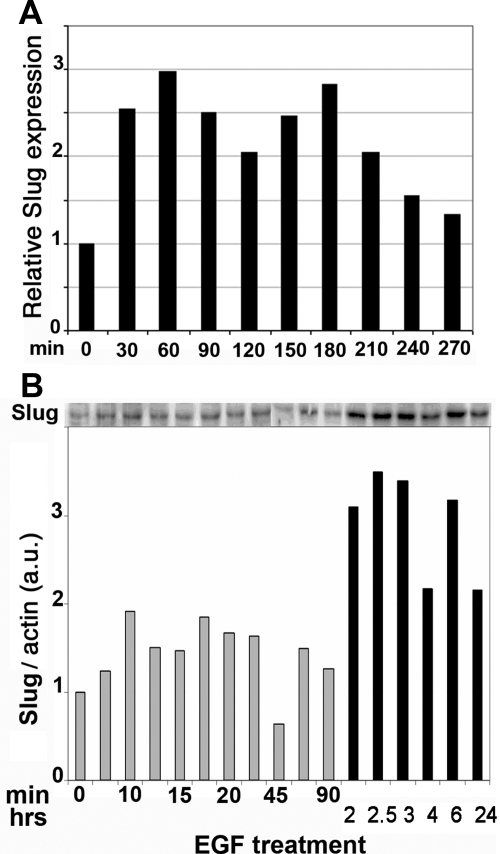
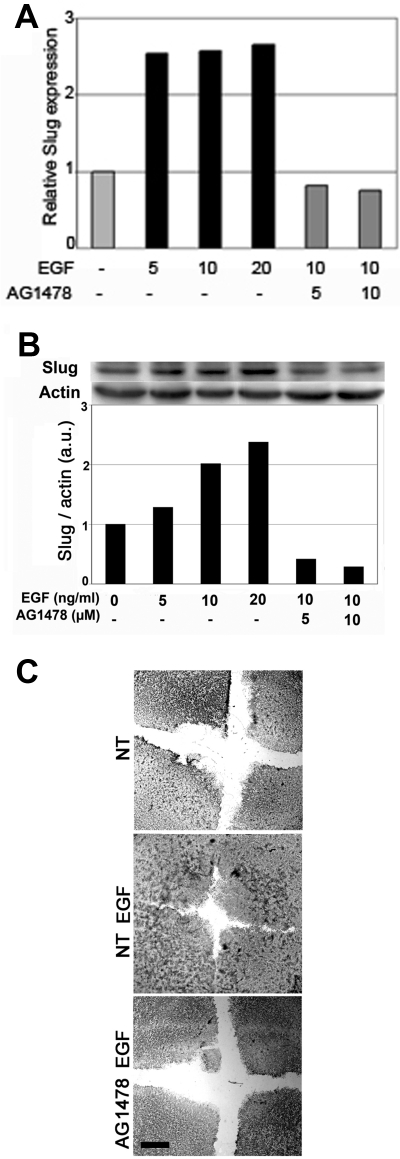
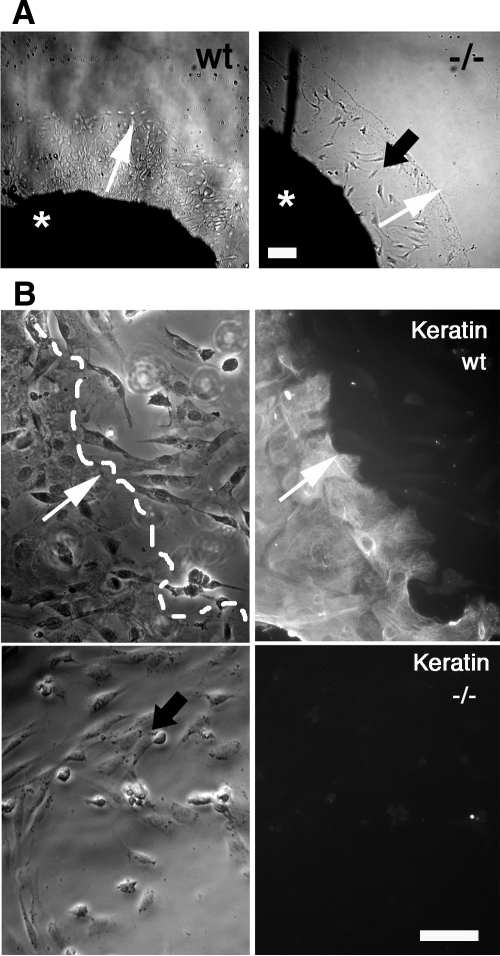
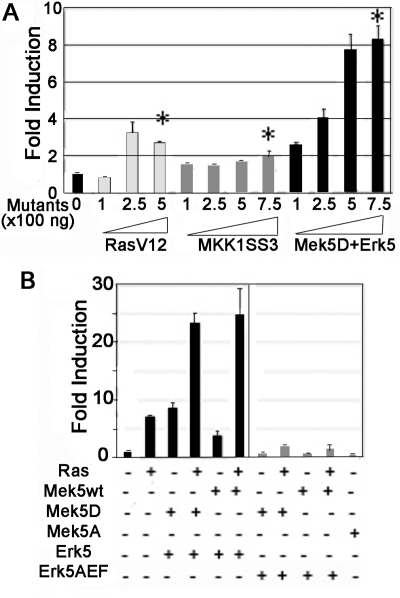
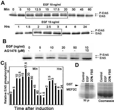
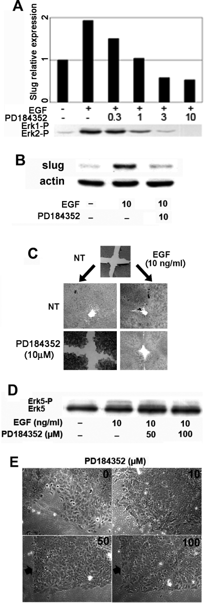
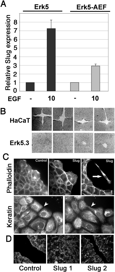
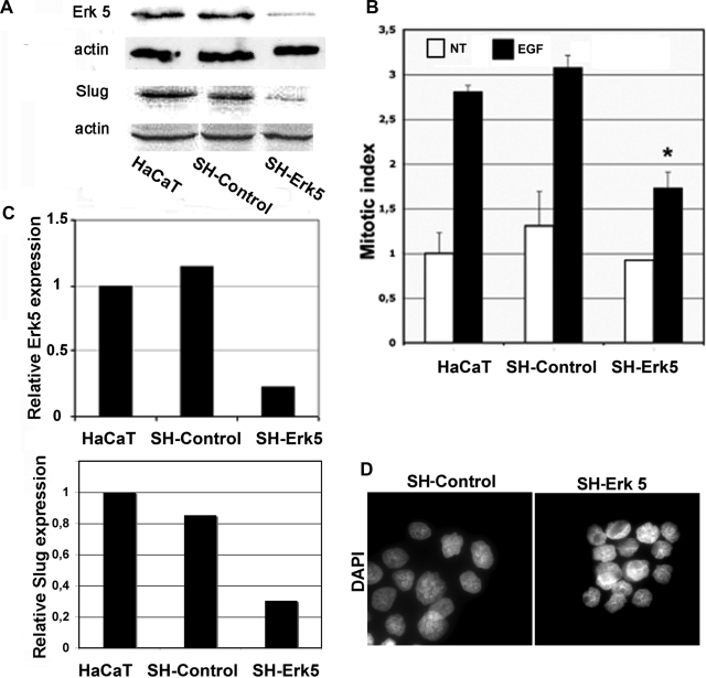
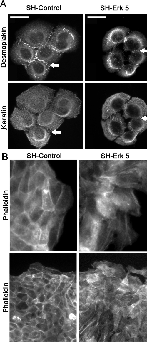
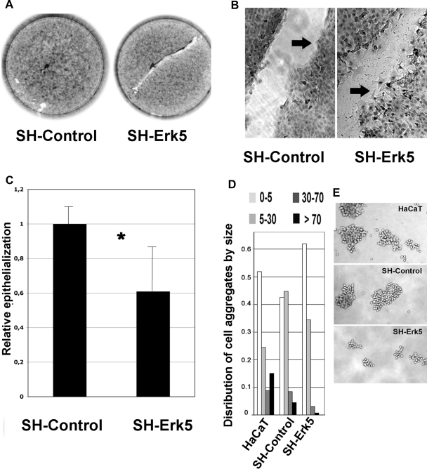
Reepithelialization during cutaneous wound healing involves numerous signals that result in basal keratinocyte activation, spreading, and migration, all linked to a loosening of cell-cell adhesion structures. The transcription factor Slug is required for this process, and EGF treatment of human keratinocytes induced activating phosphorylation of Erk5 that coincides with slug transcription. Accordingly, ectopic activation of Erk5 led to increased Slug mRNA levels and faster wound healing, whereas keratinocyte migration was totally blocked by Erk5 pathway inhibition. Expression of a shRNA specific for Erk5 strongly diminished Erk5 levels in keratinocytes and significantly decreased their motility response to EGF, along with induction of Slug expression. These Erk5-deprived keratinocytes showed an altered, more compact morphology, along with disruption of desmosome organization. Accordingly, they displayed an altered ability to form cell aggregates. These results implicate a novel EGFR/Erk5/Slug pathway in the control of cytoskeleton organization and cell motility in keratinocytes treated with EGF.


Figures

Figure 1.
Figure 1.
EGF induces Slug expression. Serum-starved HaCaT cells were stimulated with 10 ng/ml EGF for the indicated time before extracting RNA and protein. (A) RNA levels were measured by quantitative RT-PCR. Slug RNA levels were calculated relative to the gene 36B4, using the value 2ΔCT where ΔCT is the number of cycles between Slug and 36B4. Slug RNA levels are represented as fold induction relative to serum-starved cells. (B) Protein extracts (40 μg) were loaded on SDS-PAGE and transferred to nitrocellulose, and Slug was detected and quantified by immunoblotting, using β-actin as the reference for protein levels. Two gels were needed to span the kinetics. Protein levels were quantified from the films using NIH Image software. Abbreviation used: a.u., arbitrary unit.
Figure 2.
Figure 2.
The EGFR mediates EGF-induced Slug expression and reepithelialization by immortalized keratinocytes. Serum-starved cells were pretreated for 1 h with the indicated concentration of the specific EGFR inhibitor AG1478 and then stimulated with the indicated concentrations of EGF for 48 h. RNA (A) and protein (B) levels were analyzed and quantified as described in the legend to Figure 3, using β-actin as the reference for protein levels. (C) Confluent HaCaT cells were wounded, and reepithelialization was evaluated 28 h later in untreated cells (NT), cells incubated with 10 ng/ml EGF alone (NT EGF) or with AG1478 (AG 1478 EGF). Scale bar, 100 μm.
Figure 3.
Figure 3.
Keratinocytes from Slug−/− mice fail to spread and migrate from skin explants, even in the presence of EGF. (A) Skin explants from a wild-type (wt) and a Slug-LacZ homozygote mouse (−/−) were cultured ex vivo with 10 ng/ml EGF. (B) Phase-contrast micrographs and keratin (CK) immunolocalization in cells migrating from wild-type and Slug−/− skin explants. Keratinocytes are identified by keratin reactivity. The results shown are representative of four separate experiments. Black arrow, mesenchymal cells negative for keratin immunolabeling; white arrow; direction of keratinocyte migration; broken white line, keratinocyte migrating front; *, skin explant. Scale bar, 25 μm.
Figure 4.
Figure 4.
A reporter gene driven by the Slug promoter is strongly and specifically activated by the Erk5 pathway. (A) CCL39 cells were cotransfected with 250 ng of the reporter gene prSlug-Luc and expression vectors for RasV12, MK1SS3, Mek5D+Erk5 at 100, 250, 500, and 750 ng (1, 2.5, 5, and 7.5, respectively) as indicated. (B) CCL39 cells were transfected with 250 ng prSlug-Luc and the indicated mix of expression vectors (250 ng each). Erk5-AEF expresses a dominant negative mutant of Erk5. The fold induction was calculated relative to the activity of prSlug-Luc alone. The transfections were performed in duplicate and are representative of three independent experiments. *Significantly different from the control transfection (paired Student's test, p < 0.05).
Figure 5.
Figure 5.
EGF leads to a biphasic induction of Erk5-activating phosphorylation via the EGFR. (A) Serum-starved HaCaT cells were stimulated with 10 ng/ml EGF for the indicated times before extracting protein. Protein extracts (40 μg) were separated on a 7.5% SDS-PAGE and transferred to nitrocellulose, and Erk5 detected by immunoblotting. (B) Serum-starved HaCaT cells were pretreated for 1 h with AG 1478 and then induced for 15 min with increasing amounts of EGF. Erk5 was visualized from protein extracts as described in A. (C) Quantification of Erk5 phosphorylated band from A. The panel presents the time points from A. Results obtained after 1-h or more treatment were standardized to the 60-min time point to maintain proportionality between the two gels in A. (D) Kinase assay. EGF activates ERK5 kinase activity in HaCaT cells. Starved HaCaT cells were induced for 30 min with 10 ng/ml EGF (EGF) or 20% fetal bovine serum (serum). ERK5 was immunoprecipitated from cell lysates and incubated with GST-MEF2C in the presence of [γ-32P]ATP. Proteins were separated by SDS-PAGE and visualized by staining with Coomassie brilliant blue (coom; bottom panel), followed by phosphorimagery of the dried gel (32P; top panel). Both images have been trimmed and are representative of several independent experiments.
Figure 6.
Figure 6.
Pharmacological inhibition of Erks affects Slug expression and reepithelialization by HaCaT cells. (A) Serum-starved HaCaT cells were pretreated for 1 h with the indicated concentration of PD184352 (μM) and then induced for 15 min with 10 ng/ml EGF (+). Slug RNA levels were analyzed and quantified as described in the legend to Figure 2. Protein extracts were loaded on SDS-PAGE and transferred to nitrocellulose, and phosphoERK1/2 was detected by immunoblotting. (B) Serum-starved HaCaT cells were pretreated for 1 h with PD184352 and then induced for 2 h with 10 ng/ml EGF as indicated. Protein levels were analyzed and quantified as described in the legend to Figure 3, using β-actin as the reference. (C) HaCaT cells were grown on 24-well plate to high confluency in 10% FBS. Cell monolayers were equivalently wounded and pretreated with 10 μM PD184352 for 1 h followed by stimulation with 10 ng/ml EGF as indicated for 52 h. (D) Serum-starved HaCaT cells were pretreated for 1 h with 50 or 100 μM PD184352 and then induced for 15 min with 10 ng/ml EGF as indicated. Erk5 was visualized by immunoblotting as described in the legend to Figure 5. (E) HaCaT cells were prepared as in C and pretreated for 1 h with the indicated concentrations of PD184352 before the addition of 10 ng/ml EGF. Wound edge is shown after 52 h culture. When 50 or 100 μM PD184352 was applied to the cells, the wound edge remained essentially stationary (arrow), but cells remained alive.
Figure 7.
Figure 7.
Erk5 potentiates EGF-driven Slug induction and reepithelialization by HaCaT cells. (A) Pools of transfectants stably expressing Erk5 or Erk5-AEF were serum-starved overnight and then stimulated with 10 ng/ml EGF for 1 h. RNAs were extracted, and Slug RNA levels were determined by quantitative RT-PCR as described in the legend to Figure 3. (B) Wound-healing assay using wt HaCaT cells or the Erk5-overexpressing clone Erk5.3, as described in the legend to Figure 4C. Four separate experiments are shown. (C) HaCaT clones that overexpress Slug or generated with the vector alone (control) were analyzed for actin (phalloidin) and keratin mesh organization. Some cells detached from the aggregates and migrated as individual cells (arrow). Keratin organization was more homogeneous in control than in Slug-overexpressing clones (arrowhead). (D) Wound-healing assay using control or Slug-overexpressing clones Slug1 and Slug 2. Cells were treated with 10 ng/ml EGF and photographed 48 h after wounding.
Figure 8.
Figure 8.
Erk5 knockdown using a shRNA-expressing vector in HaCaT cells reduces Slug RNA and mitogenesis induction by EGF. (A) Protein extracts from normal HaCaT cells or cells stably expressing a control or Erk5-specific shRNA were immunoblotted for Erk and β-actin. Protein levels were quantified as described in the other figure legends. (B) The same three cell populations were scored for the number of mitotic figures in noninduced cells or cells treated for 48 h with 10 ng/ml EGF. (C) Normal HaCaT cell or cells stably expressing a control or Erk5-specific shRNA were serum-starved and then induced for 1 h with 10 ng/ml EGF. RNAs were extracted. Erk5 and Slug RNA levels determined by quantitative RT-PCR as described in the legend to Figure 3. (D) HaCaT cells stably expressing the shControl or SH-Erk5 RNAs were cultured on glass coverslips and then fixed and stained with DAPI to visualize nuclei. *Significantly different from the SH-Control (Student's test, p < 0.001).
Figure 9.
Figure 9.
Erk5 knockdown in HaCaT cells induces a destabilization of keratin and actin cytoskeleton. HaCaT cells stably expressing the SH-Control or SH-Erk5 RNAs were cultured on glass coverslips and then fixed and immunostained for desmoplakins, keratins (A) or incubated with fluorescein-labeled phalloidin (B), displayed at low (bottom panel) or high (top panel) magnification. Representative cell aggregates are shown here, based on three independent experiments. The arrows indicate desmosomes (Desmoplakin, keratin) or subcortical actin involved in adherens junctions (Phalloidin). Scale bar, 25 μm.
Figure 10.
Figure 10.
Erk5 knockdown in HaCaT cells hampers postwound reepithelialization and reduces cell–cell adherence. SH-Control and SH-Erk5 HaCaT cells were grown to confluency then wounded as described in Material and Methods. Wound area was measured by phase microscopy immediately after the wound (T0) and after 24-h reepithelialization (T24) (A) Low-magnification picture shows a delay in reepithelialization pattern in SH-Erk5 cells. (B) High-magnification picture shows a poor cohesiveness between front migrating SH-Erk5 cells, when SH-Control migrate as a more cohesive sheet (arrows). High-magnification picture from SH-Erk5 cells was taken after about 15-h reepithelialization, when the wound was not fully covered, as seen on A. (C) Relative reepithelialization was computed as T0T24, related to SH-Control. Each value is the average of five distinct wells for each condition. Error bars, SD (population). Statistical analysis using Student's test; *significant difference (p = 0.03) between SH-control and SH-Erk5 cells. (D) Cell–cell adherence was estimated by growing isolated cells in suspension for 120 min on a rotary shaker, to favor aggregation, according to a published method. Cell aggregates were counted and sorted into four size classes, based on the number of cells included. Percentage of cells involved in each class was calculated and reported in D. Examples of larger aggregates are shown in E.

Similar articles

Cited by

References

    1. Andree C., Swain W. F., Page C. P., Macklin M. D., Slama J., Hatzis D., Eriksson E. In vivo transfer and expression of a human epidermal growth factor gene accelerates wound repair. Proc. Natl. Acad. Sci. USA. 1994;91:12188–12192. - PMC - PubMed
    1. Arnoux V., Come C., Kusewitt D., Savagner P. Cutaneous wound healing: a partial and reversible EMT. In: Savagner P., editor. Rise and Fall of Epithelial Phenotype: Concepts of Epithelial-Mesenchymal Transition. Austin, TX: Landes Biosciences; 2005. pp. 111–134.
    1. Barros J. C., Marshall C. J. Activation of either ERK1/2 or ERK5 MAP kinase pathways can lead to disruption of the actin cytoskeleton. J. Cell Sci. 2005;118:1663–1671. - PubMed
    1. Boukamp P., Chen J., Gonzales F., Jones P. A., Fusenig N. E. Progressive stages of “transdifferentiation” from epidermal to mesenchymal phenotype induced by MyoD1 transfection, 5-aza-2′-deoxycytidine treatment, and selection for reduced cell attachment in the human keratinocyte line HaCaT. J. Cell Biol. 1992;116:1257–1271. - PMC - PubMed
    1. Brown G. L., Nanney L. B., Griffen J., Cramer A. B., Yancey J. M., Curtsinger L. J., 3rd, Holtzin L., Schultz G. S., Jurkiewicz M. J., Lynch J. B. Enhancement of wound healing by topical treatment with epidermal growth factor. N. Engl. J. Med. 1989;321:76–79. - PubMed

Publication types

MeSH terms

Substances