

The role of macrophages in optic nerve regeneration
- PMID: 18708126
- PMCID: PMC2670061
- DOI: 10.1016/j.neuroscience.2008.07.036
The role of macrophages in optic nerve regeneration
Abstract
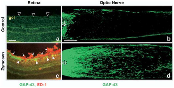
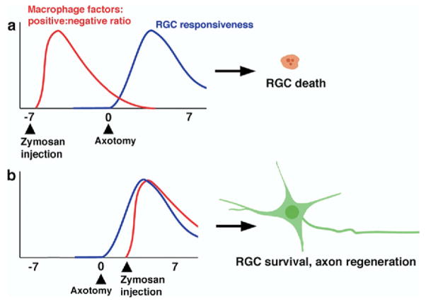
Following injury to the nervous system, the activation of macrophages, microglia, and T-cells profoundly affects the ability of neurons to survive and to regenerate damaged axons. The primary visual pathway provides a well-defined model system for investigating the interactions between the immune system and the nervous system after neural injury. Following damage to the optic nerve in mice and rats, retinal ganglion cells, the projection neurons of the eye, normally fail to regenerate their axons and soon begin to die. Induction of an inflammatory response in the vitreous strongly enhances the survival of retinal ganglion cells and enables these cells to regenerate lengthy axons beyond the injury site. T cells modulate this response, whereas microglia are thought to contribute to the loss of retinal ganglion cells in this model and in certain ocular diseases. This review discusses the complex and sometimes paradoxical actions of blood-borne macrophages, resident microglia, and T-cells in determining the outcome of injury in the primary visual pathway.
Figures
Similar articles
-
Neuroprotective and axon growth-promoting effects following inflammatory stimulation on mature retinal ganglion cells in mice depend on ciliary neurotrophic factor and leukemia inhibitory factor.J Neurosci. 2009 Nov 11;29(45):14334-41. doi: 10.1523/JNEUROSCI.2770-09.2009. J Neurosci. 2009. PMID: 19906980 Free PMC article.
-
Intravitreal macrophage activation enables cat retinal ganglion cells to regenerate injured axons into the mature optic nerve.Exp Neurol. 2005 Nov;196(1):153-63. doi: 10.1016/j.expneurol.2005.07.015. Epub 2005 Aug 19. Exp Neurol. 2005. PMID: 16112114
-
The complex role of neuroinflammation in glaucoma.Cold Spring Harb Perspect Med. 2014 Jul 3;4(8):a017269. doi: 10.1101/cshperspect.a017269. Cold Spring Harb Perspect Med. 2014. PMID: 24993677 Free PMC article. Review.
-
Multiple neuroprotective mechanisms of minocycline in autoimmune CNS inflammation.Neurobiol Dis. 2007 Mar;25(3):514-25. doi: 10.1016/j.nbd.2006.10.022. Epub 2007 Jan 18. Neurobiol Dis. 2007. PMID: 17239606
-
Neuroinflammation, Microglia and Implications for Retinal Ganglion Cell Survival and Axon Regeneration in Traumatic Optic Neuropathy.Front Immunol. 2022 Mar 4;13:860070. doi: 10.3389/fimmu.2022.860070. eCollection 2022. Front Immunol. 2022. PMID: 35309305 Free PMC article. Review.
Cited by
-
Neuroprotection and progenitor cell renewal in the injured adult murine retina requires healing monocyte-derived macrophages.J Exp Med. 2011 Jan 17;208(1):23-39. doi: 10.1084/jem.20101202. Epub 2011 Jan 10. J Exp Med. 2011. PMID: 21220455 Free PMC article.
-
Pathophysiology of the cochlear intrastrial fluid-blood barrier (review).Hear Res. 2016 Aug;338:52-63. doi: 10.1016/j.heares.2016.01.010. Epub 2016 Jan 20. Hear Res. 2016. PMID: 26802581 Free PMC article. Review.
-
Optic neuritis in neuromyelitis optica.Prog Retin Eye Res. 2013 Sep;36:159-71. doi: 10.1016/j.preteyeres.2013.03.001. Epub 2013 Mar 30. Prog Retin Eye Res. 2013. PMID: 23545439 Free PMC article.
-
Human Inner Ear Immune Activity: A Super-Resolution Immunohistochemistry Study.Front Neurol. 2019 Jul 10;10:728. doi: 10.3389/fneur.2019.00728. eCollection 2019. Front Neurol. 2019. PMID: 31354608 Free PMC article.
-
Peritoneal macrophages attenuate retinal ganglion cell survival and neurite outgrowth.Neural Regen Res. 2021 Jun;16(6):1121-1126. doi: 10.4103/1673-5374.300462. Neural Regen Res. 2021. PMID: 33269759 Free PMC article.
References
-
- Abromson-Leeman S, Hayashi M, Martin C, Sobel R, al-Sabbagh A, Weiner H, Dorf ME. T cell responses to myelin basic protein in experimental autoimmune encephalomyelitis-resistant BALB/c mice. J Neuroimmunol. 1993;45:89–101. - PubMed
-
- Aguayo AJ, Rasminsky M, Bray GM, Carbonetto S, McKerracher L, Villegas-Perez MP, Vidal-Sanz M, Carter DA. Degenerative and regenerative responses of injured neurons in the central nervous system of adult mammals. Philos Trans R Soc Lond B Biol Sci. 1991;331:337–343. - PubMed
-
- Allan SM, Rothwell NJ. Cytokines and acute neurodegeneration. Nat Rev Neurosci. 2001;2:734–744. - PubMed
-
- Andersson PB, Perry VH, Gordon S. The acute inflammatory response to lipopolysaccharide in CNS parenchyma differs from that in other body tissues. Neuroscience. 1992a;48:169–186. - PubMed
Publication types
MeSH terms
Grants and funding
LinkOut - more resources
Full Text Sources
