Combined thermotherapy and cryotherapy for efficient virus eradication: relation of virus distribution, subcellular changes, cell survival and viral RNA degradation in shoot tips
- PMID: 18705855
- PMCID: PMC6640318
- DOI: 10.1111/j.1364-3703.2007.00456.x
Combined thermotherapy and cryotherapy for efficient virus eradication: relation of virus distribution, subcellular changes, cell survival and viral RNA degradation in shoot tips
Abstract
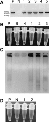
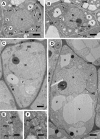
Accumulation of viruses in vegetatively propagated plants causes heavy yield losses. Therefore, supply of virus-free planting materials is pivotal to sustainable crop production. In previous studies, Raspberry bushy dwarf virus (RBDV) was difficult to eradicate from raspberry (Rubus idaeus) using the conventional means of meristem tip culture. As shown in the present study, it was probably because this pollen-transmitted virus efficiently invades leaf primordia and all meristematic tissues except the least differentiated cells of the apical dome. Subjecting plants to thermotherapy prior to meristem tip culture heavily reduced viral RNA2, RNA3 and the coat protein in the shoot tips, but no virus-free plants were obtained. Therefore, a novel method including thermotherapy followed by cryotherapy was developed for efficient virus eradication. Heat treatment caused subcellular alterations such as enlargement of vacuoles in the more developed, virus-infected cells, which were largely eliminated following subsequent cryotherapy. Using this protocol, 20-36% of the treated shoot tips survived, 30-40% regenerated and up to 35% of the regenerated plants were virus-free, as tested by ELISA and reverse transcription loop-mediated isothermal amplification. Novel cellular and molecular insights into RBDV-host interactions and the factors influencing virus eradication were obtained, including invasion of shoot tips and meristematic tissues by RBDV, enhanced viral RNA degradation and increased sensitivity to freezing caused by thermotherapy, and subcellular changes and subsequent death of cells caused by cryotherapy. This novel procedure should be helpful with many virus-host combinations in which virus eradication by conventional means has proven difficult.
Figures


Similar articles
-
Combining Thermotherapy with Cryotherapy for Efficient Eradication of Apple stem grooving virus from Infected In-vitro-cultured Apple Shoots.Plant Dis. 2018 Aug;102(8):1574-1580. doi: 10.1094/PDIS-11-17-1753-RE. Epub 2018 Jun 11. Plant Dis. 2018. PMID: 30673422
-
Improved recovery of cryotherapy-treated shoot tips following thermotherapy of in vitro-grown stock shoots of raspberry (Rubus idaeus L.).Cryo Letters. 2009 May-Jun;30(3):170-82. Cryo Letters. 2009. PMID: 19750241
-
Combining Thermotherapy with Shoot Tip Culture or Cryotherapy for Improved Virus Eradication from In Vitro Actinidia macrosperma.Plant Dis. 2024 Oct;108(10):3072-3077. doi: 10.1094/PDIS-03-24-0546-RE. Epub 2024 Sep 18. Plant Dis. 2024. PMID: 38853335
-
Cryopreservation of sweetpotato (Ipomoea batatas) and its pathogen eradication by cryotherapy.Biotechnol Adv. 2011 Jan-Feb;29(1):84-93. doi: 10.1016/j.biotechadv.2010.09.002. Epub 2010 Sep 16. Biotechnol Adv. 2011. PMID: 20851757 Review.
-
In vitro thermotherapy-based methods for plant virus eradication.Plant Methods. 2018 Oct 6;14:87. doi: 10.1186/s13007-018-0355-y. eCollection 2018. Plant Methods. 2018. PMID: 30323856 Free PMC article. Review.
Cited by
-
Characterization of virus-derived small interfering RNAs in Apple stem grooving virus-infected in vitro-cultured Pyrus pyrifolia shoot tips in response to high temperature treatment.Virol J. 2016 Oct 6;13(1):166. doi: 10.1186/s12985-016-0625-0. Virol J. 2016. PMID: 27716257 Free PMC article.
-
Thermotherapy Followed by Shoot Tip Cryotherapy Eradicates Latent Viruses and Apple Hammerhead Viroid from In Vitro Apple Rootstocks.Plants (Basel). 2022 Feb 22;11(5):582. doi: 10.3390/plants11050582. Plants (Basel). 2022. PMID: 35270052 Free PMC article.
-
Infection cycle of Artichoke Italian latent virus in tobacco plants: meristem invasion and recovery from disease symptoms.PLoS One. 2014 Jun 9;9(6):e99446. doi: 10.1371/journal.pone.0099446. eCollection 2014. PLoS One. 2014. PMID: 24911029 Free PMC article.
-
Identification and characterization of microRNAs from in vitro-grown pear shoots infected with Apple stem grooving virus in response to high temperature using small RNA sequencing.BMC Genomics. 2015 Nov 16;16:945. doi: 10.1186/s12864-015-2126-8. BMC Genomics. 2015. PMID: 26573813 Free PMC article.
-
Pathogenic seedborne viruses are rare but Phaseolus vulgaris endornaviruses are common in bean varieties grown in Nicaragua and Tanzania.PLoS One. 2017 May 25;12(5):e0178242. doi: 10.1371/journal.pone.0178242. eCollection 2017. PLoS One. 2017. PMID: 28542624 Free PMC article.
References
-
- Agrios, G.N. (2005) Plant Pathology, 5th edn. Burlington: Elsevier Academic Press.
-
- Amari, K. , Burgos, L. , Pallas, V. and Sanchez‐Pina, M.A. (2007) Prunus necrotic ringspot virus early invasion and its effects on apricot pollen grain performance. Phytopathology, 97, 892–899. - PubMed
-
- Appiano, A. and Pennazio, S. (1972) Electron microscopy of potato meristem tips infected with potato virus X. J. Gen. Virol. 14, 273–276. - PubMed
-
- Barbara, D.J. , Morton, A. , Ramcharan, S. , Cole, I.W. , Phillips, A. and Knight, V.H. (2001) Occurrence and distribution of Raspberry bushy dwarf virus in commercial Rubus plantations in England and Wales. Plant Pathol. 50, 747–754.
-
- Barnett, O.W. , Gibson, P.B. and Seo, A. (1975) A comparison of heat treatment, cold treatment and meristem tip‐culture for obtaining virus‐free plants of Trifolium repens . Plant Dis. Rep. 59, 834–837.
Publication types
MeSH terms
Substances
LinkOut - more resources
Full Text Sources
Research Materials

