. 2008 Oct 24;283(43):28944-57.
doi: 10.1074/jbc.M800685200. Epub 2008 Aug 12.

The aryl hydrocarbon receptor attenuates tobacco smoke-induced cyclooxygenase-2 and prostaglandin production in lung fibroblasts through regulation of the NF-kappaB family member RelB

Affiliations

The aryl hydrocarbon receptor attenuates tobacco smoke-induced cyclooxygenase-2 and prostaglandin production in lung fibroblasts through regulation of the NF-kappaB family member RelB

Carolyn J Baglole et al. J Biol Chem. .

Abstract

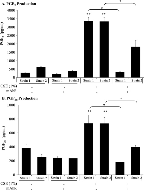
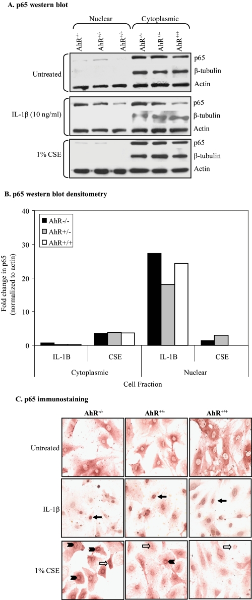
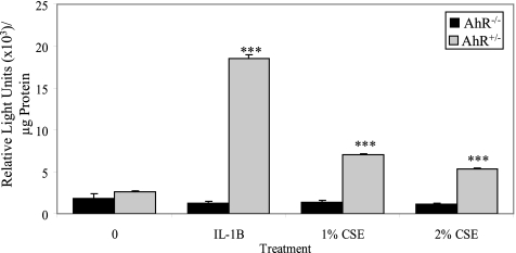
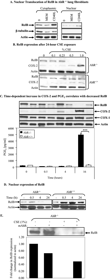
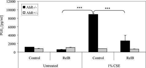
Diseases such as chronic obstructive pulmonary disease and lung cancer caused by cigarette smoke affect millions of people worldwide. The aryl hydrocarbon receptor (AhR) is a ligand-activated transcription factor that influences responses to certain environmental pollutants such as tobacco smoke. However, the physiological function(s) of the AhR is unknown. Herein we propose that the physiologic role of the AhR is to limit inflammation. We show that lung fibroblasts from AhR(-/-) mice produce a heightened inflammatory response to cigarette smoke, typified by increased levels of cyclooxygenase-2 (COX-2) and prostaglandins (PGs), when compared with wild type (AhR(+/+)) fibroblasts. This response was dependent on AhR expression as transient transfection of an AhR expression plasmid into AhR(-/-) fibroblasts significantly attenuated the smoke-induced COX-2 and PG production, confirming the anti-inflammatory role of the AhR. The AhR can interact with NF-kappaB. However, the heightened inflammatory response observed in AhR(-/-) fibroblasts was not the result of NF-kappaB (p50/p65) activation. Instead it was coupled with a loss of the NF-kappaB family member RelB in AhR(-/-) fibroblasts. Taken together, these studies provide compelling evidence that AhR expression limits proinflammatory COX-2 and PG production by maintaining RelB expression. The association between RelB and AhR may represent a new therapeutic and more selective target with which to combat inflammation-associated diseases.


Figures

FIGURE 1.
FIGURE 1.
Characterization of AhR expression and function in mouse lung fibroblasts. Whole cell lysates of fibroblasts generated from AhR+/+, AhR+/–, and AhR–/– mice were left untreated (control medium) or were treated with AhR ligands for 18 h and subjected to Western blot analysis for AhR or CYP1B1 protein expression. A, fibroblasts from both AhR+/+ and AhR+/– mice expressed AhR protein, whereas fibroblasts from two different AhR–/– mice (Strain 1 and Strain 2) lacked AhR expression. AhR expression levels were compared against Hepa cells. B, whole cell lysates of AhR+/– fibroblasts were treated with vehicle (DMSO), dioxin (1 nm), or ITE (1 μm), and AhR expression was analyzed by Western blot. Both AhR ligands resulted in loss of AhR protein expression. C, dioxin increases CYP1B1 expression in AhR-expressing lung fibroblasts (AhR+/+ and AhR+/–) but not AhR–/– fibroblasts.
FIGURE 2.
FIGURE 2.
CSE exposure induces significantly more prostaglandin production in AhR–/– mouse lung fibroblasts compared with AhR+/+ fibroblasts. Fibroblasts derived from AhR+/+, AhR+/–, and AhR–/– mice were treated with medium alone or with CSE (0.1–2%) for 24 h, and PGE2 and PGF levels were assessed by enzyme immunoassay as described under “Experimental Procedures.” Exposure to CSE significantly increased PGE2 (A) and PGF (B) production in AhR–/– lung fibroblast strains at concentrations as low as 0.25% (PGE2) and 0.5% (PGF). PG production was increased in AhR+/– at concentrations of 1.5% or greater, and PG production was not significantly increased in response to CSE in AhR+/+ mouse lung fibroblasts. ***, p < 0.0001, indicates statistical significance between cells treated with CSE relative to cells treated with medium alone. Results are presented as pg/ml ± S.E.
FIGURE 3.
FIGURE 3.
CSE induces significantly more COX-2, but not COX-1, protein expression in AhR–/– mouse lung fibroblasts compared with AhR+/+ lung fibroblasts. Equivalent amounts of whole cell extracts were prepared from mouse lung fibroblasts that were treated with CSE, and COX protein expression was examined by Western blot analysis. A, representative COX-1 Western blots. COX-1 is constitutively expressed in mouse lung fibroblasts, and its expression is not altered as a consequence of CSE exposure. There was no difference in COX-1 expression between AhR+/+ and AhR–/– lung fibroblasts. B, densitometric analysis of COX-1 protein expression. There was a slight (not significant) increase in COX-1 expression following treatment with 1% CSE. The COX-1 level of untreated AhR-expressing fibroblasts was arbitrarily set at 1. C, representative COX-2 Western blots of CSE-treated mouse lung fibroblasts. CSE induced COX-2 expression in all lung fibroblasts examined. CSE-induced COX-2 expression was higher in AhR–/– mouse lung fibroblasts compared with either AhR+/+ or AhR+/– fibroblasts. D, densitometric analysis of COX-2 expression (normalized to actin) revealed that exposure to 1% CSE for 24 h significantly induced COX-2 expression in AhR–/– mouse lung fibroblasts when compared with both untreated AhR-deficient fibroblasts and CSE-treated AhR+/+ mouse lung fibroblasts. Statistical analysis was performed on individual Western blots from three to four separate experiments involving fibroblasts derived from two AhR–/– and two AhR+/+ mice. NS, no statistical significance. Results are expressed as mean ± S.E.
FIGURE 4.
FIGURE 4.
Components of cigarette smoke increase COX-2 protein expression more robustly in AhR–/– fibroblasts compared with AhR+/+ fibroblasts. Fibroblasts were treated with B[a]P (0.1, 1, and 5 μm), HQ (0.1, 1, and 10 μm), and nicotine (0.1, 1, and 10 μm) for 24 h, and cell lysates were analyzed for COX-2 and CYP1B1 expression by Western blot. Actin was used as a loading control. A, B[a]P increased CYP1B1 expression in AhR+/+ lung fibroblasts but not in AhR-deficient fibroblasts. COX-2 was slightly increased in AhR-deficient fibroblasts at the highest concentration of B[a]P used (5 μm). B, lower concentrations of HQ increased COX-2 expression in AhR-deficient cells compared with AhR+/+ fibroblasts. Levels of CYP1B1 were not dramatically altered. C, nicotine potently induced COX-2 expression only in AhR–/– fibroblasts. Changes in CYP1B1 were marginal.
FIGURE 5.
FIGURE 5.
Reconstitution of the AhR into AhR–/– lung fibroblasts increases AhR expression and reduces CSE-induced COX-2 expression. AhR–/– mouse lung fibroblasts were transfected with a plasmid encoding AhR (mAhR) using nucleofection. Once confluent, cells were cultured in serum-free MEM and treated with 1% CSE for 24 h. AhR and COX-2 expression was assessed by Western blot analysis. A, nucleofection of the mAhR plasmid dose-dependently increased AhR expression in AhR–/– mouse lung fibroblasts as determined by Western blot. B, CSE-induced COX-2 expression was attenuated in AhR–/– lung fibroblasts nucleofected with 2 μg of mAhR. COX-2 expression is reduced to levels comparable to that in untreated cells or those that were untreated and nucleofected with mAhR. Actin was used as a loading control.
FIGURE 6.
FIGURE 6.
Reconstitution of the AhR into AhR–/– lung fibroblasts reduces CSE-induced PG production. Two strains of AhR–/– mouse lung fibroblasts were transfected with a plasmid encoding mAhR using nucleofection as described above. Once confluent, cells were cultured in serum-free MEM and treated with 1% CSE for 24 h, and PG production was determined by PG-specific enzyme immunoassay. Transfection with mAhR significantly reduced PGE2 (A) and PGF (B) levels in lung fibroblast generated from two different knock-out animals (Strain 1 and Strain 2). **, p < 0.05, indicates statistical significance between untreated/2 μg of mAhR and cells treated with 1% CSE. *, p < 0.05, indicates statistical significance between fibroblasts treated with 1% CSE and transfected cells treated with 1% CSE. Results are expressed as mean ± S.E.
FIGURE 7.
FIGURE 7.
AhR–/–, AhR+/–, and AhR+/+ lung fibroblasts show similar levels of canonical NF-κB p65 nuclear translocation after IL-1β and CSE exposure. A, cytoplasmic and nuclear protein fractions were generated from AhR–/–, AhR+/–, and AhR+/+ lung fibroblasts that were treated with IL-1β or 1% CSE for 30 min. IL-1β induced nuclear translocation of p65 as evidenced by the increase in p65 protein in the nuclear fraction. The levels of p65 in the nuclear fractions following treatment with 1% CSE were much lower and barely above levels in cells that were treated with control medium (Untreated). β-Tubulin was used to confirm the purity of the cytoplasmic and nuclear fractions, and actin was used as a loading control. B, densitometric analysis of cytoplasmic and nuclear p65 protein levels following treatment with IL-1β or 1% CSE. p65 nuclear levels increased in AhR–/–, AhR+/–, and AhR+/+ lung fibroblasts in response to IL-1β. There was minimal change in nuclear p65 levels in response to CSE (0–1.3-fold). C, immunocytochemical staining of p65. Fibroblasts cultured on glass chamber slides were treated with IL-1β (10 ng/ml) or 1% CSE for 30 min, and p65 translocation was detected using an anti-p65 antibody. IL-1β induced strong nuclear translocation of p65 (closed arrows) in both AhR-expressing and AhR–/– fibroblasts (compare with A). 1% CSE weakly induced p65 translocation to the nucleus in a few cells (open arrows). Intense perinuclear staining was also evident in the AhR-null fibroblasts treated with CSE (arrowheads); this perinuclear staining was observed in a few AhR+/– fibroblasts.
FIGURE 8.
FIGURE 8.
CSE does not induce NF-κB-dependent gene expression in AhR–/– fibroblasts. AhR+/– and AhR–/– fibroblasts were transfected with 4 μg of an NF-κB-luciferase (p50/p65) reporter gene using nucleofection. Once confluent, cells were cultured in serum-free medium and treated with 1 or 2% CSE or IL-1β (10 ng/ml) for 6 h, and cell lysates were assayed for luciferase. Treatment with IL-1β resulted in a significant induction of luciferase in AhR+/– fibroblasts but not in AhR–/– fibroblasts. When cells were treated with CSE, luciferase activity was not induced in AhR–/– lung fibroblasts but was induced in AhR+/– fibroblasts. Results are expressed as the mean ± S.E.; ***, p < 0.0001 compared with untreated. Transfection efficiency was not significantly different between AhR-expressing and AhR–/– fibroblasts (supplemental Fig. 4).
FIGURE 9.
FIGURE 9.
CSE induces loss of RelB in AhR–/–, but not AhR-expressing, lung fibroblasts. A, CD40L induces RelB nuclear translocation in AhR–/– fibroblasts. AhR–/– fibroblasts were untreated or treated with CD40L or negative control (empty insect membrane; MEM) for 24 h, and cytoplasmic and nuclear proteins were prepared as described under “Experimental Procedures.” RelB expression was analyzed by Western blot. RelB is constitutively expressed in mouse lung fibroblasts. Treatment with CD40L increased both RelB expression and nuclear accumulation. β-Tubulin was used to confirm the purity of the cytoplasmic versus nuclear extracts. Total actin was used as a loading control. B, enhanced COX-2 expression in response to CSE in AhR-deficient fibroblasts correlates with loss of RelB protein expression. AhR–/– and AhR+/+ fibroblasts were treated with increasing concentrations of CSE for 24 h, and RelB and COX-2 protein expression was assessed by Western blot. Upon exposure to CSE, expression of RelB was lost from AhR–/– fibroblasts but not from those that express the AhR (AhR+/+). This loss of RelB correlated with strong COX-2 expression. C, cytoplasmic and nuclear extracts were prepared from AhR+/+ and AhR–/– fibroblasts treated with 1% CSE for varying times, and Western blot analysis was performed for RelB, COX-1, COX-2, and total actin; analysis of PGE2 production was assessed from cell culture supernatants. There was a time-dependent increase in COX-2 expression and PGE2 production in AhR–/– fibroblasts but not AhR+/+ fibroblasts. This increase in PGE2 production in AhR–/– fibroblasts was significantly greater than untreated controls (*) as well as CSE-treated AhR-expressing cells (***). Results are expressed as mean ± S.E. The increase in COX-2 paralleled a decrease in cytoplasmic RelB only in the AhR–/– fibroblasts. COX-1 expression is unchanged with CSE exposure. D, CSE induced nuclear translocation of RelB within 30 min of treatment. In AhR–/– fibroblasts, nuclear expression of RelB is lost when cells are exposed to CSE. Nuclear RelB expression persisted in AhR-expressing fibroblasts. All samples were run on the same gel. E, CSE-induced loss of RelB is AhR-dependent. Transient transfection of AhR–/– fibroblasts with mAhR reduced the CSE-induced loss of RelB expression. Densitometric analysis of RelB revealed that re-expression of the AhR in AhR–/– fibroblasts restored RelB following CSE treatment to levels that were ≈50% of control (medium only, non-transfected). Values are normalized to actin.
FIGURE 10.
FIGURE 10.
Differential regulation of COX-2 and RelB mRNA in CSE-treated AhR–/–versus AhR+/+ lung fibroblasts. Fibroblasts from AhR–/– and AhR+/+ mice were exposed to medium only or were treated with 1% CSE for 3, 6, or 24 h, and RNA expression was analyzed by PCR as described under “Experimental Procedures.” A, reverse transcription (RT)-PCR. The induction of COX-2 and RelB mRNA was more intense in AhR+/+ fibroblasts treated with CSE compared with AhR-deficient fibroblasts. COX-1 mRNA levels were not dramatically different. B, real time PCR-COX-2. There was a significant increase in COX-2 mRNA in AhR-expressing fibroblasts treated with 1% CSE for 3 h (20.2 ± 10.1) (**, p < 0.001); these COX-2 mRNA levels were significantly higher when compared with AhR-deficient fibroblasts (3.4 ± 1.3) (*, p < 0.05). Note that basal COX-2 mRNA levels were slightly higher in AhR–/– fibroblasts when compared with AhR-expressing fibroblasts (≈4-fold). C, real time PCR-RelB. RelB mRNA levels were significantly higher in AhR+/+ fibroblasts exposed to 1% CSE for 3 h (1.6 ± 0.7) (*, p < 0.05). All values were normalized to glyceraldehyde-3-phosphate dehydrogenase, and the results are expressed as mean ± S.E. of three independent experiments.
FIGURE 11.
FIGURE 11.
CSE-induced PGE2 in AhR-deficient fibroblasts is dependent on loss of RelB. AhR-expressing (AhR+/–) and AhR-deficient fibroblasts were transfected with an empty plasmid (Control) or a plasmid that expresses mouse RelB using nucleofection as described above. PGE2 levels were measured in cell culture supernatants following treatment with medium alone (Untreated) or treated with 1% CSE for 24 h. PGE2 was significantly increased in AhR–/– cells (black bars) treated with 1% CSE (***, p < 0.0001) compared with untreated cells transfected with the control plasmid (Control). This CSE-induced PGE2 was significantly attenuated when AhR–/– fibroblasts were transfected with a plasmid encoding mouse RelB. CSE did not induce PGE2 in AhR+/– fibroblasts (gray bars). Results are based on triplicates of two independent experiments and are expressed as the mean ± S.E.

Similar articles

Cited by

References

    1. Hida, T., Yatabe, Y., Achiwa, H., Muramatsu, H., Kozaki, K., Nakamura, S., Ogawa, M., Mitsudomi, T., Sugiura, T., and Takahashi, T. (1998) Cancer Res. 58 3761–3764 - PubMed
    1. Xaubet, A., Roca-Ferrer, J., Pujols, L., Ramirez, J., Mullol, J., Marin-Arguedas, A., Torrego, A., Gimferrer, J. M., and Picado, C. (2004) Sarcoidosis Vasc. Diffuse Lung Dis. 21 35–42 - PubMed
    1. Kamiyama, M., Pozzi, A., Yang, L., DeBusk, L. M., Breyer, R. M., and Lin, P. C. (2006) Oncogene 25 7019–7028 - PubMed
    1. Subbaramaiah, K., and Dannenberg, A. J. (2003) Trends Pharmacol. Sci. 24 96–102 - PubMed
    1. Smith, W. L., DeWitt, D. L., and Garavito, R. M. (2000) Annu. Rev. Biochem. 69 145–182 - PubMed

Publication types