. 2008 Aug 15;29(2):306-17.
doi: 10.1016/j.immuni.2008.05.019.

Gr1(+) inflammatory monocytes are required for mucosal resistance to the pathogen Toxoplasma gondii

Affiliations

Gr1(+) inflammatory monocytes are required for mucosal resistance to the pathogen Toxoplasma gondii

Ildiko R Dunay et al. Immunity. .

Erratum in

  • Immunity. 2008 Oct;29(4):660

Abstract

The enteric pathogen Toxoplasma gondii is controlled by a vigorous innate T helper 1 (Th1) cell response in the murine model. We demonstrated that after oral infection, the parasite rapidly recruited inflammatory monocytes [Gr1(+) (Ly6C(+), Ly6G(-)) F4/80(+)CD11b(+)CD11c(-)], which established a vital defensive perimeter within the villi of the ileum in the small intestine. Mice deficient of the chemokine receptor CCR2 or the ligand CCL2 failed to recruit Gr1(+) inflammatory monocytes, whereas dendritic cells and resident tissue macrophages remained unaltered. The selective lack of Gr1(+) inflammatory monocytes resulted in an inability of mice to control replication of the parasite, high influx of neutrophils, extensive intestinal necrosis, and rapid death. Adoptive transfer of sorted Gr1(+) inflammatory monocytes demonstrated their ability to home to the ileum in infected animals and protect Ccr2(-/-) mice, which were otherwise highly susceptible to oral toxoplasmosis. Collectively, these findings illustrate the critical importance of inflammatory monocytes as a first line of defense in controlling intestinal pathogens.


Figures

Figure 1
Figure 1. Survival of mice following oral challenge with T. gondii depends on MCP-1 and CCR2
(A) Survival of mice after peroral infection with T. gondii. Wild type C57BL/6 mice largely survived challenge while the majority of CCR2−/− and MCP-1−/− mice succumbed by day 15. Combined results of four experiments (3–6 animals per group, n=19). (B) Tissue burdens in the ileum of infected mice. Oral challenge of CCR2−/− and MCP-1−/− mice resulted in higher numbers of T. gondii parasites compared to wild type mice. Parasites were enumerated by counting the mean number of parasites per mm2 in the ileum. Values are means ± s.d. from a representative experiment, n = 3 animals each. * P < 0.05. (A) Cytokine production in T. gondii infected mice. Cytokines were detected in serum from C57BL/6, CCR2−/− and MCP-1−/− at intervals after peroral infection. No survivors were available from CCR2−/− at day 11 (†). Values are means ± s.d. from a representative experiment, n = 3 animals each. * P ≤ 0.05.
Figure 2
Figure 2. Absence of CCR2 or MCP-1 leads to increased parasite numbers, neutrophil recruitment and intestinal necrosis following T. gondii infection
(A) Oral infection with T. gondii induced mild intestinal inflammation in wild type mice but extensive pathology in CCR2−/− and MCP-1−/− mice. Orally infected mice were examined at day 9 post-infection by H&E staining of paraffin sections. Normal ileum morphology was seen in noninfected C57BL/6. Infected C57BL/6 had slightly swollen villi and increased cells in the lamina propria. Lesions (asterisk) observed in the ileum of infected CCR2−/− and infected MCP-1−/− mice were characterized by disruption of the villus architecture, destruction of the inner circular layer of the tunica muscularis (ICM), and extensive replication of parasites (arrow and insets). Scale = 200 µm (15 µm in insets). Right panels are enlarged views. (B) High numbers of neutrophils colocalized with T. gondii in the lesion areas of CCR2−/− and MCP-1−/− mice. Orally infected mice were examined at 9 days post-infection by staining with polyclonal anti-T. gondii (green) and anti-neutrophil (Ly6G, mAb 1A8) followed by Alexa-conjugated secondary antibodies. Scale bar = 20 µm.
Figure 3
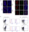
Figure 3. Recruitment of Gr1+ (Ly6C+) monocytes to the lamina propria in the ileum following T. gondii infection depends on CCR2 and MCP-1
(A) Recruitment of Gr1+ cells in the ileum of orally infected C57BL/6 mice. Gr1+ cells increased within the lamina propria and were found beneath the basement membrane, surrounding the villus core within the ileum. Gr1+ cells were also seen in noninfected wild type (C57BL/6) but not in noninfected (data not shown) or infected CCR2−/− and MCP-1−/− mice. Frozen sections of orally infected mice were examined 9 days post-infection with anti-Gr1 (RB6-8C5) followed by Alexa-594 conjugated goat anti-rat IgG. Scale bar = 25 µm. (B) Characterization of Gr1+ cells in the lamina propria of orally infected mice. Gr1+ cells stained with F4/80 but not CD11c or the neutrophil marker 1A8. Frozen sections of ileum from orally infected mice were examined after 9 days post-infection. Top panel: anti-Gr1 (RB6-8C5) directly conjugated to Alexa 488 and anti-F4/80 directly conjugated to Alexa 594; middle panel: anti-Gr1 (RB6-8C5) and anti-CD11c (N418) followed by Alexa-conjugated secondary antibodies; Bottom panel: anti-Ly6G (1A8) and anti-CD11c (N418), followed by Alexa-conjugated secondary antibodies Scale bar =25 µm. (C) Oral infection with T. gondii induced recruitment of Gr1+ (Ly6C+) monocytes to the ileum. Leukocytes were isolated from the ileum of mice on day 9 after oral infection, stained for CD11b (mAb M1/70 conjugated to PE or FITC) and F4/80 (mAb A31 conjugated to APC), and quantified by FACS. Double positive macrophages (CD11b+ F4/80+) were gated based on isotype controls and circled populations were analyzed after staining with anti-Ly6C (mAb AL-21 conjugated to FITC) or Ly6G (mAb 1A8 conjugated to PE). Resident macrophages in non-infected mice (top) were negative for both Ly6C and Ly6G. In contrast, doubly positive cells from infected mice are strongly positive for Ly6C, but did not stain for Ly6G (bottom). Results shown are representative of three or more experiments; cells were pooled from three mice in each group. (D) Analysis of peritoneal cells in noninfected (top) and infected (bottom) CCR2−/− mice. While neutrophils (CD11b+, F4/80) increased dramatically in infected mice, the population of doubly positive CD11b+ F4/80+ cells was absent.
Figure 4
Figure 4. Gr1+ (Ly6C+) inflammatory monocytes in the lamina propria express iNOS, TNFα, and IL-12 but not dendritic cell markers
(A) Induction of iNOS in the ileum of infected mice. iNOS expression was low in noninfected ileum of C57BL/6 mice, but was induced following infection in lamina propria cells. In contrast, in CCR2−/− and MCP-1−/− mice, iNOS expression was only observed at the apical region of enterocytes. Orally infected mice were examined at day 9 post-infection by staining frozen sections with rabbit anti-iNOS followed by Alex 594-conjugated secondary antibodies (red). Bar = 25 µm.\ (B) Intracellular IL-12 and TNFα expression was detected within some cell in the villus region of the Ileum in infected C57BL/6 mice. Staining for these intracellular markers was not detected in noninfected mice. Frozen sections were stained with rat anti- IL-12 (BD Pharmingen) or rat anti-TNFα (BD Pharmingen) followed by detection using Alexa-594-conjugated goat anti-rat IgG. Bar = 25 µm. (C) Leukocytes isolated from the lamina propria at day 9 after oral infection were stained with Ly6C (mAb AL-21 conjugated to FITC) and F4/80 (mAb A3-1 conjugated to APC), gated based on isotype controls, and analyzed for expression of other surface or intracellular markers (with antibodies conjugated to PE, see methods). F4/80+ Ly6C+ monocytes did not express CD11c as shown by absence of staining with mAb to p150/90. In contrast, F4/80+ Ly6C+ monocytes stained positively for iNOS, TNFα, and IL-12. Intracellular staining was performed after incubation in brefeldin A for 4 hrs. In each case, the positive cells are plotted as the blue line vs. isotype control in the red line. Representative of three similar experiments.
Figure 5
Figure 5. Adoptive transfer of Gr1+ (Ly6C+) inflammatory monocytes protects against death in CCR2−/− mice
(A) High numbers of Gr1+ F4/80+ monocytes were recruited to the peritoneal cavity of mice following low dose T. gondii infection. Cell populations were characterized by flow cytometry using anti-Gr1 (mAb RB6-8C5 conjugated to Cy5) and anti-F4/80 (mAb A3-1 conjugated to PE). Double positive cells (Gr1+ F4/80+) were sorted for adoptive transfer (upper circle, right plot) Single positive F4/80+ cells were used for control transfers (lower circle, left plot). Results are representative of four experiments performed. (B) Characterization of peritoneal monocytes elicited in T. gondii infected C57BL6 mice by staining for CD11b (mAb M1/70 conjugated to PE or FITC) and F4/80 (mAb A3-1 conjugated to APC). Doubly positive cells were gated based on isotype controls and the circled population of cells analyzed for expression of Ly6C (mAb AL-21 conjugated to FITC) and Ly6G (mAb 1A8 conjugated to PE). Inflammatory monocytes (CD11b, F4/80) strongly expressed Ly6C but did not stain for Ly6G. Representative results from three or more similar experiments. In each case, the positive cells are plotted as the blue line vs. isotype control in the red line. (C) Adoptive transfer of Gr1+ F4/80+ inflammatory monocytes protect infected CCR2−/− mice from death. Three days after oral infection of mice with T. gondii, mice received sorted cells (see A) by i.v. injection. All infected CCR2−/− mice died on day 14 after infection, while mice that received the F4/80+ single positive cell population (negative control) died on day 18 after infection. Approximately half of C57BL/6 wild type mice and the CCR2−/− mice that received Gr1+ F4/80+cells survived the infection. Results are mean values of three separate experiments with three to four mice per group. (D) Adoptive transfer of inflammatory monocytes into noninfected or infected C57BL/6 mice. Monocyte populations were isolated from the peritoneal cavity of T. gondii infected mice and separated by FACS after staining for Gr1 (mAb RB6-8C5 conjugated to Cy5) and F4/80 (mAb A3-1 conjugated to PE) (similar to A). Cell populations were labeled with CFSE in vitro and administered by i.v. inoculation (106 cells per animals) into wild type mice that were either noninfected, or animals that were infected 3 days previously by oral challenge. Inflammatory monocytes (Gr1+ Ly6C+ F4/80+) were recruited to the ileum in infected C57BL/6 mice (black bars) but not in noninfected mice (white bar). Gr1 Ly6C F4/80+ macrophages were also recruited to the intestine of infected mice, albeit at much lower levels. CFSE+ cells were counted by examining frozen sections by fluorescence microscopy. Values represent the average number of cells per 20 microscope fields (40X) and were obtained from 2 separate experiments with 2 animals each group. Mean ± S.D.
Figure 6
Figure 6. Adoptively transferred Gr1+ (Ly6C+) monocytes home to the intestine and prevent extensive pathology in CCR2−/− mice
(A) Sorted cells were adoptively transferred to infected CCR2−/− mice on day 3 post-infection and the ilea were examined at day 9 by H&E staining of paraffin sections. Typical lesions (asterisk) were observed in the ileum of infected CCR2−/− mice that did not receive cells (Control) or received F4/80+ singly positive cells. CCR2−/− mice that received Gr1+ (Ly6C+) F4/80+ cells show well-preserved architecture in the ileum, similar to wild type mice. Representative of two similar experiments containing 2 animals per group. Scale bar = 200 µm (15 µm inset). Right panels are enlarged views. (B) Adoptively transferred Gr1+ (Ly6C+) monocytes home to the ileum of infected mice. Sorted, CFSE labeled Gr1+ (Ly6C+) F4/80+ monocytes i.v. inoculated into C57BL/6 mice on day 3 after oral infection. One day following adoptive transfer (day 4 after oral infection), ilea were collected and frozen sections were examined by fluorescent microscopy. Recruitment of CFSE labeled Gr1+ (Ly6C+) monocytes was observed in the lamina propria region in the villi and in the crypt region, which contained lymphocytic aggregations. Control represents C57BL/6 mouse that was infected but did not receive CFSE labeled cells. Scale bar = 20 µm.
Figure 7
Figure 7. Recruitment from the bone marrow and trafficking of inflammatory monocytes in wild type and CCR2−/− mice
(A) Characterization of bone marrow cells stained with Ly6C (mAb AL-21 conjugated to FITC) and CD11b (mAb M1/70 conjugated to Cy5). In wild type mice (top), infection with T. gondii increased the number of doubly positive cells by 2 fold (boxed numbers represent the gates and are given in %). The number of doubly positive cells was elevated in the absence of infection and increased by ~2 fold in CCR2−/− mice following infection with T. gondii (lower). Representative of five similar experiments. (B) Trafficking of Ly6C+ CD11b+ inflammatory monocytes following i.v. transfer into CCR2−/− mice. Adoptively transferred inflammatory monocytes from the bone marrow of wild type (C57BL/6) or CCR2−/− mice were able to home with comparable efficiency to the small intestine of CCR2−/− recipient mice that were infected with T. gondii. Bone marrow cells were stained as in A, sorted by FACS, and labeled in vitro using CFSE. Approximately 106 labeled cells were injected i.v. into mice that had been infected 3 days previously by oral inoculation with T. gondii. CFSE+ cells were counted by examining frozen sections by fluorescence microscopy. Values represent the average number of cells per 20 microscope fields (40X) and were obtained from 2 separate experiments with 2 animals each group. Mean ± S.D. (C) Representative images of CFSE+ Ly6C+ CD11b+ monocytes in frozen sections of the small intestines of mice that had received donor cells from wild type (CCR2+/+) vs. CCR2 deficient (CCR2−/−) animals. Bar = 25 microns.

Similar articles

Cited by

References

    1. Aliberti J, Sousa CR, Schito M, Hieny S, Wells T, Huffnagle GB, Sher A. CCR5 provides a signal for microbial induced production of IL-12 by CD8-α+ dendritic cells. Nat. Immunol. 2000;1:83–87. - PubMed
    1. Auffray C, Fogg D, Garfa M, Elain G, Join-Lambert O, Kayal S, Sarnacki S, Cumano A, Lauvau G, Geissmann F. Monitoring of blood vessels and tissues by a population of monocytes with patrolling behavior. Science. 2007;317:666–670. - PubMed
    1. Barragan A, Sibley LD. Migration of Toxoplasma gondii across biological barriers. Trends Microbiol. 2003;11:426–430. - PubMed
    1. Bliss SK, Gavrilescu LC, Alcaraz A, Denkers EY. Neutrophil depletion during Toxoplasma gondii infection leads to impaired immunity and lethal systemic pathology. Infect. Immun. 2001;69:4898–4905. - PMC - PubMed
    1. Bliss SK, Marshall AJ, Zhang Y, Denkers EY. Human polymorphonuclear leukocytes produce IL-12, TNF-α, and the chemokines macrophage-inflammatory protein-1α and -1β in response to Toxoplasma gondii antigens. J. Immunol. 1999;162:7369–7375. - PubMed

Publication types

MeSH terms