. 2008 Oct;7(10):1685-98.
doi: 10.1128/EC.00146-08. Epub 2008 Aug 8.

PKC1 is essential for protection against both oxidative and nitrosative stresses, cell integrity, and normal manifestation of virulence factors in the pathogenic fungus Cryptococcus neoformans

Affiliations

PKC1 is essential for protection against both oxidative and nitrosative stresses, cell integrity, and normal manifestation of virulence factors in the pathogenic fungus Cryptococcus neoformans

Kimberly J Gerik et al. Eukaryot Cell. 2008 Oct.

Abstract

Cell wall integrity is crucial for fungal growth, survival, and pathogenesis. Responses to environmental stresses are mediated by the highly conserved Pkc1 protein and its downstream components. In this study, we demonstrate that both oxidative and nitrosative stresses activate the PKC1 cell integrity pathway in wild-type cells, as measured by phosphorylation of Mpk1, the terminal protein in the PKC1 phosphorylation cascade. Furthermore, deletion of PKC1 shows that this gene is essential for defense against both oxidative and nitrosative stresses; however, other genes involved directly in the PKC1 pathway are dispensable for protection against these stresses. This suggests that Pkc1 may have multiple and alternative functions other than activating the mitogen-activated protein kinase cascade from a "top-down" approach. Deletion of PKC1 also causes osmotic instability, temperature sensitivity, severe sensitivity to cell wall-inhibiting agents, and alterations in capsule and melanin. Furthermore, the vital cell wall components chitin and its deacetylated form chitosan appear to be mislocalized in a pkc1Delta strain, although this mutant contains wild-type levels of both of these polymers. These data indicate that loss of Pkc1 has pleiotropic effects because it is central to many functions either dependent on or independent of PKC1 pathway activation. Notably, this is the first time that Pkc1 has been implicated in protection against nitrosative stress in any organism.


Figures
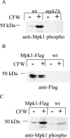
FIG. 1.
FIG. 1.
FLAG-tagged Mpk1 allows for the detection of total Mpk1, the terminal kinase in the PKC1 pathway, independent of phosphorylation. (A) Western blot analysis was done using the anti-Mpk1 phospho-antibody to probe a blot containing total protein from wild-type or mpk1Δ lysates, which were made from cells that were either uninduced or induced with calcofluor white (CFW). The amount of the 50-kDa band increased in the wild-type strain in response to CFW, and the absence of the band in the mpk1Δ lysate indicated that the band in the wild-type lysate was Mpk1. (B) The gene construct encoding the Mpk1 protein fused to the Flag epitope at the carboxy terminus replaced the wild-type MPK1 locus in KN99α. The panel shows Western blot analysis using an antibody against the Flag tag and lysates from the Mpk1-Flag tagged strain and the wild-type strain either induced or uninduced with CFW. Bands of the appropriate size were detected in the Mpk1-Flag strain but not the wild-type strain, suggesting that the protein detected was the Mpk1-Flag fusion. (C) The same blot from panel B probed with an antibody specific for the phosphorylated form of Mpk1. Phosphorylation of both the fusion protein and the wild-type protein was induced by CFW.
FIG. 2.
FIG. 2.
Mpk1 is induced by both oxidative and nitrosative stresses. The Mpk1-Flag tagged strain (KN99α background) was grown overnight in YNB (pH 4.0) at 25°C and diluted the next morning to an OD650 of 0.2. The cells were grown for an additional 2.5 h and then treated with 0.5 mM diamide for 30, 60, 90, and 120 min; with 1 mM H2O2 for 30, 60, and 90 min; or with 1 mM NaNO2 for 30 and 60 min. The uninduced control (U) for each was harvested at 60 min. Blots containing 50 μg of total protein from cell lysates were analyzed by probing sequentially with anti-Mpk1 phospho-antibody and then anti-Flag antibody after stripping the membranes.
FIG. 3.
FIG. 3.
Deletion of PKC1 requires osmotic stabilization, is temperature sensitive, and is complemented by reintroduction of the PKC1 gene. (A) Strains with PKC1 deletions in two genetic backgrounds, KN99a or cku80Δ (KN99α), were streaked onto YPD plates with or without 1 M sorbitol and grown at 25, 30, or 37°C for 5 days. The pkc1Δ strains failed to grow in the absence of sorbitol at any temperature and in the presence of sorbitol at 37°C. The parental strains (KN99a or cku80Δ in the KN99α background) were also plated as controls. wt, wild type. (B) Temperature sensitivity of pkc1Δ is rescued in the complemented strain. The pkc1Δ strain in the KN99a background (KN99a) and the complemented pkc1Δ::PKC1 strain were grown overnight in YPD plus 1 M sorbitol and then diluted to an OD650 of 1.0. Tenfold serial dilutions were plated onto YPD plates containing 1 M sorbitol and grown for 5 days at 30, 37, or 39°C.
FIG. 4.
FIG. 4.
PKC1 is important for protection against both oxidative and nitrosative stresses. The wild-type KN99a, the pkc1Δ, and the complemented pck1Δ::PKC1 strains were grown overnight in YPD containing 1 M sorbitol at 30°C and then diluted to 5 × 10⁷ cells/ml in PBS containing 1 M sorbitol. Five-microliter portions of 10-fold serial dilutions were plated onto YNB plates containing 1 M sorbitol plus the indicated oxidative or nitrosative stressor and grown for 3 days at 30°C. All plates were at pH 4.0.
FIG. 5.
FIG. 5.
PKC1 is necessary for Mpk1 phosphorylation in response to both nitrosative and oxidative stresses. The pkc1Δ strain in the Mpk1 Flag-tagged background and the Mpk1-Flag control strain were grown overnight in YPD plus 1 M sorbitol at 25°C and diluted the next morning to an OD650 of 0.05. The cells were grown for an additional 3 h and then treated with 1 mM NaNO2 for 30 min or 1 mM H2O2 for 60 min. The uninduced control (U) for each was harvested at 10 min. Blots containing 50 μg of total protein from cell lysates were analyzed by probing sequentially with anti-Mpk1 phospho-antibody then anti-Flag antibody after stripping the membrane.
FIG. 6.
FIG. 6.
PKC1 is vital for protection against cell wall stress. Cultures of KN99a and the pkc1Δ and complemented pck1Δ::PKC1 strains were grown overnight in YPD containing 1 M sorbitol at 30°C and then diluted to an OD650 of 1.0. Five-microliter portions of 10-fold serial dilutions were plated onto YPD plates containing 1 M sorbitol plus the indicated inhibitor and grown for 3 days at 30°C. CFW, calcofluor white.
FIG. 7.
FIG. 7.
Deletion of PKC1 causes altered capsule on capsule induction medium. (A) Strain KN99a, a strain with pkc1Δ in the KN99a background, the pkc1Δ::PKC1 strain, and the capsule-deficient cap59Δ strain in the KN99 background were streaked onto DME plates containing 1 M sorbitol and allowed to grow vertically for 5 days at 30°C in the presence of 5% CO2. The pkc1Δ strain was shinier and runnier than the parental or the complemented strain, and the strain lacking CAP59 appeared duller and drier. (B) India ink staining of the pkc1Δ strain is abnormal compared to that of the wild type. Strains were grown on DME plus 1 M sorbitol plates for 5 days at 30°C in the presence of 5% CO2. Individual isolates were resuspended in India ink-dH2O at a 1:4 ratio and viewed at a magnification of ×1,000. A representative isolate for each strain is shown. (C) Strains were grown on DME plus 1 M sorbitol plates for 5 days at 30°C in the presence of 5% CO2, and then the packed cell volume divided by total cell suspension (cryptocrit) was calculated for the KN99a, pkc1Δ, pkc1Δ::PKC1, and cap59Δ strains and shown as a percentage. All strains were analyzed in triplicate. Error bars indicate standard deviations. (D) The pellet cell volumes for each strain were calculated. These were determined from the average diameters, excluding capsule, of 50 cells each of the KN99a, pkc1Δ, pkc1Δ::PKC1, and cap59Δ strains (which were calculated to be 6.61, 4.57, 5.88, and 5.41 μm, respectively). These data suggest that although pkc1Δ cells are smaller than wild type, the increase in cryptocrit indicates that they produce more capsule than KN99a cells.
FIG. 8.
FIG. 8.
Deletion of PKC1 results in decreased melanin production. Deletion strains with both the KN99a and cku80Δ (KN99α) backgrounds were grown overnight in YPD containing 1 M sorbitol and then diluted to an OD650 of 1.0. Five-microliter portions of 10-fold serial dilutions were plated onto l-DOPA plates containing 1 M sorbitol and allowed to grow for 21 days at 30°C. A lac1Δ lac2Δ strain in the KN99 background was plated as a negative control; it produces no melanin.
FIG. 9.
FIG. 9.
Eosin Y and pontamine staining of the pkc1Δ strain are aberrant compared to wild type. The KN99a, pkc1Δ, and pkc1Δ::PKC1 strains were grown for 72 h in YPD containing 1 M sorbitol at 25°C. Cells were stained with eosin Y in addition to Trypan blue (A) and with pontamine in addition to trypan blue (B) as described in Materials and Methods. Eosin Y and pontamine staining were visualized using fluorescence microscopy (excitation wavelengths of 488 nm and 548 nm, respectively), and trypan blue staining of the same fields was visualized using light microscopy. Eosin Y and pontamine stainings are seen as uniform rings surrounding the cells containing a wild-type PKC1 gene and as more polarized and diffuse staining, respectively, for the pkc1Δ cells. Arrows represent dead wild-type cells (calculated to be ∼0.5% of the population [data not shown]) that have taken up eosin Y or pontamine as well as trypan blue. Arrowheads indicate mutant cells that have taken up either eosin Y or pontamine but not trypan blue. (C) Confocal images from the midsection of the Z series (see Fig. S2 in the supplemental material) for each strain. For all images, excitation wavelengths were set at 488 nm for eosin Y and 548 nm for pontamine.
FIG. 10.
FIG. 10.
Model for PKC1 in signal transduction of various stresses. Solid arrows represent functions in which signaling via the PKC1 pathway and its components is required. Dotted arrows denote functions in which PKC1 is important but pathway components are dispensable. Dashed arrows indicate that a specific gene and function are linked.

Similar articles

Cited by

References

    1. Alspaugh, J. A., R. Pukkila-Worley, T. Harashima, L. M. Cavallo, D. Funnell, G. M. Cox, J. R. Perfect, J. W. Kronstad, and J. Heitman. 2002. Adenylyl cyclase functions downstream of the Gα protein Gpa1 and controls mating and pathogenicity of Cryptococcus neoformans. Eukaryot. Cell 175-84. - PMC - PubMed
    1. Baker, L. G., C. A. Specht, M. J. Donlin, and J. Lodge. 2007. Chitosan, the deacetylated form of chitin, is necessary for cell wall integrity in Cryptococcus neoformans. Eukaryot. Cell 6855-867. - PMC - PubMed
    1. Banks, I. R., C. A. Specht, M. J. Donlin, K. J. Gerik, S. M. Levitz, and J. K. Lodge. 2005. A chitin synthase and its regulator protein are critical for chitosan production and growth of the fungal pathogen Cryptococcus neoformans. Eukaryot. Cell 41902-1912. - PMC - PubMed
    1. Bartnicki-Garcia, S. 2006. Chitosomes: past, present and future. FEMS Yeast Res. 6957-965. - PubMed
    1. Brown, S. M., L. T. Campbell, and J. K. Lodge. 2007. Cryptococcus neoformans, a fungus under stress. Curr. Opin. Microbiol. 10320-325. - PMC - PubMed

Publication types

MeSH terms

LinkOut - more resources