. 2008 Oct;28(20):6290-301.
doi: 10.1128/MCB.00142-08. Epub 2008 Aug 4.

CSIG inhibits PTEN translation in replicative senescence

Affiliations

CSIG inhibits PTEN translation in replicative senescence

Liwei Ma et al. Mol Cell Biol. 2008 Oct.

Abstract

Using a suppressive subtractive hybridization system, we identified CSIG (cellular senescence-inhibited gene protein; RSL1D1) that was abundant in young human diploid fibroblast cells but declined upon replicative senescence. Overexpression or knockdown of CSIG did not influence p21(Cip1) and p16(INK4a) expressions. Instead, CSIG negatively regulated PTEN and p27(Kip1) expressions, in turn promoting cell proliferation. In PTEN-silenced HEK 293 cells and PTEN-deficient human glioblastoma U87MG cells, the effect of CSIG on p27(Kip1) expression and cell division was abolished, suggesting that PTEN was required for the role of CSIG on p27(Kip1) regulation and cell cycle progression. Investigation into the underlying mechanism revealed that the regulation of PTEN by CSIG was achieved through a translational suppression mechanism. Further study showed that CSIG interacted with PTEN mRNA in the 5' untranslated region (UTR) and that knockdown of CSIG led to increased luciferase activity of a PTEN 5' UTR-luciferase reporter. Moreover, overexpression of CSIG significantly delayed the progression of replicative senescence, while knockdown of CSIG expression accelerated replicative senescence. Knockdown of PTEN diminished the effect of CSIG on cellular senescence. Our findings indicate that CSIG acts as a novel regulatory component of replicative senescence, which requires PTEN as a mediator and involves in a translational regulatory mechanism.


Figures

FIG. 1.
FIG. 1.
The tissue expression patterns, senescence-associated expression and subcellular localization of CSIG. (A) Schematic representation of human CSIG. (B) Multiple-tissue expression of CSIG. Results of Northern blot analysis using human multiple-tissue blot (Clontech). (C) Representative confocal images for indirect immunofluorescence of CSIG (green) in 2BS (top two rows) and HeLa (bottom two rows) cells. Cells were immunolabeled for endogenous CSIG with antisera against CSIG. Nucleoli (red) were immunolabeled with antinucleolin antibody, and nuclei (blue) were counterstained with DAPI. Right panels show merged confocal images. (D) Age-dependent decrease of CSIG. Results of Western blot analysis of expression of CSIG in young (Y; ∼28 pdl), middle-aged (M; ∼45 pdl), and senescent (S; ∼58 pdl) 2BS cells. β-Actin was served as a loading Ctrl. Relative abundances of CSIG were assessed by densitometry and are expressed as increases relative to the CSIG level in young 2BS cells.
FIG. 2.
FIG. 2.
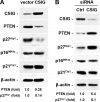
Influence of CSIG levels on PTEN expression. 2BS cells were stably transfected with pIRES-CSIG (A) or pSilencer-CSIG (B) or the respective empty vectors. Whole-cell lysates were prepared and subjected to Western blot analysis to determine PTEN, p27Kip1, p16INK4a, and p21Cip1 levels, and β-actin was served as a loading Ctrl. Relative abundances of PTEN and p27Kip1 were measured by densitometry and are expressed as increases relative to their respective levels in empty-vector-transfected cells.
FIG. 3.
FIG. 3.
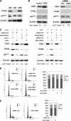
PTEN is required for CSIG to regulate p27Kip1 expression. (A) Results of Western blot analysis of endogenous PTEN and p27Kip1 expression in HEK 293, U87, and 2BS cells. (B) U87 cells were transfected with pIRES-CSIG (CSIG) versus empty vector (vector) (left panel) or with 20 nM siRNA targeting CSIG (siRNA CSIG) versus Ctrl siRNA (siRNA Ctrl) (right panel) for 48 h. Expressions of CSIG, PTEN, and p27Kip1 were assessed by Western blot analysis; β-actin served as a loading Ctrl. Relative p27Kip1abundances are expressed as increases relative to its level in control cells. (C) HEK 293 cells were transiently transfected with siRNA (20 nM) targeting PTEN (PTEN siRNA) (lanes 3 and 4) or siRNA Ctrl (lanes 1 and 2). Twenty-four hours later, cells were further transfected with pIRES-CSIG (CSIG) (left panel, lanes 2 and 4) versus an empty vector (vector; left panel, lanes 1 and 3) or with siRNA (20 nM) targeting CSIG (CSIG siRNA) (right panel, lanes 2 and 4) versus siRNA Ctrl (right panel, lanes 1 and 3). Cells were then cultured for an additional 48 h. Expression of CSIG, PTEN, and p27Kip1 as well as the relative abundances of p27Kip1 were evaluated as described for panel B. (D and E) FACS analysis of cells used in the right panel of panel C (D) and in the right panel of panel B (E).
FIG. 4.
FIG. 4.
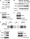
CSIG expression regulates PTEN in the translational level without influencing PTEN mRNA levels. (A and B) Ectopic intervention of CSIG expression was achieved by transiently transfecting HEK 293 cells either with pIRES-CSIG along with an empty vector (A) or with siRNA targeting CSIG (20 nM [lane 3] and 50 nM [lane 4]) along with a Ctrl siRNA (20 nM [lane 1] and 50 nM [lane 2]) for 48 h. (B) CSIG and PTEN expression were detected by Western blot analysis; β-actin was served as a loading Ctrl. Relative PTEN abundances were estimated by densitometry and are expressed as increases relative to PTEN levels in control cells. (C and D) Results of Northern blot analysis of PTEN mRNA levels after CSIG overexpression (C) or CSIG knockdown (D); GADPH served as a loading Ctrl. The PTEN mRNA abundance is expressed as the increase relative to PTEN mRNA levels in Ctrl cells. (E) Analysis of PTEN translation in CSIG-silenced HEK 293 cells. Newly translated PTEN was measured by incubating cells with l-[35S]methionine and l-[35S]cysteine for 20 min, followed by IP using either anti-PTEN antibody, anti-GAPDH antibody, or IgG; resolving immunoprecipitated samples by SDS-PAGE; and transferring for visualization of signals by using a PhosphorImager. (F) Results of Western blot analysis of PTEN and p27Kip1 (left panel), and results of Northern blot analysis (panel right) of PTEN in young (∼28 pdl) and senescent (∼58 pdl) 2BS cells. β-Actin and GADPH served as loading Ctrls for Western or Northern blot analysis, respectively.
FIG. 5.
FIG. 5.
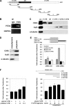
Binding of CSIG to PTEN mRNA and influence of PTEN 5′ UTR on the expression of a chimeric luciferase reporter construct after CSIG silencing. (A) Schematic presentation of the full-length PTEN cDNA and various transcripts derived from the 5′ UTR, CR, and 3′ UTR used in this study. (B) Whole-cell lysates (100 μg) were prepared from HEK 293 cells, and endogenous target transcripts were detected by RT-PCR assay of the corresponding IP materials; PCR products corresponding to PTEN mRNA were visualized on agarose gel. The PCR product of GAPDH served as a negative Ctrl. (C) Results of a pulldown assay using biotinylated fragments to detect bound CSIG by Western blotting. A 10-μg portion of whole-cell lysates (Lys.) and binding of HuR (positive Ctrl) and α-tubulin (negative Ctrl) to PTEN mRNA were included. (D) A polysomal fraction from HEK 293 cell lysates was prepared and subjected to Western blot analysis to evaluate the presence of CSIG. (E) pGL3, pGL3-5′ UTR, pGL3-CR, and pGL3-3′ UTR-2 plasmid (0.1 μg/ml) were transiently transfected into HEK 293 cells along with the pRL-CMV reporter (5 ng/ml) as a Ctrl. Twenty-four hours later, transfected cells were cotransfected either with CSIG siRNA to silence CSIG or with a Ctrl siRNA for 48 h; firefly luciferase activities were determined and normalized against Renilla luciferase activity. Values represent means ± standard errors of the means (SEM) of the results for five independent experiments. (F) Left panel: pGL3-5′ UTR (0.1 μg/ml), pRL-CMV (5 ng/ml) plasmids, and a different dose of pcDNA-5′ UTR [0 μg/ml (−), 2 μg/ml (+), 4 μg/ml (++), or 6 μg/ml (+++)] were transiently cotransfected into HEK 293 cells for 48 h. Right panel: pGL3-5′ UTR (0.1 μg/ml), pRL-CMV (5 ng/ml) plasmids, and pcDNA vectors expressing PTEN 5′ UTR fragments A, B, C, or D (6 μg/ml) were transiently cotransfected into HEK 293 cells for 48 h; firefly luciferase activities then were determined and normalized against Renilla luciferase activity. Values represent means ± SEMs of the results for five independent experiments.
FIG. 6.
FIG. 6.
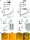
Influences of CSIG levels on replicative senescence. (A) Early passage 2BS (∼28 pdl) cells were transfected with either pIRES-CSIG (CSIG) versus empty vector (vector) (left panel) or pSilencer-CSIG (CSIG siRNA) versus vector expressing Ctrl siRNA (siRNA Ctrl) (right panel) and then selected by G418 for 3 weeks. Expression of CSIG was monitored by Western blot analysis; β-actin served as a loading Ctrl. Relative CSIG abundances were assessed by densitometry and are expressed as increases relative to CSIG levels in cells transfected with the empty vector. (B) Effect of overexpression (left panel) or silencing (right panel) of CSIG on cell growth. Transfected cells were seeded 2 × 10³ per well in 96-well plates, and cell number was assessed by MTT (methyl thiazolyldiphenyl-tetrazolium bromide) method at the times indicated. Values represent the means ± SEMs of the results for three independent experiments. (C) Transfected cells were subjected to FACS analysis to evaluate the effect of ectopic intervention to either overexpress (left panel) or knock down (right panel) CSIG on cell cycle distribution. Bars represent the means ± SEMs of the results for three independent experiments. (D) Influence of CSIG levels on SA-β-gal activity. Transfected 2BS cells that either elevate (left panel) or reduce (right panel) CSIG levels were stained to assess SA-β-gal activity. Data are representative of the results for three independent experiments.
FIG. 7.
FIG. 7.
PTEN is required for CSIG to regulate cell proliferation and senescence. 2BS cells were stably transfected with vectors expressing Ctrl siRNA (siRNA Ctrl) or CSIG siRNA and were cotransfected with vectors expressing Ctrl siRNA plus PTEN siRNA (siRNA Ctrl+PTEN siRNA) or CSIG siRNA plus PTEN siRNA (CSIG siRNA+PTEN siRNA). (A) Expression of CSIG, PTEN, and p27Kip1 was monitored by Western blot analysis; β-actin served as a loading Ctrl. (B) Transfected cells were seeded 2 × 10³ per well in 96-well plates, and cell numbers were assessed at the times indicated by the MTT method. Values represent the means ± SEMs of the results for three independent experiments. (C) Transfected cells were stained to assess SA-β-gal activity. Data were representative of the results for three independent experiments.

Similar articles

Cited by

References

    1. Alexander, K., and P. W. Hinds. 2001. Requirement for p27(KIP1) in retinoblastoma protein-mediated senescence. Mol. Cell. Biol. 213616-3631. - PMC - PubMed
    1. Baker, S. J. 2007. PTEN enters the nuclear age. Cell 12825-28. - PubMed
    1. Bayreuther, K., H. P. Rodemann, R. Hommel, K. Dittmann, M. Albiez, and P. I. Francz. 1988. Human skin fibroblasts in vitro differentiate along a terminal cell lineage. Proc. Natl. Acad. Sci. USA 855112-5116. - PMC - PubMed
    1. Bringold, F., and M. Serrano. 2000. Tumor suppressors and oncogenes in cellular senescence. Exp. Gerontol. 35317-329. - PubMed
    1. Campisi, J. 2001. Cellular senescence as a tumor-suppressor mechanism. Trends Cell Biol. 11S27-S31. - PubMed

Publication types

MeSH terms