On the origin of the beta cell
- PMID: 18676806
- PMCID: PMC2735346
- DOI: 10.1101/gad.1670808
On the origin of the beta cell
Abstract
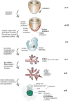
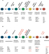
The major forms of diabetes are characterized by pancreatic islet beta-cell dysfunction and decreased beta-cell numbers, raising hope for cell replacement therapy. Although human islet transplantation is a cell-based therapy under clinical investigation for the treatment of type 1 diabetes, the limited availability of human cadaveric islets for transplantation will preclude its widespread therapeutic application. The result has been an intense focus on the development of alternate sources of beta cells, such as through the guided differentiation of stem or precursor cell populations or the transdifferentiation of more plentiful mature cell populations. Realizing the potential for cell-based therapies, however, requires a thorough understanding of pancreas development and beta-cell formation. Pancreas development is coordinated by a complex interplay of signaling pathways and transcription factors that determine early pancreatic specification as well as the later differentiation of exocrine and endocrine lineages. This review describes the current knowledge of these factors as they relate specifically to the emergence of endocrine beta cells from pancreatic endoderm. Current therapeutic efforts to generate insulin-producing beta-like cells from embryonic stem cells have already capitalized on recent advances in our understanding of the embryonic signals and transcription factors that dictate lineage specification and will most certainly be further enhanced by a continuing emphasis on the identification of novel factors and regulatory relationships.
Figures


Similar articles
-
Depressing time: Waiting, melancholia, and the psychoanalytic practice of care.In: Kirtsoglou E, Simpson B, editors. The Time of Anthropology: Studies of Contemporary Chronopolitics. Abingdon: Routledge; 2020. Chapter 5. In: Kirtsoglou E, Simpson B, editors. The Time of Anthropology: Studies of Contemporary Chronopolitics. Abingdon: Routledge; 2020. Chapter 5. PMID: 36137063 Free Books & Documents. Review.
-
Qualitative evidence synthesis informing our understanding of people's perceptions and experiences of targeted digital communication.Cochrane Database Syst Rev. 2019 Oct 23;10(10):ED000141. doi: 10.1002/14651858.ED000141. Cochrane Database Syst Rev. 2019. PMID: 31643081 Free PMC article.
-
Unlocking data: Decision-maker perspectives on cross-sectoral data sharing and linkage as part of a whole-systems approach to public health policy and practice.Public Health Res (Southampt). 2024 Nov 20:1-30. doi: 10.3310/KYTW2173. Online ahead of print. Public Health Res (Southampt). 2024. PMID: 39582242
-
Comparison of Two Modern Survival Prediction Tools, SORG-MLA and METSSS, in Patients With Symptomatic Long-bone Metastases Who Underwent Local Treatment With Surgery Followed by Radiotherapy and With Radiotherapy Alone.Clin Orthop Relat Res. 2024 Dec 1;482(12):2193-2208. doi: 10.1097/CORR.0000000000003185. Epub 2024 Jul 23. Clin Orthop Relat Res. 2024. PMID: 39051924
-
Trends in Surgical and Nonsurgical Aesthetic Procedures: A 14-Year Analysis of the International Society of Aesthetic Plastic Surgery-ISAPS.Aesthetic Plast Surg. 2024 Oct;48(20):4217-4227. doi: 10.1007/s00266-024-04260-2. Epub 2024 Aug 5. Aesthetic Plast Surg. 2024. PMID: 39103642 Review.
Cited by
-
GATA factors in endocrine neoplasia.Mol Cell Endocrinol. 2016 Feb 5;421:2-17. doi: 10.1016/j.mce.2015.05.027. Epub 2015 May 28. Mol Cell Endocrinol. 2016. PMID: 26027919 Free PMC article. Review.
-
Analysis of Half a Billion Datapoints Across Ten Machine-Learning Algorithms Identifies Key Elements Associated With Insulin Transcription in Human Pancreatic Islet Cells.Front Endocrinol (Lausanne). 2022 Mar 23;13:853863. doi: 10.3389/fendo.2022.853863. eCollection 2022. Front Endocrinol (Lausanne). 2022. PMID: 35399953 Free PMC article.
-
A Supportive Role of Mesenchymal Stem Cells on Insulin-Producing Langerhans Islets with a Specific Emphasis on The Secretome.Biomedicines. 2023 Sep 18;11(9):2558. doi: 10.3390/biomedicines11092558. Biomedicines. 2023. PMID: 37761001 Free PMC article. Review.
-
Inhibition of LSD1 promotes the differentiation of human induced pluripotent stem cells into insulin-producing cells.Stem Cell Res Ther. 2020 May 19;11(1):185. doi: 10.1186/s13287-020-01694-8. Stem Cell Res Ther. 2020. PMID: 32430053 Free PMC article.
-
Resveratrol potentiates glucose-stimulated insulin secretion in INS-1E beta-cells and human islets through a SIRT1-dependent mechanism.J Biol Chem. 2011 Feb 25;286(8):6049-60. doi: 10.1074/jbc.M110.176842. Epub 2010 Dec 16. J Biol Chem. 2011. PMID: 21163946 Free PMC article.
References
-
- Ahlgren U., Jonsson J., Edlund H. The morphogenesis of the pancreatic mesenchyme is uncoupled from that of the pancreatic epithelium in IPF1/PDX1-deficient mice. Development. 1996;122:1409–1416. - PubMed
-
- Alanentalo T., Chatonnet F., Karlen M., Sulniute R., Ericson J., Andersson E., Ahlgren U. Cloning and analysis of Nkx6.3 during CNS and gastrointestinal development. Brain Res. Gene Expr. Patterns. 2006;6:162–170. - PubMed
-
- Ang S.L., Rossant J. HNF-3 β is essential for node and notochord formation in mouse development. Cell. 1994;78:561–574. - PubMed
-
- Ang S.L., Wierda A., Wong D., Stevens K.A., Cascio S., Rossant J., Zaret K.S. The formation and maintenance of the definitive endoderm lineage in the mouse: Involvement of HNF3/forkhead proteins. Development. 1993;119:1301–1315. - PubMed
Publication types
MeSH terms
Grants and funding
LinkOut - more resources
Full Text Sources
Other Literature Sources
