Review
. 2008 Sep;155(2):152-69.
doi: 10.1038/bjp.2008.252. Epub 2008 Jul 21.

Annexin-A1: a pivotal regulator of the innate and adaptive immune systems

Affiliations
Review

Annexin-A1: a pivotal regulator of the innate and adaptive immune systems

F D'Acquisto et al. Br J Pharmacol. 2008 Sep.

Abstract

The glucocorticoids are the most potent anti-inflammatory drugs that we possess and are effective in a wide variety of diseases. Although their action is known to involve receptor mediated changes in gene transcription, the exact mechanisms whereby these bring about their pleiotropic action in inflammation are yet to be totally understood. Whilst many different genes are regulated by the glucocorticoids, we have identified one particular protein-annexin A1 (Anx-A1)-whose synthesis and release is strongly regulated by the glucocorticoids in many cell types. The biology of this protein, as revealed by studies using transgenic animals, peptide mimetics and neutralizing antibodies, speaks to its role as a key modulator of both of the innate and adaptive immune systems. The mechanism whereby this protein exerts its effects is likely to be through the FPR receptor family-a hitherto rather enigmatic family of G protein coupled receptors, which are increasingly implicated in the regulation of many inflammatory processes. Here we review some of the key findings that have led up to the elucidation of this key pathway in inflammatory resolution.


Figures
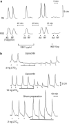
Figure 1
Figure 1
Detection and subsequent characterization of gluocorticoid-induced protein with eicosanoid suppressive actions. (a) An experiment in which the outflow from a guinea pig-isolated perfused lung was passed over a rabbit aortic (RA) or rat stomach strip (RSS) to detect the release TxA2. Arachidonic acid (AA; 10 μg) and ‘releasing factor' (RF; 5 U—later identified as a mixture of leukotrienes) both release substantial amounts of TxA2 from this preparation. When dexamethasone (Dex; 1 μg mL−1) is infused for 45 min into the perfused lung, the release of TxA2 by RF is selectively inhibited. This effect decays about 45 min after the infusion is terminated. Administration of a cyclooxygenase inhibitor (Ind; 10 μg) blocks the generation of eicosanoids triggered by either stimulus. The experiment demonstrates that glucocorticoids do not block the cyclooxygenase directly, but rather inhibit some process leading to the liberation of the substrate arachidonic acid. The nature of the soluble factor that brought this about was subsequently identified as Anx-A1 (redrawn from Nijkamp et al. (1976)). (b) The first demonstration of the biological activity of hu-r-Anx-A1. Once again the rabbit aortic strip was used as a detecting organ to monitor the release of TxA2 from the guinea pig-isolated perfused lung. Injections of LTC4 were made at 30 min intervals. During the time periods indicated by the horizontal bar, human recombinant Anx-A1 (0.4 μg mL−1) was infused into the preparation, blocking the release of TxA2 in response to 2 ng (top panel) or 10 ng (middle panel) of LTC4 in a manner strongly reminiscent of the glucocorticoids themselves. The bottom panel shows that a sham preparation, prepared from Escherichia coli transfected with a dummy construct for control purposes, was without effect (taken from Cirino et al. (1987)). Anx-A1, annexin A1; hu-r-Anx-A1, human recombinant annexin A1; LTC4, leukotriene C4; TxA2, thromboxane A2.
Figure 2
Figure 2
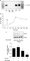
Regulation of Anx-A1 expression by glucocorticoids in three different systems. (a) This experiment shows the induction by glucocorticoids of Anx-A1 in resident rat peritoneal macrophages as assessed by western blotting. Con: injection of saline vehicle for 30 min before collection of cells. Con+RU: rats were pretreated with 2 × 20 mg kg−1 RU486 18 and 2 h before the saline vehicle. Dex+RU: 0.08 mg kg−1 dexamethasone injected after pretreatment with RU486 for 1 h. Dex: 0.08 mg kg−1 dexamethasone injected 30 min before collection. HCT: injection of 1 mg kg−1 hydrocortisone 30 min before collection of cells. The experiment demonstrates that Anx-A1 is present in ‘resting' resident peritoneal macrophages, presumably because of the endogenous glucocorticoid drive since administration of RU486, the glucocorticoid antagonist, reduces this below detectable limits. Both dexamethasone and hydrocortisone further induce Anx-A1 in these cells, but the effects are blocked by a preadministration of the glucocorticoid receptor antagonist (taken from Peers et al. (1993)). (b) The injection of 100 mg hydrocortisone into a human volunteer results, within 30 min, in a rise of Anx-A1 expression on the surface of human peripheral blood leukocytes as measured by an ELISA assay (redrawn from Goulding et al. (1990a)). (c) In T cells, unlike most other leukocytes, glucocorticoid treatment downregulates Anx-A1 gene synthesis and transcription. Upper panel shows western blot and lower panel shows real-time PCR for Anx-A1 protein or mRNA in human peripheral blood CD4+ T cells incubated for 12 h with dexamethasone for different times (taken from D'Acquisto et al., 2008a). Anx-A1, annexin A1; HCT, hydrocortisone. *P<0.05; **P<0.01.
Figure 3
Figure 3
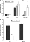
Attenuated effect of dexamethasone in wild-type and Anx-A1 null animals shown in vitro and ex vivo. (a) Lung fibroblasts obtained from Anx-A1 wild-type, heterozygote or null animals were cultured such that the generation of PGE2 could be measured in the supernatant. The figure shows the increase in PGE2 release after the addition of FCS and the inhibitory action of dexamethasone (1 μM). There is marked increase in PGE2 generated by cells derived from the wild-type animals, but this is superseded in cells from both the heterozygote, and especially the Anx-A1 null, animals. Conversely, dexamethasone exerts a profound inhibition of PGE2 release in the wild-type cells, much less in the heterozygote, and exhibits no appreciable inhibition at all in the Anx-A1 null cells (redrawn from Croxtall et al. (2003)). *P<0.05 relative to control PGE2 synthesis. (b) Inhibition of phagocytosis by hydrocortisone. In this experiment, phagocytosis of aggregated IgG was assessed in murine peritoneal macrophages using the dihydrorhodamine assay. Hydrocortisone (10 μM) exerts a moderate inhibitory effect on this process in the Anx-A1 wild-type animals, but much less in the Anx-A1 null animals. However, the N-terminal peptide N-acetyl 2–26 (100 μg mL−1) is equally inhibitory in both phenotypes (redrawn from Yona et al. (2005)). *P<0.05; ***P<0.001 relative to control phagocytosis. Anx-A1, annexin A1; FCS, foetal calf serum; PGE2, prostaglandin E2.
Figure 4
Figure 4
Schematic representation of the role of the Anx-A1/FPRL-1 system in T-cell signalling. (a) In basal conditions, both Anx-A1 and FPRL-1 are present at very low abundance on the membrane of naive T cells. Stimulation of T cells via the TCR leads to the externalization of FPRL-1 and the release of Anx-A1. The activation of FPRL-1 by Anx-A1 modulates the strength of TCR signalling by increasing levels of transcription factors such as AP-1, NF-κB and NFAT. In pathological conditions, such as in rheumatoid arthritis, the increased expression of endogenous Anx-A1 might contribute to the basal hyperactivated state of these cells and to the increase of transcription factors that play a key role in the regulation of the expression of several inflammatory genes. (b) Proposed model for the role of Anx-A1 in the differentiation of T-helper cells. In physiological conditions, naive T cells differentiate in Th1 or Th2 effector cells depending on the microenvironment in which this process occurs. The presence of high levels of Anx-A1 promotes a protective Th1 immune response or might, in a pathological context, exacerbate autoimmune diseases such as rheumatoid arthritis. When T cells express low levels of Anx-A1, they preferentially become Th2 cells and this might be responsible for the occurrence of allergic reactions or promote a protective humoral response. Glucocorticoids, by reducing Anx-A1 synthesis by these cells, promote a Th2 phenotype. Anx-A1, annexin A1; AP-1, activator protein-1; FPRL-1, formyl peptide-like receptor-1; NF-κB, nuclear factor-κB; NFAT, nuclear factor of activated T cells; TCR, T-cell receptor.
Figure 5
Figure 5
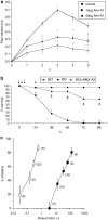
Potent anti-inflammatory effects of hu-r-Anx-A1 in three models of inflammation. (a) Anx-A1 or vehicle was injected together with carrageenin into the rat paw and the ensuing oedema measured over the next 5 h. Both 10 and 50 μg Anx-A1 produced a striking inhibition of all phases of the oedema (Cirino et al., 1989). *P<0.05 relative to control response. (b) Mortality in the Anx-A1 null mouse induced by LPS and its rescue by Anx-A1. Escherichia coli LPS, 10 mg kg−1, was injected into the mice at time 0 h. This produced a negligible mortality in the wild-type population, but was 100% fatal in the Anx-A1 null animals within 48 h. The arrows indicate points (0, 4 and 8 h) at which hu-r-Anx-A1 (10 ng) was injected into the third group. This substantially reversed the mortality caused by the LPS allowing approximately 75% survival (Damazo et al., 2005). *P<0.05 relative to wild-type controls; P<0.05 relative to Anx-A1 null mice. (c) Inhibition of PMN migration into the murine IL-1-induced air pouch model showing the graded inhibition produced by increasing doses of Anx-A1 (open circles) injected i.v., 1 h before the IL-1 injection, and also the striking effect of the N-terminal peptide N-acetyl 2–26 (filled circles). It should be noted that the dose–response curve of the latter agent is parallel to that of the intact molecule although it is clearly some two orders of magnitude less potent (Perretti et al., 1993a). Anx-A1, annexin A1; hu-r-Anx-A1, human recombinant annexin A1; IL, interleukin; LPS, lipopolysaccharide; PMN, polymorphonuclear leukocyte.
Figure 6
Figure 6
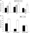
Exacerbation of the acute inflammatory response in Anx-A1 null mice and their lack of response to dexamethasone. (a) Zymosan-induced peritonitis produces a brisk neutrophil infiltration into the peritoneal cavity. In comparison to the wild-type controls, this is elevated in both the heterozygote and the homozygote Anx-A1 null animals at the 2 and 4 h time point and at 24 h in the Anx-A1 homozygote null animal (redrawn from Hannon et al. (2003)). *P<0.05 relative to control migration. (b) Zymosan peritonitis was again used as a measure of acute inflammation. Here, dexamethasone (0.5 mg kg−1) produced a striking inhibition of the control migration, but was without any effect in the Anx-A1 null animals. However, the N-terminal peptide N-acetyl 2–26 (100 μg) was equi-active in both genotypes (unpublished—but see Hannon et al. (2003) for analogous experimental data). *P<0.05; ***P<0.001 relative to control migration. Anx-A1, annexin A1.
Figure 7
Figure 7
A schematic diagram showing the integration of glucocorticoid ‘tone' with the Anx-A1 system. The diagram shows three glucocorticoid ‘states'—when levels are low, normal or high, and the ensuing events in the innate and adaptive immune systems. It illustrates that, as glucocorticoids rise, the increasing amounts of Anx-A1 within the innate immune system suppress the activation of cells such as PMN, monocytes and mast cells, while decreasing levels of Anx-A1 within the adaptive immune system tend skewing any T-cell responses towards a Th2 ‘anti-inflammatory' phenotype. Anx-A1, annexin A1; PMN, polymorphonuclear leukocyte.

Similar articles

Cited by

References

    1. Acosta-Rodriguez EV, Napolitani G, Lanzavecchia A, Sallusto F. Interleukins 1beta and 6 but not transforming growth factor-beta are essential for the differentiation of interleukin 17-producing human T helper cells. Nat Immunol. 2007;8:942–949. - PubMed
    1. Ahn SH, Sawada H, Ro JY, Nicolson GL. Differential expression of annexin I in human mammary ductal epithelial cells in normal and benign and malignant breast tissues. Clin Exp Metastasis. 1997;15:151–156. - PubMed
    1. Akama H, Tanaka H, Kawai S. Possible mechanisms of glucocorticoid—unresponsive pyrexia. Defect in lipocortin 1. Mater Med Pol. 1995;27:75–78. - PubMed
    1. Aksentijevich S, Whitfield HJ, Jr, Young WS, III, Wilder RL, Chrousos GP, Gold PW, et al. Arthritis-susceptible Lewis rats fail to emerge from the stress hyporesponsive period. Brain Res Dev Brain Res. 1992;65:115–118. - PubMed
    1. Allcock GH, Allegra M, Flower RJ, Perretti M. Neutrophil accumulation induced by bacterial lipopolysaccharide: effects of dexamethasone and annexin 1. Clin Exp Immunol. 2001;123:62–67. - PMC - PubMed

Publication types