The multifunctional FUS, EWS and TAF15 proto-oncoproteins show cell type-specific expression patterns and involvement in cell spreading and stress response
- PMID: 18620564
- PMCID: PMC2478660
- DOI: 10.1186/1471-2121-9-37
The multifunctional FUS, EWS and TAF15 proto-oncoproteins show cell type-specific expression patterns and involvement in cell spreading and stress response
Abstract
Background: FUS, EWS and TAF15 are structurally similar multifunctional proteins that were first discovered upon characterization of fusion oncogenes in human sarcomas and leukemias. The proteins belong to the FET (previously TET) family of RNA-binding proteins and are implicated in central cellular processes such as regulation of gene expression, maintenance of genomic integrity and mRNA/microRNA processing. In the present study, we investigated the expression and cellular localization of FET proteins in multiple human tissues and cell types.
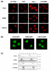
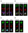
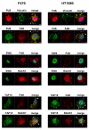
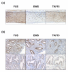
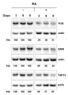
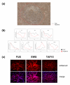
Results: FUS, EWS and TAF15 were expressed in both distinct and overlapping patterns in human tissues. The three proteins showed almost ubiquitous nuclear expression and FUS and TAF15 were in addition present in the cytoplasm of most cell types. Cytoplasmic EWS was more rarely detected and seen mainly in secretory cell types. Furthermore, FET expression was downregulated in differentiating human embryonic stem cells, during induced differentiation of neuroblastoma cells and absent in terminally differentiated melanocytes and cardiac muscle cells. The FET proteins were targeted to stress granules induced by heat shock and oxidative stress and FUS required its RNA-binding domain for this translocation. Furthermore, FUS and TAF15 were detected in spreading initiation centers of adhering cells.
Conclusion: Our results point to cell-specific expression patterns and functions of the FET proteins rather than the housekeeping roles inferred from earlier studies. The localization of FET proteins to stress granules suggests activities in translational regulation during stress conditions. Roles in central processes such as stress response, translational control and adhesion may explain the FET proteins frequent involvement in human cancer.
Figures

Similar articles
-
Gene expression responses to FUS, EWS, and TAF15 reduction and stress granule sequestration analyses identifies FET-protein non-redundant functions.PLoS One. 2012;7(9):e46251. doi: 10.1371/journal.pone.0046251. Epub 2012 Sep 25. PLoS One. 2012. PMID: 23049996 Free PMC article.
-
Characterization and expression analysis in the developing embryonic brain of the porcine FET family: FUS, EWS, and TAF15.Gene. 2012 Feb 1;493(1):27-35. doi: 10.1016/j.gene.2011.11.038. Epub 2011 Nov 28. Gene. 2012. PMID: 22143032
-
Post-transcriptional regulation of FUS and EWS protein expression by miR-141 during neural differentiation.Hum Mol Genet. 2017 Jul 15;26(14):2732-2746. doi: 10.1093/hmg/ddx160. Hum Mol Genet. 2017. PMID: 28453628
-
FET proteins in frontotemporal dementia and amyotrophic lateral sclerosis.Brain Res. 2012 Jun 26;1462:40-3. doi: 10.1016/j.brainres.2011.12.010. Epub 2011 Dec 13. Brain Res. 2012. PMID: 22261247 Review.
-
TLS, EWS and TAF15: a model for transcriptional integration of gene expression.Brief Funct Genomic Proteomic. 2006 Mar;5(1):8-14. doi: 10.1093/bfgp/ell015. Epub 2006 Feb 23. Brief Funct Genomic Proteomic. 2006. PMID: 16769671 Review.
Cited by
-
NY-ESO-1 is a ubiquitous immunotherapeutic target antigen for patients with myxoid/round cell liposarcoma.Cancer. 2012 Sep 15;118(18):4564-70. doi: 10.1002/cncr.27446. Epub 2012 Feb 22. Cancer. 2012. PMID: 22359263 Free PMC article.
-
Distinct cytoplasmic and nuclear functions of the stress induced protein DDIT3/CHOP/GADD153.PLoS One. 2012;7(4):e33208. doi: 10.1371/journal.pone.0033208. Epub 2012 Apr 9. PLoS One. 2012. PMID: 22496745 Free PMC article.
-
Structure-function based molecular relationships in Ewing's sarcoma.Biomed Res Int. 2015;2015:798426. doi: 10.1155/2015/798426. Epub 2015 Jan 22. Biomed Res Int. 2015. PMID: 25688366 Free PMC article. Review.
-
Molecular hallmarks of ageing in amyotrophic lateral sclerosis.Cell Mol Life Sci. 2024 Mar 2;81(1):111. doi: 10.1007/s00018-024-05164-9. Cell Mol Life Sci. 2024. PMID: 38430277 Free PMC article. Review.
-
JAK-STAT signalling controls cancer stem cell properties including chemotherapy resistance in myxoid liposarcoma.Int J Cancer. 2019 Jul 15;145(2):435-449. doi: 10.1002/ijc.32123. Epub 2019 Jan 29. Int J Cancer. 2019. PMID: 30650179 Free PMC article.
References
Publication types
MeSH terms
Substances
LinkOut - more resources
Full Text Sources

