. 2008 Jul 23;27(14):1953-62.
doi: 10.1038/emboj.2008.128. Epub 2008 Jul 3.

Mre11-Rad50-Nbs1-dependent processing of DNA breaks generates oligonucleotides that stimulate ATM activity

Affiliations

Mre11-Rad50-Nbs1-dependent processing of DNA breaks generates oligonucleotides that stimulate ATM activity

Ali Jazayeri et al. EMBO J. .

Abstract

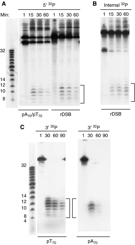
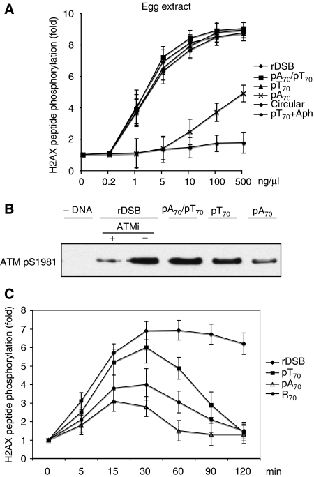
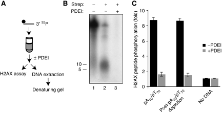
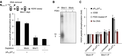
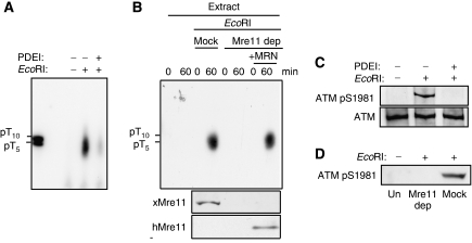
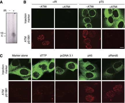
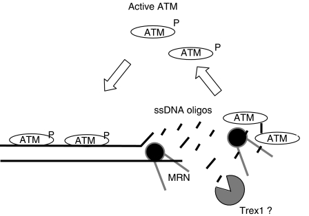
DNA double-strand breaks (DSBs) can be processed by the Mre11-Rad50-Nbs1 (MRN) complex, which is essential to promote ataxia telangiectasia-mutated (ATM) activation. However, the molecular mechanisms linking MRN activity to ATM are not fully understood. Here, using Xenopus laevis egg extract we show that MRN-dependent processing of DSBs leads to the accumulation of short single-stranded DNA oligonucleotides (ssDNA oligos). The MRN complex isolated from the extract containing DSBs is bound to ssDNA oligos and stimulates ATM activity. Elimination of ssDNA oligos results in rapid extinction of ATM activity. Significantly, ssDNA oligos can be isolated from human cells damaged with ionizing radiation and injection of small synthetic ssDNA oligos into undamaged cells also induces ATM activation. These results suggest that MRN-dependent generation of ssDNA oligos, which constitute a unique signal of ongoing DSB repair not encountered in normal DNA metabolism, stimulates ATM activity.


Figures

Figure 1
Figure 1
DNA end processing leads to generation of ssDNA oligos. (A) Different DNA structures such as rDSB and pA70/pT70 were stoichiometrically labelled at the 5′ (5′ 32P). After incubation for the indicated times in the egg extract, the DNA was recovered and ran on a 22% denaturing acrylamide gel. In (B) rDSBs were internally labelled (internal 32P) at the thirty-fifth nucleotide from the 5′ end and in (C) pT70 and pA70 were labelled at the 3′ end (3′ 32P) before incubation.
Figure 2
Figure 2
ATM activation by different DNA structures. (A) Histone H2AX carboxy-terminal peptide phosphorylation in the presence of increasing amounts of rDSB, pA70/pT70, pT70, pA70, M13 circular ssDNA or pT70 plus 100 μM aphidicolin (Aph). Activity was measured 15 min after incubation of DNA in the extract at 22°C and is expressed as fold induction over the activity measured in the untreated extract. (B) ATM serine 1981 phosphorylation in the untreated (−DNA) egg extract or extract supplemented with rDSB in the absence or presence of 10 μM Ku55933 (ATMi), 2.5 ng/μl pA70/pT70, pT70 or pA70 or after 30 min of incubation. Western blot was performed with Advance ECL for enhanced sensitivity. (C) Histone H2AX peptide phosphorylation in the presence of egg extract containing no DNA, 2.5 ng/μl rDSB, pT70 or 25 ng/μl pA70 and R70 measured at the indicated time points after DNA addition. Activity is expressed as fold induction. In all graphs, the values are average of three independent experiments and the error bars represent standard deviation.
Figure 3
Figure 3
ssDNA oligos sustain ATM activity. (A) Experimental procedure: biotinylated pA70/pT70 labelled with 32P on the 3′ end was incubated for 30 min in the extract and then removed with streptavidin beads. After pA70/pT70 removal, histone H2AX peptide phosphorylation was monitored. (B) Residual DNA left in the extract was isolated and run on a 15% TBE–urea denaturing gel. DNA was isolated from the extract incubated with biotinylated and labelled pA70/pT70 for 30 min (lane 1) and from extracts in which biotinylated pA70/pT70 was incubated for 30 min and then removed with streptavidin beads (Strep) (lanes 2 and 3). The extracts were untreated (lane 2) or supplemented with 0.001 U/μl PDEI (lane 3). (C) Histone H2AX peptide phosphorylation induced by 5 ng/μl biotinylated pA70/pT70 before and after pA70/pT70 removal in the absence (black bar) or in the presence (grey bar) of 0.001 U/μl PDEI. PDEI was added 30 min after DNA addition and was incubated for 10 min at 22°C. Activity is expressed as fold induction over the untreated extract (no DNA).
Figure 4
Figure 4
ssDNA oligos form DNA–protein complexes with MRN that are able to induce ATM activation. (A) Biotinylated labelled pA70/pT70 was incubated in the extract for 30 min and following DNA removal with streptavidin beads, Mre11 was depleted and ATM activity was monitored. Activity is expressed as fold induction over the untreated samples. (B) 32P-labelled DNA isolated from Mre11 or mock immunoprecipitations was run on a 15% TBE–urea denaturing gel. Immunoprecipitates were untreated or treated with 0.001 U/μl PDEI. (C) Histone H2AX peptide phosphorylation induced by Mre11 immunoprecipitated from an untreated or a pA70/pT70-treated extract and transferred to an extract supplemented with low concentrations of pA70/pT70 (0.2 ng/μl) (black bar), rDSB (0.2 ng/μl) (grey bar) or no DNA (red bar). Immunoprecipitates were also treated with 0.001 U/μl PDEI for 10 min at 22°C before transfer into the extract supplemented with low concentrations of DNA (white bar).
Figure 5
Figure 5
ssDNA oligos are generated from MRN-dependent processing of chromosomal DSBs. (A) Sperm nuclei were incubated in the extract for 30 min. The extract was then supplemented with 0.2 U/μl of EcoRI in the presence or absence of 0.001 U/μl of PDEI and the samples were incubated for a further 60 min. Sperm nuclei were permeabilized and processed to isolate soluble low molecular weight DNA. DNA was labelled with TdT in the presence of 32P-alpha-ddATP and ran on a 15% TBE–urea denaturing gel. (B) Upper panel: sperm nuclei were incubated in the extract for 30 min, after which the extract was supplemented with 0.2 U/μl EcoRI and immediately processed to isolate low molecular weight DNA (0 min) or incubated for a further 60 min. The extracts were untreated, mock depleted (mock) or Mre11-depleted (Mre11 dep) and supplemented with recombinant MRN complex (+MRN) (250 nM). Lower panels: egg extract treated as indicated in the upper panel was probed with anti-Xenopus (xMre11) and anti-human Mre11 (hMre11) antibodies. (C) Sperm nuclei were incubated in the extract in the presence or absence of 0.2 U/μl of EcoRI or 0.001 U/μl of PDEI for 60 min. ATM was immunoprecipitated and immunoblotted as indicated. (D) Sperm nuclei were incubated in the extracts that were untreated (Un), mock depleted (mock) or Mre11 depleted (Mre11 dep) in the presence or absence of 0.2 U/μl of EcoRI for 60 min. ATM was immunoprecipitated and immunoblotted as indicated.
Figure 6
Figure 6
ssDNA oligos are generated following induction of DSBs in human cells and promote ATM activation when injected in undamaged cells. (A) Human U2OS cells were irradiated with 10 Gy of IR. Following a short recovery, cells were permeabilized and processed to isolate soluble low molecular weight DNA. DNA was labelled with TdT in the presence of 32P-alpha-ddATP and ran on a 15% TBE–urea denaturing gel. (B) U2OS cells were co-microinjected with 5 μg/μl pT5 and anti-goat IgG conjugated with Alexa Fluor 488. After 30 min incubation, cells were fixed and stained with anti-ATM phospho-serine 1981 antibody. Where indicated cells were treated with 10 Gy of IR and after 1 h were fixed and immunostained. The ATM inhibitor (ATMi) was used at 10 μM for 1 h prior to irradiation or microinjection. (C) U2OS cells were injected as indicated. All DNA concentrations were at 5 μg/μl and dTTP was 100 μM. After 30 min incubation, cells were fixed and stained with anti-ATM phospho-serine 1981 antibody. All the images were acquired under identical microscope settings. Scale bar is 10 mm.
Figure 7
Figure 7
Proposed model for ssDNA oligo action. ssDNA oligos generated by MRN-dependent processing of DSBs stimulates ATM activity by forming MRN–DNA oligo complexes. Elimination of ssDNA oligos by single-stranded DNA exonucleases such as Trex1 (see Discussion) might indirectly control ATM activity.

Similar articles

Cited by

References

    1. Bakkenist CJ, Kastan MB (2003) DNA damage activates ATM through intermolecular autophosphorylation and dimer dissociation. Nature 421: 499–506 - PubMed
    1. Barlow JH, Lisby M, Rothstein R (2008) Differential regulation of the cellular response to DNA double-strand breaks in G1. Mol Cell 30: 73–85 - PMC - PubMed
    1. Berkovich E, Monnat RJ Jr, Kastan MB (2007) Roles of ATM and NBS1 in chromatin structure modulation and DNA double-strand break repair. Nat Cell Biol 9: 683–690 - PubMed
    1. Bhaskara V, Dupre A, Lengsfeld B, Hopkins BB, Chan A, Lee JH, Zhang X, Gautier J, Zakian V, Paull TT (2007) Rad50 adenylate kinase activity regulates DNA tethering by Mre11/Rad50 complexes. Mol Cell 25: 647–661 - PMC - PubMed
    1. Costanzo V, Paull T, Gottesman M, Gautier J (2004a) Mre11 assembles linear DNA fragments into DNA damage signaling complexes. PLoS Biol 2: E110. - PMC - PubMed

Publication types

MeSH terms