Expression and secretion of cathelicidin LL-37 in human epithelial cells after infection by Mycobacterium bovis Bacillus Calmette-Guérin
- PMID: 18579695
- PMCID: PMC2546677
- DOI: 10.1128/CVI.00178-08
Expression and secretion of cathelicidin LL-37 in human epithelial cells after infection by Mycobacterium bovis Bacillus Calmette-Guérin
Abstract
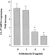
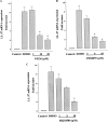
The antimicrobial cathelicidin LL-37 is considered to play an important role in the innate immune response to tuberculosis infection. However, little is known about the induction and secretion of this antimicrobial peptide in A549 epithelial cells after infection with Mycobacterium bovis bacillus Calmette-Guérin (BCG), the world's most widely used tuberculosis vaccine. In this study, we investigated the effect of M. bovis BCG on LL-37 mRNA levels in A549 cells by real-time PCR and on protein levels by Western blotting. Treatment of cells with M. bovis BCG upregulates LL-37 mRNA expression in a dose- and time-dependent manner. The quantitative analysis of LL-37 gene expression correlated with our Western blotting results. Moreover, our results demonstrated that treatment of cells with the transcriptional inhibitor actinomycin D effectively inhibited in a concentration-dependent manner the ability of M. bovis BCG to induce LL-37 mRNA expression. Finally, inhibition of the MEK1/2 and p38 mitogen-activated protein kinase (MAPK) signaling pathways reduced M. bovis BCG-mediated LL-37 mRNA expression, a reduction that correlated with the observed high level of downregulation of LL-37 protein induction. Thus, these results indicate that the MEK1/2 and p38 MAPK signaling pathways play a critical role in the regulation of inducible LL-37 gene expression in A549 cells infected with M. bovis BCG.
Figures


Similar articles
-
Role of reactive oxygen species (ROS) in Mycobacterium bovis bacillus Calmette Guérin-mediated up-regulation of the human cathelicidin LL-37 in A549 cells.Microb Pathog. 2009 Nov;47(5):252-7. doi: 10.1016/j.micpath.2009.08.006. Epub 2009 Sep 1. Microb Pathog. 2009. PMID: 19729059
-
Mycobacterium bovis Bacillus Calmette-Guérin (BCG) stimulates IL-10 production via the PI3K/Akt and p38 MAPK pathways in human lung epithelial cells.Cell Immunol. 2008 Jan;251(1):37-42. doi: 10.1016/j.cellimm.2008.03.002. Cell Immunol. 2008. PMID: 18423589
-
Mouse Bone Marrow Sca-1+ CD44+ Mesenchymal Stem Cells Kill Avirulent Mycobacteria but Not Mycobacterium tuberculosis through Modulation of Cathelicidin Expression via the p38 Mitogen-Activated Protein Kinase-Dependent Pathway.Infect Immun. 2017 Sep 20;85(10):e00471-17. doi: 10.1128/IAI.00471-17. Print 2017 Oct. Infect Immun. 2017. PMID: 28739828 Free PMC article.
-
Prospects in Mycobacterium bovis Bacille Calmette et Guérin (BCG) vaccine diversity and delivery: why does BCG fail to protect against tuberculosis?Vaccine. 2015 Sep 22;33(39):5035-41. doi: 10.1016/j.vaccine.2015.08.033. Epub 2015 Aug 28. Vaccine. 2015. PMID: 26319069 Free PMC article. Review.
-
Molecular mechanisms of LL-37-induced receptor activation: An overview.Peptides. 2016 Nov;85:16-26. doi: 10.1016/j.peptides.2016.09.002. Epub 2016 Sep 5. Peptides. 2016. PMID: 27609777 Review.
Cited by
-
Short-Term versus Long-Term Culture of A549 Cells for Evaluating the Effects of Lipopolysaccharide on Oxidative Stress, Surfactant Proteins and Cathelicidin LL-37.Int J Mol Sci. 2020 Feb 9;21(3):1148. doi: 10.3390/ijms21031148. Int J Mol Sci. 2020. PMID: 32050475 Free PMC article.
-
Innate immunity in the respiratory epithelium.Am J Respir Cell Mol Biol. 2011 Aug;45(2):189-201. doi: 10.1165/rcmb.2011-0011RT. Epub 2011 Feb 17. Am J Respir Cell Mol Biol. 2011. PMID: 21330463 Free PMC article. Review.
-
Antimicrobial Activity of Mesenchymal Stem Cells: Current Status and New Perspectives of Antimicrobial Peptide-Based Therapies.Front Immunol. 2017 Mar 30;8:339. doi: 10.3389/fimmu.2017.00339. eCollection 2017. Front Immunol. 2017. PMID: 28424688 Free PMC article. Review.
-
Molecular analysis of non-specific protection against murine malaria induced by BCG vaccination.PLoS One. 2013 Jul 4;8(7):e66115. doi: 10.1371/journal.pone.0066115. Print 2013. PLoS One. 2013. PMID: 23861742 Free PMC article.
-
Molecular Markers of Early Immune Response in Tuberculosis: Prospects of Application in Predictive Medicine.Int J Mol Sci. 2023 Aug 26;24(17):13261. doi: 10.3390/ijms241713261. Int J Mol Sci. 2023. PMID: 37686061 Free PMC article. Review.
References
-
- Agerberth, B., J. Charo, J. Werr, B. Olsson, F. Idali, L. Lindbom, R. Kiessling, H. Jörnvall, H. Wigzell, and G. H. Gudmundsson. 2000. The human antimicrobial and chemotactic peptides LL-37 and alphadefensins are expressed by specific lymphocyte and monocyte populations. Blood 96:3086-3093. - PubMed
-
- Ashitani, J., I. Mukae, T. Hiratsuka, M. Nakazato, K. Kumamoto, and S. Matsakura. 2002. Elevated levels of α-defensins in plasma and BAL fluid of patients with active pulmonary tuberculosis. Chest 121:519-526. - PubMed
-
- Bowdish, D. M., D. J. Davidson, Y. E. Lau, K. Lee, M. G. Scott, and R. E. Hancock. 2005. Impact of LL-37 of anti-infective immunity. J. Leukoc. Biol. 77:451-459. - PubMed
-
- Bowdish, D. M. E., D. J. Davidson, and R. E. W. Hancock. 2005. A re-evaluation of the role of host defence peptides in mammalian immunity. Curr. Protein Pept. Sci. 6:35-51. - PubMed
Publication types
MeSH terms
Substances
LinkOut - more resources
Full Text Sources
Miscellaneous

