. 2008 Aug;135(15):2637-48.
doi: 10.1242/dev.022244. Epub 2008 Jun 25.

Temporal requirements of the fragile X mental retardation protein in the regulation of synaptic structure

Affiliations

Temporal requirements of the fragile X mental retardation protein in the regulation of synaptic structure

Cheryl L Gatto et al. Development. 2008 Aug.

Abstract

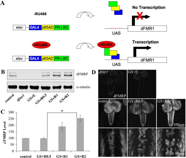
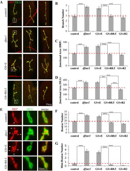
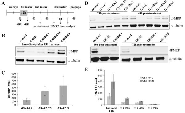
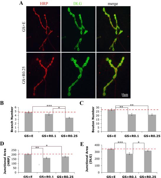
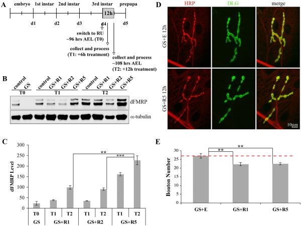
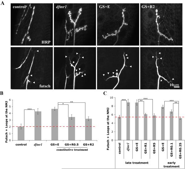
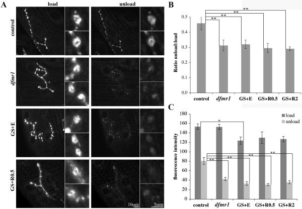
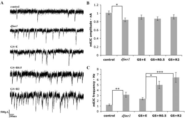
Fragile X syndrome (FraX), caused by the loss-of-function of one gene (FMR1), is the most common inherited form of both mental retardation and autism spectrum disorders. The FMR1 product (FMRP) is an mRNA-binding translation regulator that mediates activity-dependent control of synaptic structure and function. To develop any FraX intervention strategy, it is essential to define when and where FMRP loss causes the manifestation of synaptic defects, and whether the reintroduction of FMRP can restore normal synapse properties. In the Drosophila FraX model, dFMRP loss causes neuromuscular junction (NMJ) synapse over-elaboration (overgrowth, overbranching, excess synaptic boutons), accumulation of development-arrested satellite boutons, and altered neurotransmission. We used the Gene-Switch method to conditionally drive dFMRP expression to define the spatiotemporal requirements in synaptic mechanisms. Constitutive induction of targeted neuronal dFMRP at wild-type levels rescues all synaptic architectural defects in Drosophila Fmr1 (dfmr1)-null mutants, demonstrating a presynaptic requirement for synapse structuring. By contrast, presynaptic dFMRP expression does not ameliorate functional neurotransmission defects, indicating a postsynaptic dFMRP requirement. Strikingly, targeted early induction of dFMRP effects nearly complete rescue of synaptic structure defects, showing a primarily early-development role. In addition, acute dFMRP expression at maturity partially alleviates dfmr1-null defects, although rescue is not as complete as either early or constitutive dFMRP expression, showing a modest capacity for late-stage structural plasticity. We conclude that dFMRP predominantly acts early in synaptogenesis to modulate architecture, but that late dFMRP introduction at maturity can weakly compensate for early absence of dFMRP function.


Figures

Figure 1
Figure 1. Gene-Switch system drives targeted dFMRP expression in neurons
A) Cartoon of Gene-Switch system. The GAL4 DNA-binding domain is fused to the p65 activation domain and a mutated progesterone receptor ligand-binding domain. In the absence of RU486, the Gene-Switch is “off.” In the presence of RU486, the hormone-responsive GAL4 drives dFMRP transcription downstream of the UAS regulatory sequence. This approach allows spatial and temporal control of dFMRP expression in the dfmr1 null background. B) Western blot of isolated 3rd instar CNS. Genotype as indicated: w1118 (control), homozygous dfmr150M null allele (dfmr1) and dfmr150M, elav-GSG-301/dfmr150M, UAS-9557-3 (GS). Treatment as indicated: GS fed ethanol vehicle (GS+E) and RU486 at X μg/mL (GS+RX). Blot probed for dFMRP and α-tubulin, illustrating RU486 dosage-responsiveness. C) Quantification of western blot dFMRP levels. Individual band intensities were determined normalized to α-tubulin and expressed as percent of control. Bars indicate mean±s.e.m. Significance level: p<0.05 (*). D) dFMRP immunohistochemistry of wandering 3rd instar CNS. Note RU486 dosage dependence of dFMRP expression.
Figure 2
Figure 2. Targeted presynaptic dFMRP rescues all dfmr1 NMJ structure defects
A) Representative images of wandering 3rd instar NMJs in genotypes and treatments shown. NMJs co-labeled for HRP (presynaptic marker) and DLG (postsynaptic marker), with three examples of each condition shown. B-D) Quantification of NMJ synaptic branch number and area, defined for presynaptic area (HRP domain) and postsynaptic area (DLG domain). Null dfmr1 terminals display over-growth and over-elaboration. GS+E phenocopies dfmr1, and complete rescue occurs with RU486 constitutively driving presynaptic dFMRP expression. E) Representative images of mini/satellite boutons in genotypes and treatments shown. High magnification images of boxed regions in (A) showing mini-boutons (arrowheads) in both dfmr1 and GS+E genotypes and their absence following RU486 induction. F, G) Quantification of normal (>2 μm diameter) and mini-bouton (<2 μm diameter, attached to a normal bouton) number per NMJ in genotypes and treatments shown. n=12 animals for all conditions. Bars indicate mean±s.e.m. Dashed red lines highlight control level quantifications. Significance levels are represented as p<0.01 (**) and p<0.001 (***).
Figure 3
Figure 3. Temporal control of dFMRP expression by acute early RU486 treatment
A) Depiction of time line employed for early larval induction of dFMRP indicating points of dFMRP protein analyses. B) Representative western blot of 1st instar CNS probed for dFMRP and α-tubulin. Samples were taken immediately after 12 hour treatment with RU486 as indicated. C) Quantification of western blot dFMRP levels. Individual band intensities were determined normalized to α-tubulin and expressed as percent of control. Bars indicate mean±s.e.m. D) Representative western blots of 2nd and 3rd instar CNS. dFMRP levels progressively diminish and are minimally detectable 60 hours post-treatment. E) Quantification of dFMRP levels as a function of age. Individual band intensities were determined normalized to α-tubulin and expressed as percent of control. Bars indicate mean±s.e.m.
Figure 4
Figure 4. Early presynaptic dFMRP rescues dfmr1 NMJ architectural phenotypes
A) Representative images showing NMJs from animals either vehicle- (GS+E) or experimentally treated (GS+RU486 at 0.25 mg/mL, RU+0.25) for 12 hours immediately post-hatching and then analyzed as 3rd instars (108 hours AEL), co-labeled with HRP and DLG. B-E) Quantification of NMJ structure. Statistically significant rescue occurs for synaptic branch number (B), synaptic bouton number (C), presynaptic junctional area (D), and postsynaptic junction area (E). n=12-14 animals for each condition. Bars indicate mean±s.e.m. Dashed red lines highlight dfmr1 null conditions. Significance levels are represented as p<0.05 (*); p<0.01 (**); p<0.001 (***).
Figure 5
Figure 5. Acute late presynaptic dFMRP partially rescues dfmr1 NMJ structure
A) Depiction of time line employed for late larval stage intervention. Larvae were raised to 96 hours AEL, transferred to RU486-containing food for 12 hours, and then immediately processed. B) Representative western blot of 3rd instar CNS probed for dFMRP and α-tubulin. C) Quantification of western blot dFMRP levels. Individual band intensities were determined normalized to α-tubulin and expressed as percent of control. Bars indicate mean±s.e.m. Significance levels are represented as p<0.01 (**) and p<0.001 (***). D) Representative NMJ images from animals treated during the late 12 hour time period, co-labeled with HRP and DLG. Acute dFMRP expression reduces excess number of boutons observed in the dfmr1 null. E) Quantification of synaptic bouton number. Late 12 hour treatment with RU486 to induce dFMRP effects partial rescue of NMJ structural alterations. n=10-12 animals for each condition. Bars indicate mean±s.e.m. Dashed red line highlights the dfmr1 null condition. Significance levels are represented as p<0.01 (**).
Figure 6
Figure 6. Targeted presynaptic dFMRP rescue dfmr1 NMJ cytoskeletal defects
A) Representative images of NMJs from wandering 3rd instars probed for HRP and Futsch/MAP1B. GS animals were constitutively fed either EtOH vehicle or RU486 at 2 μg/mL. Note the increased number of Futsch loops present throughout the dfmr1 null synaptic terminals. B) Quantification of Futsch loops. In wandering 3rd instars, constitutive treatment with RU486 partially rescues the cytoskeletal alteration of null mutant. n=11-13 animals for each condition. C) Quantification of futsch loops after either early and late 12 hour treatment with RU486 (taken at 108 hours AEL). Age-matched control and dfmr1 nulls are presented. n=10-15 animals for each condition. Bars indicate mean±s.e.m. Dashed red line highlights control level quantifications. Significance levels are represented as p<0.05 (*); p<0.01 (**); p<0.001(***).
Figure 7
Figure 7. Constitutive presynaptic dFMRP does not rescue dfmr1 FM1-43 function defect
A) Representative images of wandering 3rd instar NMJs loaded with FM1-43 and then subsequently unloaded with high K+ depolarizing saline. Note the elevated relative fluorescence retained in control versus dfmr1 null and GS mutants. Representative synaptic boutons shown at higher magnification. B) Quantification of the FM1-43 unload:load fluorescence intensity ratio. Sample sizes: n=8 each control and dfmr1; n=13 GS+E, n=6 GS+R0.5, and n=9 GS+RU2. C) Quantification of average fluorescence intensity per bouton in loaded and unloaded conditions. Sample sizes: n=5 for each control, dfmr1, GS+E, GS+R0.5, and GS+RU2. Bars indicate mean±s.e.m. Significance levels are represented as p<0.05 (*), p<0.01 (**) and p<0.01 (**).
Figure 8
Figure 8. Constitutive presynaptic dFMRP does not rescue dfmr1 mEJC function defect
A) Sample mEJC traces from wandering 3rd instar NMJs showing 3 seconds of recording from control, dfmr1, GS+E, GS+R0.5, and GS+RU2. Note the increased number of events in dfmr1 null compared to control, and further increase upon dFMRP induction. B) Quantification of mEJC peak amplitude. C) Quantification of mEJC frequency. Bars indicate mean±s.e.m; n=10 for each category. Significance levels represented as p<0.05 (*); p<0.01 (**); p<0.001 (***).

Similar articles

Cited by

References

    1. Antar LN, Afroz R, Dictenberg JB, Carroll RC, Bassell GJ. Metabotropic glutamate receptor activation regulates fragile x mental retardation protein and FMR1 mRNA localization differentially in dendrites and at synapses. J Neurosci. 2004;24:2648–55. - PMC - PubMed
    1. Ashley J, Packard M, Ataman B, Budnik V. Fasciclin II signals new synapse formation through amyloid precursor protein and the scaffolding protein dX11/Mint. J Neurosci. 2005;25:5943–55. - PMC - PubMed
    1. Bailey DB, Jr., Hatton DD, Skinner M, Mesibov G. Autistic behavior, FMR1 protein, and developmental trajectories in young males with fragile X syndrome. J Autism Dev Disord. 2001a;31:165–74. - PubMed
    1. Bailey DB, Jr., Hatton DD, Tassone F, Skinner M, Taylor AK. Variability in FMRP and early development in males with fragile X syndrome. Am J Ment Retard. 2001b;106:16–27. - PubMed
    1. Bear MF, Huber KM, Warren ST. The mGluR theory of fragile X mental retardation. Trends Neurosci. 2004;27:370–7. - PubMed

Publication types

MeSH terms

Substances

LinkOut - more resources