. 2008 Sep;82(17):8316-29.
doi: 10.1128/JVI.00665-08. Epub 2008 Jun 25.

CD81 is a central regulator of cellular events required for hepatitis C virus infection of human hepatocytes

Affiliations

CD81 is a central regulator of cellular events required for hepatitis C virus infection of human hepatocytes

Michela Brazzoli et al. J Virol. 2008 Sep.

Abstract

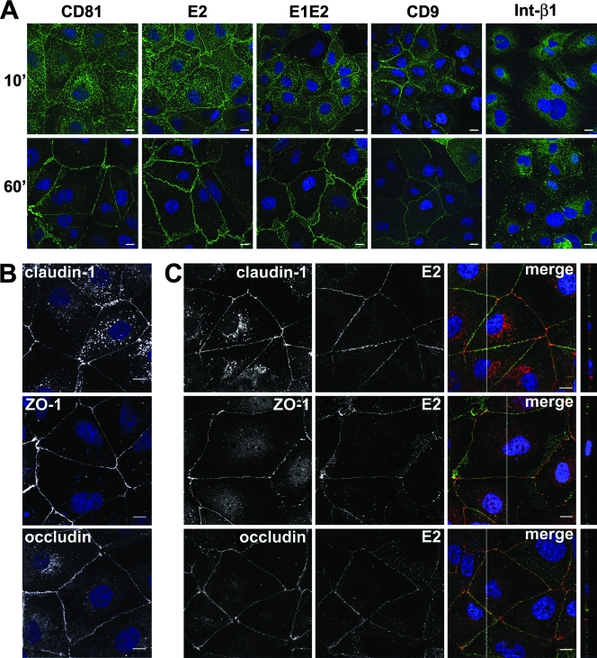
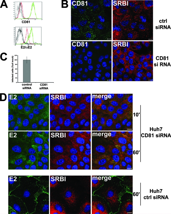
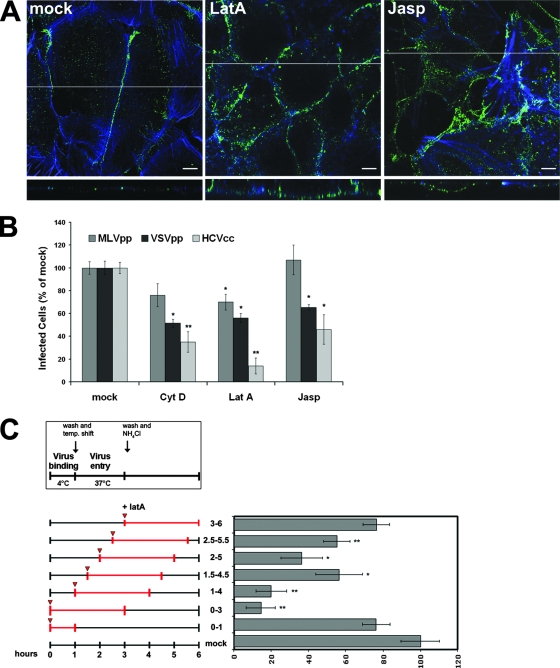
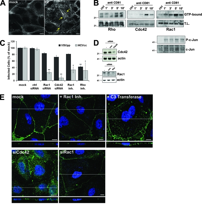
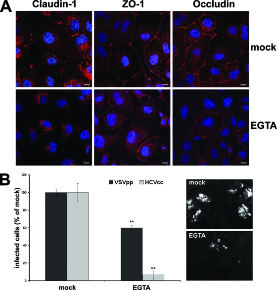
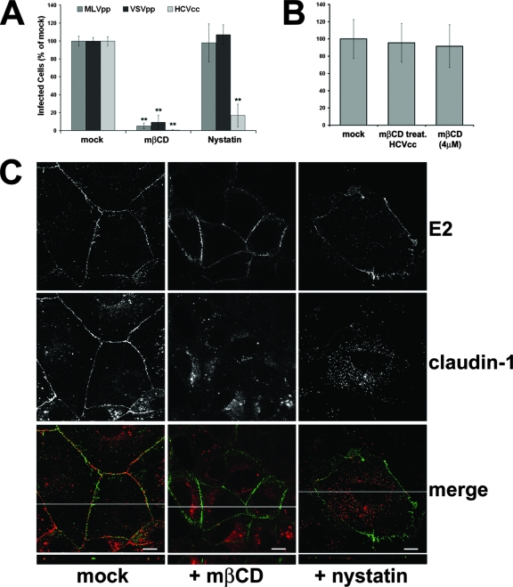
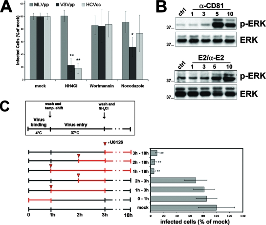
Infection with hepatitis C virus (HCV) is still a major public health problem, and the events leading to hepatocyte infection are not yet fully understood. Combining confocal microscopy with biochemical analysis and studies of infection requirements using pharmacological inhibitors and small interfering RNAs, we show here that engagement of CD81 activates the Rho GTPase family members Rac, Rho, and Cdc42 and that the block of these signaling pathways drastically reduces HCV infectivity. Activation of Rho GTPases mediates actin-dependent relocalization of the HCV E2/CD81 complex to cell-cell contact areas where CD81 comes into contact with the tight-junction proteins occludin, ZO-1, and claudin-1, which was recently described as an HCV coreceptor. Finally, we show that CD81 engagement activates the Raf/MEK/ERK signaling cascade and that this pathway affects postentry events of the virus life cycle. In conclusion, we describe a range of cellular events that are manipulated by HCV to coordinate interactions with its multiple coreceptors and to establish productive infections and find that CD81 is a central regulator of these events.


Figures

FIG. 1.
FIG. 1.
Upon engagement, CD81 relocalizes to areas of cell-cell contact. (A) Huh-7 monolayers grown on polylysine-coated slides were incubated with an anti-CD81 MAb (JS-81), recombinant HCV E1E2 or HCV E2 glycoprotein plus anti-E2 MAb 291, the anti-β1 integrin MAb, or the anti-CD9 MAb for the indicated time (minutes) at 37°C. Cells were then fixed and stained with an anti-mouse-Alexa Fluor 488 secondary antibody (green). Nuclei were visualized with To-Pro3 iodide (blue). The samples were then examined by confocal microscopy. The images provided represent one z section from deconvolved z stack images. Bars, 10 μm. (B) Nonstimulated Huh-7 monolayers were fixed, permeabilized, and stained with rabbit polyclonal antibodies against occludin, claudin-1, and ZO-1. (C) rE2 and anti-E2 MAb 291 were allowed to bind to Huh-7 cells at 37°C for 60 min. Cells were then fixed, permeabilized, and stained with rabbit polyclonal antibodies against occludin, claudin-1, and ZO-1, followed by a rabbit-Alexa Fluor 568 secondary antibody (red) and an anti-mouse-Alexa Fluor 488 secondary antibody (green), as indicated. Areas of colocalization appear yellow. In merged confocal images, the white line indicates the position of the y-z cross section. The images provided are one z section from deconvolved z stack images. Bars, 10 μm.
FIG. 2.
FIG. 2.
Silencing of CD81 reduces HCV E2 binding to Huh-7 cells, abolishes its relocalization, and greatly impairs susceptibility to HCV infection. (A) Flow cytometry analysis. Huh-7 cells transfected with control (green line) or CD81 (red line) siRNA were incubated with anti-CD81 MAb or with E2 glycoprotein plus anti-E2 MAb and then treated with a fluorescein isothiocyanate-conjugate secondary antibody. Black lines indicate the isotype control. (B) Immunofluorescence analysis. Huh-7 cells transfected with control (ctrl) or CD81 siRNA were fixed, permeabilized, and stained with an anti-CD81 MAb and an anti-SR-BI rabbit polyclonal antibody. The images provided are one z section from deconvolved z stack images. Bars, 10 μm. (C) Huh-7 cells transfected with control or CD81 siRNA were infected with 100 TCID50 HCVcc. Infected cells were visualized 72 h postinfection by staining with anti-HCV core MAb 3G1-1 plus an anti-mouse-Alexa Fluor 488 secondary antibody and counted under a fluorescence microscope. Infectivity is expressed relative to the values obtained for the control as the mean percentage ± the SD of four replicates in three independent experiments (** P < 0.001). (D) Huh-7 cells transfected with CD81 or control siRNA were incubated with sE2 plus anti-E2 MAb 291 at 37°C for the indicated time (minutes), fixed, permeabilized, and stained with anti-SR-BI rabbit polyclonal antibody, followed by an anti-rabbit-Alexa Fluor 568 secondary antibody (red) and an anti-mouse-Alexa Fluor 488 secondary antibody (green). The images provided represent merged z stacks. Bars, 10 μm.
FIG. 3.
FIG. 3.
An intact actin cytoskeleton is required for CD81 relocalization to the TJ and for HCVcc infection. (A) Huh-7 cells pretreated for 60 min with LatA (1 μM) or jasplakinolide (Jasp; 500 nM) or mock treated (dimethyl sulfoxide) were allowed to bind rE2 plus anti-E2 MAb 291 at 37°C for 60 min in the continuous presence of compounds. Cells were then fixed, permeabilized, and stained with an anti-mouse-Alexa Fluor 488 secondary antibody (green) and phalloidin (blue). The images shown here represent one z section from deconvolved z stack images. The white line indicates the position of the x-z cross section. Bars, 10 μm. (B) Huh-7 cells were mock treated or incubated for 1 h with medium containing 1 μM LatA, 5 μM cytochalasin D (Cyt D), or 500 nM Jasp and infected with different viruses for 3 h in the presence or absence of the drugs. After washing, cells were left for 72 h in medium alone and then fixed, permeabilized, and stained with anti-HCV core MAb 3G1-1. Positive cells were counted under a fluorescence microscope. Infectivity is expressed relative to the values obtained in the absence of the drugs as the mean percentage ± the SD of four replicates in three independent experiments (*, P < 0.05; **, P < 0.001). As a control, infections with retroviral pseudoparticles bearing VSV-G or Abelson murine leukemia virus envelope proteins were performed under the same conditions and the level of infectivity was determined by FACS analysis of GFP expression. (C) Kinetics of LatA inhibitory activity. Virus was allowed to bind to cells for 1 h at 4°C in the absence or presence of LatA (1 μM). Subsequently, cells were washed and shifted to 37°C to allow entry. As indicated in the scheme, LatA was added directly or 30, 60, 90, or 120 min after the temperature (temp.) shift and left for 3 h thereafter. NH4Cl was added 2 h after warming to block further infection. Efficiency of infection was determined 72 h later as the percentage of HCV core-positive cells relative to that of control infections performed in the same way but always without the drug. Mean values and SD of four replicates in three independent experiments are given (*, P < 0.05; **, P < 0.001).
FIG. 4.
FIG. 4.
CD81 triggers the Rho family GTPases Rho, Rac, and Cdc42. (A) Phalloidin staining of the actin cytoskeleton in Huh-7 cells mock treated or exposed to rE2 (10 μg/ml) plus anti-E2 MAb 291 (10 μg/ml) for 60 min at 37°C. Yellow arrows indicate actin stress fibers. (B) Huh-7 cells were incubated with an anti-CD81 MAb or an isotype control (ctrl) and then cross-linked with GαM for the indicated times (minutes) at 37°C (5 min for the isotype control). After lysis, GTP-bound Rho, Cdc42, or Rac was affinity precipitated and immunoblotted with specific antibodies as described in Materials and Methods. Whole-cell lysate (T.L.) was also blotted to evaluate total proteins and the level of phosho-c-Jun (P-c-Jun) and total c-Jun. (C) Mock-treated Huh-7 cells or cells treated with Rac1 inhibitor (inh.) NSC23766 or C3 exoenzyme or transfected with Cdc42, Rac, or control siRNA were infected with 100 TCID50 HCVcc for 3 h in the presence or absence of the drugs. Cells were then left in medium alone for an additional 72 h before fixation and staining with anti-HCV core MAb 3G1-1. Positive cells were counted under a fluorescence microscope. Infectivity is expressed relative to the values obtained in the absence of the drugs as the mean percentage ± the SD of four replicates of three independent experiments (*, P < 0.05; **, P < 0.001). As a control, infections with retroviral pseudoparticles bearing VSV-G were performed under the same conditions and the level of infectivity was determined by FACS analysis of GFP expression. (D) Lysates of Huh-7 cells transfected with control (con), Cdc42, and Rac siRNAs or not transfected were immunoblotted with Cdc42-, Rac-, and actin-specific antibodies. (E) Huh-7 cells transfected with Cdc42 or Rac1 siRNA or treated with the Rac1 inhibitor NSC23766 or the C3 exoenzyme or mock treated were exposed to E2 plus anti-E2 for 1 h at 37°C and then fixed, permeabilized, and stained with anti-mouse-Alexa Fluor 488 secondary antibody (green). Images shown here represent one z section from deconvolved z stack images. The white line indicates the position of the x-z cross section. Nuclei were stained in blue with To-Pro3 iodide. Bars, 10 μm. The values to the left of the gels in panels B and D are molecular sizes in kilodaltons.
FIG. 5.
FIG. 5.
Disruption of TJs inhibits viral entry. (A) Confluent Huh-7 cells were mock treated or exposed to 2.5 mM EGTA for 3 h, followed by fixation, permeabilization, and immunofluorescent staining of endogenous claudin-1, ZO-1, and occludin. (B) Untreated Huh-7 cells or cells treated for 3 h with 2.5 mM EGTA were infected with 100 TCID50 HCVcc for 2 h in the presence or absence of EGTA. Cells were then washed and left in medium alone for an additional 72 h before fixation and staining with the anti-HCV core MAb 3G1-1. Positive cells or foci were counted under a fluorescence microscope. Infectivity is expressed relative to the values obtained in the absence of the drugs as the mean percentage ± the SD of four replicates (**, P < 0.001). As a control, infection with retroviral pseudoparticles bearing the VSV-G envelope protein was performed under the same conditions and the level of infectivity was determined by FACS analysis of GFP expression.
FIG. 6.
FIG. 6.
Effects of cholesterol-depleting drugs. (A) Huh-7 cells were mock treated or incubated for 1 h with medium containing MβCD or nystatin and infected with different viruses for 3 h in the presence or absence of the drugs. Cells were then washed and left for 72 h in the presence of medium alone. Infectivity was determined by counting HCV core-positive cells under a fluorescence microscope. Infectivity is expressed relative to the values obtained in the absence of the drugs as the mean percentage ± the SD of four replicates in three different experiments (**, P < 0.001). As a control, infections with retroviral pseudoparticles bearing VSV-G or Abelson murine leukemia virus envelope proteins were performed as described for Fig. 3B. (B) HCVcc particles were pretreated for 1 h at 4°C with 1 mM MβCD or mock treated; virus was then diluted in fresh medium to reach an MβCD concentration of 4 μM when viral particles were put on cells. As a control, an aliquot of mock-treated HCV particles was diluted to 4 μM MβCD just before infection. Infectivity was determined by counting HCV core-positive cells under a fluorescence microscope and is expressed relative to the values obtained in the absence of the drugs as the mean percentage ± the SD of four replicates in three independent experiments. (C) Huh-7 cells treated with MβCD or nystatin or mock treated were exposed to rE2 plus anti-E2 for 1 h at 37°C and then stained for E2 (green) and claudin-1 (red). In merged confocal images, areas of colocalization appear yellow. The images provided represent one z section from deconvolved z stack images. The white line in each merged image indicates the position of the x-z cross section. Bars, 10 μm.
FIG. 7.
FIG. 7.
Postentry requirements for HCV infection. (A) Huh-7 cells were mock treated or treated for 1 h with medium containing NH4Cl (10 mM), wortmannin (1 μM), or nocodazole (1 μM) and infected with different viruses for 3 h in the presence or absence of the drugs. Cells were then washed and left for 72 h in medium alone. Infectivity was determined by counting HCV core-positive cells under a fluorescence microscope. Infectivity is expressed relative to the values obtained in the absence of the drugs as the mean percentage ± the SD of four replicates in three independent experiments (*, P < 0.05; **, P < 0.001). As a control, infections with retroviral pseudoparticles bearing VSV-G or Abelson murine leukemia virus envelope proteins were performed as described for Fig. 3B. (B) Huh-7 cells were incubated with anti-CD81, rE2 plus anti-E2 MAb, or an isotype control for 5 min at 37°C and then cross-linked with GαM for the indicated times (minutes) at 37°C (5 min for the isotype control). Lysates were immunoblotted with anti-phospho-p42/p44 MAPK (p-Erk) antibody, stripped, and then reprobed with a rabbit polyclonal p42/p44 MAPK (Erk) antibody. (C) The Raf/MEK/ERK signaling pathway is required for a postentry event in HCV infection. Virus binding to cells was performed for 1 h at 4°C in the absence or presence of U0126 (1 μM). Subsequently, cells were washed and shifted to 37°C to allow entry. U0126 was added as indicated in the scheme; 2 h after warming, NH4Cl was added to block further infection (virus inactivation). Efficiency of infection was determined 72 h later as the percentage of HCV core-positive cells relative to control infections. Mean values and SD of four replicates in three independent experiments are shown (**, P < 0.001). The values to the left of the gels in panel B are molecular sizes in kilodaltons.

Similar articles

Cited by

References

    1. Agnello, V., G. Abel, M. Elfahal, G. B. Knight, and Q.-X. Zhang. 1999. Hepatitis C virus and other Flaviviridae viruses enter cells via low density lipoprotein receptor. Proc. Natl. Acad. Sci. USA 9612766-12771. - PMC - PubMed
    1. Bartosch, B., A. Vitelli, C. Granier, C. Goujon, J. Dubuisson, S. Pascale, E. Scarselli, R. Cortese, A. Nicosia, and F. L. Cosset. 2003. Cell entry of hepatitis C virus requires a set of co-receptors that include the CD81 tetraspanin and the SR-B1 scavenger receptor. J. Biol. Chem. 27841624-41630. - PubMed
    1. Berditchevski, F. 2001. Complexes of tetraspanins with integrins: more than meets the eye. J. Cell Sci. 1144143-4151. - PubMed
    1. Bernards, A., and J. Settleman. 2004. GAP control: regulating the regulators of small GTPases. Trends Cell Biol. 14377-385. - PubMed
    1. Blanchard, E., S. Belouzard, L. Goueslain, T. Wakita, J. Dubuisson, C. Wychowski, and Y. Rouille. 2006. Hepatitis C virus entry depends on clathrin-mediated endocytosis. J. Virol. 806964-6972. - PMC - PubMed