

The facilitative glucose transporter GLUT3: 20 years of distinction
- PMID: 18577699
- PMCID: PMC2519757
- DOI: 10.1152/ajpendo.90388.2008
The facilitative glucose transporter GLUT3: 20 years of distinction
Abstract
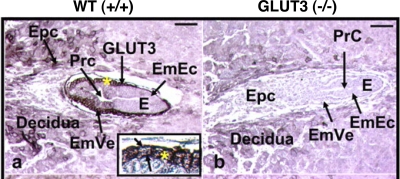
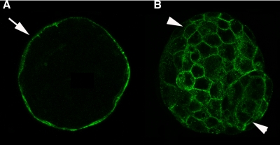
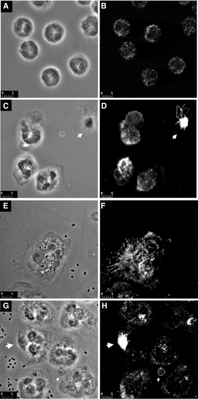
Glucose metabolism is vital to most mammalian cells, and the passage of glucose across cell membranes is facilitated by a family of integral membrane transporter proteins, the GLUTs. There are currently 14 members of the SLC2 family of GLUTs, several of which have been the focus of this series of reviews. The subject of the present review is GLUT3, which, as implied by its name, was the third glucose transporter to be cloned (Kayano T, Fukumoto H, Eddy RL, Fan YS, Byers MG, Shows TB, Bell GI. J Biol Chem 263: 15245-15248, 1988) and was originally designated as the neuronal GLUT. The overriding question that drove the early work on GLUT3 was why would neurons need a separate glucose transporter isoform? What is it about GLUT3 that specifically suits the needs of the highly metabolic and oxidative neuron with its high glucose demand? More recently, GLUT3 has been studied in other cell types with quite specific requirements for glucose, including sperm, preimplantation embryos, circulating white blood cells, and an array of carcinoma cell lines. The last are sufficiently varied and numerous to warrant a review of their own and will not be discussed here. However, for each of these cases, the same questions apply. Thus, the objective of this review is to discuss the properties and tissue and cellular localization of GLUT3 as well as the features of expression, function, and regulation that distinguish it from the rest of its family and make it uniquely suited as the mediator of glucose delivery to these specific cells.
Figures
Similar articles
-
GLUT3 inhibitor discovery through in silico ligand screening and in vivo validation in eukaryotic expression systems.Sci Rep. 2022 Jan 26;12(1):1429. doi: 10.1038/s41598-022-05383-9. Sci Rep. 2022. PMID: 35082341 Free PMC article.
-
Glucose transporter 3 in neuronal glucose metabolism: Health and diseases.Metabolism. 2021 Oct;123:154869. doi: 10.1016/j.metabol.2021.154869. Epub 2021 Aug 21. Metabolism. 2021. PMID: 34425073 Review.
-
Neurodevelopment Is Dependent on Maternal Diet: Placenta and Brain Glucose Transporters GLUT1 and GLUT3.Nutrients. 2024 Jul 21;16(14):2363. doi: 10.3390/nu16142363. Nutrients. 2024. PMID: 39064806 Free PMC article. Review.
-
Developmental regulation of glucose transporters GLUT3, GLUT4 and GLUT8 in the mouse cerebellar cortex.J Anat. 2010 Nov;217(5):616-23. doi: 10.1111/j.1469-7580.2010.01291.x. Epub 2010 Aug 30. J Anat. 2010. PMID: 20819112 Free PMC article.
-
Insulin regulates neuronal glucose uptake by promoting translocation of glucose transporter GLUT3.Exp Neurol. 2006 Mar;198(1):48-53. doi: 10.1016/j.expneurol.2005.10.035. Epub 2005 Dec 9. Exp Neurol. 2006. PMID: 16337941
Cited by
-
SLC2A3 promotes macrophage infiltration by glycolysis reprogramming in gastric cancer.Cancer Cell Int. 2020 Oct 12;20:503. doi: 10.1186/s12935-020-01599-9. eCollection 2020. Cancer Cell Int. 2020. PMID: 33061855 Free PMC article.
-
Glucose transporters in brain in health and disease.Pflugers Arch. 2020 Sep;472(9):1299-1343. doi: 10.1007/s00424-020-02441-x. Epub 2020 Aug 13. Pflugers Arch. 2020. PMID: 32789766 Free PMC article. Review.
-
Hepatic expression and cellular distribution of the glucose transporter family.World J Gastroenterol. 2012 Dec 14;18(46):6771-81. doi: 10.3748/wjg.v18.i46.6771. World J Gastroenterol. 2012. PMID: 23239915 Free PMC article. Review.
-
Integrative landscape of dysregulated signaling pathways of clinically distinct pancreatic cancer subtypes.Oncotarget. 2018 Jun 26;9(49):29123-29139. doi: 10.18632/oncotarget.25632. eCollection 2018 Jun 26. Oncotarget. 2018. PMID: 30018740 Free PMC article.
-
Emerging Roles in the Biogenesis of Cytochrome c Oxidase for Members of the Mitochondrial Carrier Family.Front Cell Dev Biol. 2019 Jan 31;7:3. doi: 10.3389/fcell.2019.00003. eCollection 2019. Front Cell Dev Biol. 2019. PMID: 30766870 Free PMC article. Review.
References
-
- Aghajanian GK, Bloom FE. The formation of synaptic junctions in developing rat brain: a quantitative electron microscopic study. Brain Res 6: 716–727, 1967. - PubMed
-
- Akkerman JW, Holmsen H. Interrelationships among platelet responses: studies on the burst in proton liberation, lactate production, and oxygen uptake during platelet aggregation and Ca2+ secretion. Blood 57: 956–966, 1981. - PubMed
-
- Akkerman JWN Platelet Responses and Metabolism, edited by Holmsen H. Boca Raton, FL: CRC, 1987, p. 190–206.
-
- Angulo C, Rauch MC, Droppelmann A, Reyes AM, Slebe JC, Delgado-Lopez F, Guaiquil VH, Vera JC, Concha II. Hexose transporter expression and function in mammalian spermatozoa: cellular localization and transport of hexoses and vitamin C. J Cell Biochem 71: 189–203, 1998. - PubMed
-
- Apelt J, Mehlhorn G, Schliebs R. Insulin-sensitive GLUT4 glucose transporters are colocalized with GLUT3-expressing cells and demonstrate a chemically distinct neuron-specific localization in rat brain. J Neurosci Res 57: 693–705, 1999. - PubMed
Publication types
MeSH terms
Substances
Grants and funding
LinkOut - more resources
Full Text Sources
Other Literature Sources