Differential expression of GADD45beta in normal and osteoarthritic cartilage: potential role in homeostasis of articular chondrocytes
- PMID: 18576389
- PMCID: PMC3950332
- DOI: 10.1002/art.23504
Differential expression of GADD45beta in normal and osteoarthritic cartilage: potential role in homeostasis of articular chondrocytes
Abstract
Objective: Our previous study suggested that growth arrest and DNA damage-inducible protein 45beta (GADD45beta) prolonged the survival of hypertrophic chondrocytes in the developing mouse embryo. This study was undertaken, therefore, to investigate whether GADD45beta plays a role in adult articular cartilage.
Methods: Gene expression profiles of cartilage from patients with late-stage osteoarthritis (OA) were compared with those from patients with early OA and normal controls in 2 separate microarray analyses. Histologic features of cartilage were graded using the Mankin scale, and GADD45beta was localized by immunohistochemistry. Human chondrocytes were transduced with small interfering RNA (siRNA)-GADD45beta or GADD45beta-FLAG. GADD45beta and COL2A1 messenger RNA (mRNA) levels were analyzed by real-time reverse transcriptase-polymerase chain reaction, and promoter activities were analyzed by transient transfection. Cell death was detected by Hoechst 33342 staining of condensed chromatin.
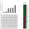
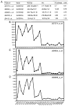
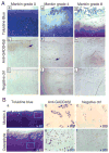
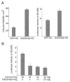
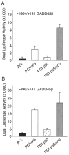
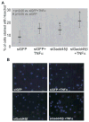
Results: GADD45beta was expressed at higher levels in cartilage from normal donors and patients with early OA than in cartilage from patients with late-stage OA. All chondrocyte nuclei in normal cartilage immunostained for GADD45beta. In early OA cartilage, GADD45beta was distributed variably in chondrocyte clusters, in middle and deep zone cells, and in osteophytes. In contrast, COL2A1, other collagen genes, and factors associated with skeletal development were up-regulated in late OA, compared with early OA or normal cartilage. In overexpression and knockdown experiments, GADD45beta down-regulated COL2A1 mRNA and promoter activity. NF-kappaB overexpression increased GADD45beta promoter activity, and siRNA-GADD45beta decreased cell survival per se and enhanced tumor necrosis factor alpha-induced cell death in human articular chondrocytes.
Conclusion: These observations suggest that GADD45beta might play an important role in regulating chondrocyte homeostasis by modulating collagen gene expression and promoting cell survival in normal adult cartilage and in early OA.
Figures
Similar articles
-
CCAAT/enhancer binding protein β (C/EBPβ) regulates the transcription of growth arrest and DNA damage-inducible protein 45 β (GADD45β) in articular chondrocytes.Pathol Res Pract. 2016 Apr;212(4):302-9. doi: 10.1016/j.prp.2016.01.009. Epub 2016 Jan 27. Pathol Res Pract. 2016. PMID: 26896926
-
Senescence of chondrocytes in aging articular cartilage: GADD45β mediates p21 expression in association with C/EBPβ in senescence-accelerated mice.Pathol Res Pract. 2011 Apr 15;207(4):225-31. doi: 10.1016/j.prp.2011.01.007. Epub 2011 Feb 24. Pathol Res Pract. 2011. PMID: 21353395 Free PMC article.
-
A novel role for GADD45beta as a mediator of MMP-13 gene expression during chondrocyte terminal differentiation.J Biol Chem. 2005 Nov 18;280(46):38544-55. doi: 10.1074/jbc.M504202200. Epub 2005 Sep 2. J Biol Chem. 2005. PMID: 16144844 Free PMC article.
-
Reactive oxygen species, aging and articular cartilage homeostasis.Free Radic Biol Med. 2019 Feb 20;132:73-82. doi: 10.1016/j.freeradbiomed.2018.08.038. Epub 2018 Aug 31. Free Radic Biol Med. 2019. PMID: 30176344 Free PMC article. Review.
-
Epigenetic Mechanisms Underlying the Aging of Articular Cartilage and Osteoarthritis.Gerontology. 2019;65(4):387-396. doi: 10.1159/000496688. Epub 2019 Apr 10. Gerontology. 2019. PMID: 30970348 Free PMC article. Review.
Cited by
-
IKKα/CHUK regulates extracellular matrix remodeling independent of its kinase activity to facilitate articular chondrocyte differentiation.PLoS One. 2013 Sep 2;8(9):e73024. doi: 10.1371/journal.pone.0073024. eCollection 2013. PLoS One. 2013. PMID: 24023802 Free PMC article.
-
MiR-146b is down-regulated during the chondrogenic differentiation of human bone marrow derived skeletal stem cells and up-regulated in osteoarthritis.Sci Rep. 2017 Apr 24;7:46704. doi: 10.1038/srep46704. Sci Rep. 2017. PMID: 28436462 Free PMC article.
-
Dual regulation of metalloproteinase expression in chondrocytes by Wnt-1-inducible signaling pathway protein 3/CCN6.Arthritis Rheum. 2012 Jul;64(7):2289-99. doi: 10.1002/art.34411. Arthritis Rheum. 2012. PMID: 22294415 Free PMC article.
-
Roles of inflammatory and anabolic cytokines in cartilage metabolism: signals and multiple effectors converge upon MMP-13 regulation in osteoarthritis.Eur Cell Mater. 2011 Feb 24;21:202-20. doi: 10.22203/ecm.v021a16. Eur Cell Mater. 2011. PMID: 21351054 Free PMC article.
-
The essential anti-angiogenic strategies in cartilage engineering and osteoarthritic cartilage repair.Cell Mol Life Sci. 2022 Jan 14;79(1):71. doi: 10.1007/s00018-021-04105-0. Cell Mol Life Sci. 2022. PMID: 35029764 Free PMC article. Review.
References
-
- Hunter DJ, Zhang Y, Niu J, Tu X, Amin S, Goggins J, et al. Structural factors associated with malalignment in knee osteoarthritis: the Boston osteoarthritis knee study. J Rheumatol. 2005;32:2192–9. - PubMed
-
- Alexopoulos LG, Williams GM, Upton ML, Setton LA, Guilak F. Osteoarthritic changes in the biphasic mechanical properties of the chondrocyte pericellular matrix in articular cartilage. J Biomech. 2005;38:509–17. - PubMed
-
- Pritzker KP, Gay S, Jimenez SA, Ostergaard K, Pelletier JP, Revell PA, et al. Osteoarthritis cartilage histopathology: grading and staging. Osteoarthritis Cartilage. 2006;14:13–29. - PubMed
-
- Bau B, Gebhard PM, Haag J, Knorr T, Bartnik E, Aigner T. Relative messenger RNA expression profiling of collagenases and aggrecanases in human articular chondrocytes in vivo and in vitro. Arthritis Rheum. 2002;46:2648–57. - PubMed
-
- Hermansson M, Sawaji Y, Bolton M, Alexander S, Wallace A, Begum S, et al. Proteomic analysis of articular cartilage shows increased type II collagen synthesis in osteoarthritis and expression of inhibin βA (activin A), a regulatory molecule for chondrocytes. J Biol Chem. 2004;279:43514–21. - PubMed
Publication types
MeSH terms
Substances
Grants and funding
LinkOut - more resources
Full Text Sources
Other Literature Sources
Medical

