. 2008 Jun 20;30(6):678-88.
doi: 10.1016/j.molcel.2008.06.001.

JNK1-mediated phosphorylation of Bcl-2 regulates starvation-induced autophagy

Affiliations

JNK1-mediated phosphorylation of Bcl-2 regulates starvation-induced autophagy

Yongjie Wei et al. Mol Cell. .

Abstract

Starvation induces autophagy to preserve cellular homeostasis in virtually all eukaryotic organisms. However, the mechanisms by which starvation induces autophagy are not completely understood. In mammalian cells, the antiapoptotic protein, Bcl-2, binds to Beclin 1 during nonstarvation conditions and inhibits its autophagy function. Here we show that starvation induces phosphorylation of cellular Bcl-2 at residues T69, S70, and S87 of the nonstructured loop; Bcl-2 dissociation from Beclin 1; and autophagy activation. In contrast, viral Bcl-2, which lacks the phosphorylation site-containing nonstructured loop, fails to dissociate from Beclin 1 during starvation. Furthermore, the stress-activated signaling molecule, c-Jun N-terminal protein kinase 1 (JNK1), but not JNK2, mediates starvation-induced Bcl-2 phosphorylation, Bcl-2 dissociation from Beclin 1, and autophagy activation. Together, our findings demonstrate that JNK1-mediated multisite phosphorylation of Bcl-2 stimulates starvation-induced autophagy by disrupting the Bcl-2/Beclin 1 complex. These findings define a mechanism that cells use to regulate autophagic activity in response to nutrient status.


Figures
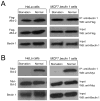
Figure 1
Figure 1. Starvation Regulates the Interaction between Cellular but not Viral Bcl-2 and Beclin 1
(A) Co-immunoprecipitation of Flag epitope-tagged KSHV vBcl-2 with Beclin 1 in HeLa cells or MCF7.beclin 1 cells transfected with a plasmid expressing Flag-vBcl-2, and grown either in normal media (normal) or in HBSS for four hours (starvation). (B) Co-immunoprecipitation of Myc epitope-tagged cellular Bcl-2 with Beclin 1 in HeLa cells or MCF7.beclin 1 cells transfected with a plasmid expressing Myc-Bcl-2, and grown either in normal media (normal) or in HBSS for four hours (starvation).
Figure 2
Figure 2. Starvation Stimulates Bcl-2 Multi-Site Phosphorylation
(A) Structure-based sequence alignment of human Bcl-2 and KSHV vBcl-2. Residue numbers for each sequence are indicated in alignment block. Yellow, pink and red backgrounds indicate increasing sequence conservation, with identical residues highlighted in bold white letter. α-helices and coil secondary structures, color-coded by BH domain are indicated above each alignment. Red triangles indicate phosphorylation sites within the non-structured loop of human Bcl-2. (B–C) Detection by P32 metabolic labeling of phosphorylated endogenous Bcl-2 in HeLa cells and MCF7.beclin 1 cells (B) or phosphorylated Bcl-2 in MCF7.beclin 1 cells transfected with wild-type or mutant forms of Myc-Bcl-2 (C). Cells were grown in normal media (starvation -) or HBSS for four hours (starvation +) prior to lysis, immunoprecipitated with anti-Bcl-2 (B) or anti-Myc (C), separated by SDS-PAGE, and subjected to either autoradiography or Western blot analysis with the indicated antibody. (D) Detection of phosphorylated Bcl-2 using a phospho-specific anti-human Bcl-2 (Ser70) antibody (top panel) in MCF7.beclin 1 cells transfected with wild-type or mutant forms of Myc-Bcl-2. Cells were grown in normal media (starvation -) or HBSS for four hours (starvation +) prior to lysis. The faster migration band represents mono-site phosphorylation on serine 70 of Bcl-2 and the slower migration band (arrow) indicates multi-site phosphorylation.
Figure 3
Figure 3. Phosphorylation Site Mutations in Bcl-2 Alter the Regulation of Bcl-2/Beclin 1 Binding
(A and B) Co-immunoprecipitation of Bcl-2 with Beclin 1 in MCF7.beclin 1 cells (A) or HeLa cells (B) transfected with plasmids expressing wild-type or designated mutant Myc-tagged Bcl-2 and grown in normal media (starvation -) or HBSS for four hours (starvation +). (C) Co-immunoprecipitation of chimeric viral/cellular Bcl-2 constructs with Beclin 1 in MCF7.beclin 1 cells transfected with plasmids expressing chimeras with wild-type or indicated mutant cellular Bcl-2 loop inserts and grown either in normal media (starvation -) or in HBSS for four hours (starvation +).
Figure 4
Figure 4. Phosphorylation Site Mutations in Bcl-2 Alter the Regulation of Autophagy
(A) Representative images of GFP-LC3 staining during starvation in MC7.beclin 1 cells co-transfected with GFP-LC3 and indicated plasmid. Arrows denote representative cells containing GFP-LC3 dots (i.e. autophagosomal structures). (B and C) Light microscopic quantitation of autophagy in MCF7.beclin 1 cells co-transfected with GFP-LC3 and plasmid indicated below x axis. In (B) plasmids expressed wild-type or indicated mutant cellular Bcl-2. In (C) plasmids express a chimeric viral/cellular Bcl-2 in which amino acids 24-34 of vBcl-2 are replaced with amino acids 25-95 from wild-type or indicated mutant cellular Bcl-2. Results shown represent mean ± SEM for triplicate samples of greater than 100 cells per sample. Similar results were obtained in three independent experiments.
Figure 5
Figure 5. Endogenous jnk1 is Required for Starvation-Induced Bcl-2 Phosphorylation, Dissociation of the Bcl-2/Beclin 1 Complex, and Autophagy
(A and B) Western blot analysis of active JNK (A) and active P38 (B) in MCF7.beclin 1 cells grown in normal media (starvation -) or in HBSS for four hours (starvation +), Active JNK was detected by immunoprecipitation using a polyclonal goat anti-JNK antibody, followed by Western blot analysis with a rabbit polyclonal Thr183/Tyr185 phosphorylation specific JNK antibody (A, upper panel). Active P38 was detected by a rabbit polyclonal phospho-P38 MAP kinase (Thr180/Tyr182) antibody (B, upper panel). Total JNK and P38 (A–B, lower panels) were detected by rabbit polyclonal JNK and p38 MAP kinase antibody, respectively. (C) Comparison of Beclin 1 co-immunoprecipitation with Bcl-2 (top panel), Bcl-2 phosphorylation (fourth panel) and JNK phosphorylation (fifth panel) in wild-type (WT), jnk1−/− and jnk2−/− MEFS during growth in normal media (starvation -) or in HBSS for four hours (starvation +). Other panels represent the total amount of Beclin 1 (second panel), Bcl-2 (third panel), JNK1 (sixth panel) and JNK2 (bottom panel) detected in cell lysates by Western blot analysis with the indicated antibody. (D) Comparison of p62/SQSTM1 levels in WT, jnk1−/− and jnk2−/− MEFS during growth in normal media (starvation -) or in HBSS for four hours (starvation +). Actin is shown as a loading control.
Figure 6
Figure 6. Constitutively Active and Dominant Negative JNK1 Regulate Bcl-2 Phosphorylation, Bcl-2 Binding to Beclin 1 and Starvation-Induced Autophagy
(A) Effects of constitutively active JNK1 and dominant negative JNK1 on Bcl-2 phosphorylation (upper panel) and Bcl-2 co-immunoprecipitation with Beclin 1 (second panel) in MCF7.beclin 1 cells during growth in normal media (starvation -) or in HBSS for four hours (starvation +). Lower panels represent total amount of Beclin 1 (third panel), Bcl-2 (fourth panel), MKK7-JNK1 (fifth panel) or active MKK-JNK1 (MKK-P-JNK1) (sixth panel) detected in cell lysates by Western blot analysis with indicated antibody. (B) Representative images of GFP-LC3 staining in MCF7.beclin 1 cells co-transfected with GFP-LC3 and indicated plasmid. Arrows denote representative cells containing GFP-LC3 dots (i.e. autophagosomal structures). (C and D) Light microscopic quantitation of autophagy in MCF7.beclin 1 cells co-transfected with GFP-LC3 and plasmid(s) indicated below x axis. Results shown represent mean ± SEM for triplicate samples of greater than 100 cells per sample. Similar results were obtained in three independent experiments.
Figure 7
Figure 7. Starvation and JNK1 Regulate Autophagy Through Multi-Site Phosphorylation of the ER-Localized Pool of Bcl-2 and Disruption of the ER Bcl-2/Beclin 1 Complex
(A) Effects of nutrient conditions and constitutively active JNK1 on Bcl-2 phosphorylation (fifth panel) and Beclin 1 co-immunoprecipitation with Bcl-2 (upper panel) in WT Bcl-2 MEFs or AAA Bcl-2 MEFs. Growth in normal media is represented as “starvation-“ and growth in HBSS is represented as “starvation+”. Other panels represent the total amount of Beclin 1 (second panel), Bcl-2 (third panel), or Flag-MKK7-JNK1 (sixth panel) detected in cell lysates by Western blot analysis with the indicated antibody or the total amount of Bcl-2 immunoprecipitated by anti-Bcl-2 (fourth panel). (B) Light microscopic quantitation of autophagy in WT bcl-2 MEFs and AAA Bcl-2 MEFs co-transfected with GFP-LC3 and plasmid indicated below x axis. Solid bars represent growth in normal media and open bars represent growth in HBSS for four hours (nutrient starvation). Results shown represent mean ± SEM for triplicate samples of greater than 100 cells per sample. Similar results were obtained in three independent experiments. (C) Effects of nutrient conditions and constitutively active JNK1 on Bcl-2 phosphorylation (top panel) and Beclin 1 co-immunoprecipitation with Bcl-2 (second panel) in heavy membrane (HM, mitochondria), light membrane (LM, ER) or cytosol (Cyto) fractions in WT Bcl-2 MEFs. Growth in normal media is represented as “starvation-“ and growth in HBSS is represented as “starvation+”. Other panels represent the total amount of Beclin 1 (third panel), Bcl-2 (fourth panel), Flag-MKK7-JNK1 (bottom panel), Calreticulin (ER marker, fifth panel) or Tim 23 (mitochondria marker, sixth panel) detected in cell lysates by Western blot analysis with the indicated antibody.

Similar articles

Cited by

References

    1. Bassik MC, Scorrano L, Oakes SA, Pozzan T, Korsmeyer SJ. Phosphorylation of BCL-2 regulates ER Ca2+ homeostasis and apoptosis. EMBO J. 2004;23:1207–1216. - PMC - PubMed
    1. Blagosklonny MV. Unwinding the loop of Bcl-2 phosphorylation. Leukemia. 2001;15:869–874. - PubMed
    1. Bogoyevitch MA. The isoform-specific functions of the c-Jn N-terminal Kinases (JNKs): differences revealed by gene targeting. BioEssays. 2006;28:923–934. - PubMed
    1. Borsello T, Croquelois K, Hornung JP, Clarke PG. N-methyl-D-aspartate-triggered neuronal death in organotypic hippocampal clutures is endocytic, autophagic and mediated by the c-Jun N-terminal kinase pathway. Eur J Neurosci. 2003;18:473–485. - PubMed
    1. Danial NN, Korsmeyer SJ. Cell death: Critical control points. Cell. 2004;116:205–219. - PubMed

Publication types

MeSH terms